Protist Unrevised Notes B1


Algae and Fungal-like Protists


Characteristics:

  •  Algae are autotrophic protists that have chloroplasts and produce their own carbohydrates by photosynthesis
  • In the past, algae was classified in the plant Kingdom, however, algae lack tissue differentiation and have no true roots, stems, or leaves
  • The reproductive structures of algae also differ from those of plants, because they form gametes in single-celled gametangia, or gamete chambers
  • Often times, algal cells contain pyrenoids, organelles that synthesize and store starch.

Structure:

  • The body portion of an alga is called a thallus; the thallus is usually haploid
  • Four types of algae are recognized: unicellular, colonial, filamentous, and multicellular
    ·        Unicellular algae have a structure that consists of a single cell; most unicellular algae are aquatic organisms that compose the phytoplankton, a population of photosynthetic organisms that forms the foundation of aquatic food chains.
    ·        Colonial algae, such as Volvox, have a structure that consists of groups of cells acting in a coordinated manner.
    ·        Some of the cells in colonial algae become specialized; this allows them to move, feed, and reproduce efficiently.
    ·        Filamentous algae, such as Spirogyra, have a slender, rod-shaped thallus composed of rows of cells joined end to end; other species of filamentous algae have specialized structures that anchor the thallus to the ocean bottom.
    ·        Multinuclear algae often have a large, complex thallus; Macrocystis is among the largest multicellular algae.

Classification
·        Algae are classified into 7 phyla, based on color, type of chlorophyll, form of food-storage substance, and cell wall composition.

Reproduction
·        Many species of algae reproduce sexually and asexually
·    Sexual reproduction in algae is often triggered by environmental stress
·        During asexual reproduction, the algae first absorbs its flagellum, then the haploid cell divides mitotically up to three times, and from two to eight haploid flagellated cells called zoospores develop within the parent cell, lastly, the asexual reproductive cells break out of the parent cell, disperse, and eventually grow to full size.
·        Sexual reproduction begins by haploid cells dividing mitotically to produce either “plus” or “minus” gametes.
·        A plus gamete and a minus gamete come into contact with one another and shed their cell walls, then they fuse and form a diploid zygote, which develops a thick protective wall; this resting stage of a zygote is called a zygospore.
·        A zygospore can withstand bad environmental conditions; during the bad environmental condition, the thick wall opens and the living zoospore emerges.

Reproduction in Multicellular Algae
·        The male unicellular gametangium, called an antheridium, produces sperm and the female unicellular gametangium, called an oogonium, produces an egg.
·        The antheridium releases sperm into the surrounding water, where they swim to the female egg and enter through small spores.
·        After fertilization, the resulting zygote is released from the female egg and forms a thick-walled, resting spore; the diploid undergoes meiosis, forming zoospores that are released into the water; the zoospore settles and divides to form a rootlike holdfast, and the others divide and form a new filament.
·        The leaflike algae Ulva has a sexual reproductive cycle that is characterized by a pattern called alternation of generations; a life cycle that exhibits alternation of generations has two distinct multicellular phases- a haploid, gamete-producing phase called a gametophyte and a diploid, spore-producing phase called a sporophyte.
·        The adult sporophyte has reproductive cells called sporangia, which produce haploid zoospore by meiosis.

Algal-Like Protists

Phylum Chlorophyta
·        The phylum Chlorophyta contains more than 7,000 identified species of organisms called green algae and members of this phylum have an amazing number of forms and reproductive methods and their body structures range from single cells and colonial forms to multicellular filaments and sheets.

Phylum Phaeophyta
·        The phylum Phaeophyta contains 1,500 species of organisms called brown algae; brown algae is mostly marine and plantlike organisms called seaweed’s and kelps, they are common along rocky coasts where ocean water is cool.
·        The brown algae contain chlorophylls a and c and a large amount of pigment called fucoxanthin, which give the algae its brown color.
·        The food brown algae produces are stored as laminarin, a carbohydrate with glucose units that are linked differently than those in starch.
·        All brown algae are multicellular; the largest brown alga is the Macrocystis.
·        The thallus is anchored to the ocean bottom by a rootlike holdfast; the stemlike portion of the alga is called the stipe and the leaflike region, modified to capture sunlight for photosynthesis is called the blade.
·        The cell walls of the Macrocystis contain alginate, an alginic acid that is used in cosmetics and various drugs, as food, and as a stabilizer in most ice creams.

Phylum Rhodophyta
·        The phylum Rhodophyta contains 4,000 species of organisms called red algae.
·        Red algae contain chlorophyll a and pigments called phycobilins, which play an important role in absorbing light for photosynthesis.
·        Phycobilins can absorb the wavelengths of light that penetrate deep into the water; they make it possible for red algae to live in depths where alga pigments cannot survive.
·        Certain species of red algae have cell walls that are coated with a sticky substance called carageenan, which is a polysaccharide.
·        Agar, which is used as a gel-forming base for culturing microbes, is also extracted from the cell wall of red algae.

Bacillariophyta
·        The phylum Bacillariophyta contains 11,500 species of organisms called diatoms.
·        Diatoms are abundant in both freshwater and marine environments; the cell wall, called shells, of the diatoms contains two pieces that fit together like a box; each half is called a valve.
·        Centric diatoms have circular or triangular shells and are most abundant in marine environments.
·        Pennate diatoms have rectangular shells and are most abundant in freshwater ponds and lakes; some pennate diatoms by secreting threads that attach to the surface of the water.
·        Diatoms are an abundant component of phytoplankton and are important producers in freshwater and marine food webs, along with being an essential source of nutrients for microscopic heterotrophs, and they release an abundance of oxygen.
·        When diatoms die their shells sink and accumulate in large numbers, forming a layer of material called diatomaceous earth.

Phylum Dinoflagellata
·      The phylum Dinoflagellata contains 1,100 species of organisms called dinoflagellates.
·        Dinoflagellates are small, usually unicellular organisms, photosynthetic, but a few are colorless and heterotrophic, and they are the major producers of organic matter in marine environments.
·      Photosynthetic dinoflagellates usually have a yellowish green to brown color due to large amounts of pigments called carotenoids and chlorophylls a and c.
·      Some species of dinoflagellates, such as Noctiluca, can produce bioluminescence, a display of sparkling light often seen in the ocean water at night.
·      When other species produce toxins and red pigments that explode, a resulting phenomenon is the red tide.

Phylum Chrysophyta
·        The phylum Chrysophyta contains about 850 species of organisms called golden algae, which live in freshwater, but few are found in marine environments.
·        Most of the species placed in this phylum are some shade of yellow or brown due to the presence of large amounts of carotenoids.
·        Golden algae store much of their surplus energy as oil and are important in the formation of petroleum deposits.

Phylum Euglenophyta
·        The phylum Euglenophyta contains 1,000 species of flagellated unicellular algae called euglenoids.
·        Euglenoids show both plantlike and animal-like characteristics; they are plantlike in that they have chlorophyll and are photosynthetic and they are animal-like in that they lack a cell wall and are highly motile.
·        Euglena is abundant in freshwater, especially in water polluted by excess nutrients.
·        Euglena lacks a cell wall and therefore is able to change its shape as it swims about.

Fungal-like Protists

Slime Molds
·        Slime molds spend half their life in a mobile, amoeba-like feeding form, engulfing organic matter and bacteria, like protozoa.
·        Slime molds produce funguslike reproductive structures, which is why they were once classified as fungi.
·        Slime molds are typically found growing on damp soil, rotting logs, decaying leaves, or other decomposing organic matter in moist areas.
·        During reproduction, slime molds produce a spore-bearing structure called a fruiting body.

Phylum Acrasiomycota
·        The phylum Acrasiomycota comprises about 65 species of cellular slime molds.
·        Cellular slime molds live as individual haploid cells that move about like amoebas; each cell moves as an independent organism, creeping over rotting logs and soil or swimming in fresh water, ingesting bacteria and other food.
·        A pseudoplasmodium is a coordinated colony of individual cells that resembles a slug, and it leaves a slimy trail as it crawls over decaying logs, leaves, and twigs.
·        Eventually a pseudoplasmodium will settle and form a fruiting body where spore will develop, then once the fruiting body breaks open, and the wind disperses the spores to new locations.

Phylum Myxomycota
·        450 species of plasmodial slime molds compose the phylum Myxomycota.
·        During the feeding stage of its life cycle, a plasmodial slime mold is a mass of cytoplasm called a plasmodium, and it may be as large as several square meters.
·        Each plasmodium is multinucleate or it contains thousands of nuclei.
·        The spores of a plasmodium are resistant to adverse conditions; in favorable conditions, they crack open and give rise to haploid reproductive cells.

Water Molds
·        A water mold is a funguslike organism composed of branched filaments of cells.
·        Water molds are aquatic and are commonly found in bodies of freshwater.

Phylum Oomycota
·        The phylum Oomycota includes a number of organisms that are pathogenic to plants.
·        Blight is a disease of plants characterized by quickly developing decay and discoloring leaves, stems, and flowers.
·        Water molds reproduce asexually and sexually.
·        During asexual reproduction, they produce motile, flagellated reproductive zoospores, which accumulate to form a matlike mass.
·        During sexual reproduction, the cells of the water mold develops egg-containing and sperm-containing structures, then tubes grow between the two types of structures letting the sperm cells to fertilize haploid egg cells to form diploid zygotes.

Phylum Chytridiomycota
·        It is approximately 750 protists species in the phylum Chytridiomycota.
·        The chytrids are primarily aquatic protists characterized by gametes and zoospores with a single, posterior flagellum.

Back


Koch Postulates & Fungal Disease


Koch’s Postulates 


Introduction: 

    In the late nineteenth century, German scientist Robert Koch established a set of procedures to isolate and identify the causative agent of a particular microbial disease. The following four steps, which are still used today, are known as Koch’s Postulates.

  1. A specific organism must be always be observed in association with the disease.
  2.  The organism must be isolated from an infected host and grown in pure culture in the laboratory.
  3. When organisms from the pure culture are inoculated into a susceptible host organism, it must cause the disease.
  4. The infectious organism must be re-isolated from the diseased organism and grown in pure culture.

Objective:

In this investigation, your group will demonstrate Koch’s Postulates by using oranges as the host organisms. The infectious agent will be Penicillium notatum, a mold. You will isolate the culture on petri dishes of Potato Dextrose Agar.


Materials:

Penicillium notatum mold,  3 oranges, incubator, 10% bleach solution, apron, gloves, paper towels, detergent, small scrub brush, wide-mouth jar, portable burner, dissecting needle, large Ziplock bags, permanent marker, petri dish, potato dextrose agar, sterile swab


Click here for Aseptic Techniques


Procedure – Part A:     


Postulate 1.  A specific organism must be always be observed in association with the disease.


1. Disinfect the work area.


2. Obtain an orange and wash it thoroughly in cool, soapy water, scrubbing with a scrub brush. Rinse well.


3. Place the orange in a jar and cover with a 10% bleach solution. Let it stand for 10 minutes.


4. Rinse the orange for 10 minutes.


5. Flame a dissecting needle and allow it to cool. Then pierce the skin of the orange three or four times with the needle.


6. Flame the mouth of the tube of fungus and, using a sterile swab, aseptically remove a small sample and smear it over the puncture wounds in the orange.


7. Place the orange in a Ziploc bag. Label with your group number and date. The bag will be allowed to remain at room temperature or in an incubator at 25oC for about a week.


7. Prepare a data chart (Figure 1) to record daily observations. The chart should have places for the date, room temperature or incubator temperature, description of changes in the orange, and sketches.


8. Each day, record in a data chart your observations of the orange and the progress of the infection.


FIGURE 1:

Date Room/Incubator Temperature Observations


Procedure – Part B  


Postulate 2. The organism must be isolated from an infected host and grown in pure culture in the laboratory.


During the week or so of incubation, you should see a white powdery spore mass on the orange that soon changes to a greenish color. When the green appears, it is time to isolate the pathogen.


1. Disinfect work area.


2. Obtain a petri dish of Potato Dextrose Agar. Label the bottom of the plate with your group number and the date.


3. With a sterile swab, aseptically transfer some of the spore mass to the plate of Potato Dextrose Agar. Streak across the plate in parallel lines.


4. Incubate the plates upside down at room temperature or in an incubator at 25oC for 5 – 7 days until the mold produces spores.


5. Make another data chart (Figure 2) to record observations of the growth on the petri dish.


6. Each day, record in a data chart your observations of the growth in the petri dish. (Do not remove the cover of the dish when making observations.)


FIGURE 2:

Date Room/Incubator Temperature Observations


Procedure – Part C:  


Postulate 3. When organisms from the pure culture are inoculated into a susceptible host organism, it must cause the disease.


Once the culture in the petri dish has produced spores, you can inoculate susceptible organisms.


1. Disinfect work area.


2. Obtain two oranges and scrub them thoroughly in cool, soapy water. Rinse well.


3. Place the oranges in a jar and cover with a 10% bleach solution. Let stand for 10 minutes.


4. Rinse the oranges for 10 minutes.


5. Flame a dissecting needle and allow it to cool. Then pierce the skin of each orange three or four times with the needle.


6. Using a sterile swab, aseptically remove a small sample of mold spores from the petri dish. Smear it over the puncture wounds in one of the oranges.


7. Place the oranges in separate Ziploc bags. Label with your group number and date. Label the orange that is NOT inoculated, “CONTROL.” The bags will be allowed to remain at room temperature or in an incubator at 25oC for about a week.


8. Prepare a data chart (Figure 3) to record daily observations.


9. Each day, record in the data chart your observations of the oranges and the progress of the infection.


FIGURE 3:

Date Room/Incubator Temperature Observations


Procedure – Part D:  


Postulate 4. The infectious organism must be re-isolated from the diseased organism and grown in pure culture.


When the spore mass appears on the inoculated orange, it is time to re-isolate the culture.


1. Disinfect work area.


2. Aseptically transfer a sample of the spores from the inoculated orange from Procedure 3 to a petri dish of Potato Dextrose Agar. Label the plate.


3. Incubate for the same length of time that you incubated in Procedure 2.


4. Make a data chart (Figure 4) to record your observations.


6. Each day, record in the data chart your observations of the growth in the petri dish.


FIGURE 4:

Date Room/Incubator Temperature Observations


Analysis:


1. What is the importance of Koch’s Postulates?


2. Why have Koch’s Postulates remained unchanged for over a century?


3. Why were oranges and a mold used in this investigation?


4. Why were you instructed to scrub the oranges with a brush?


5. What was the reason you punctured the control orange?


6. What led you to the conclusion that the same organism caused the infection each time? Be sure that your data sheets support your answer.


7. Other than observations of appearance, what further investigations might have been done to prove that the organism that grew on the plates in Procedure 4 was the same one that you started with in Procedure 1?


Fungi


Fungi
All Materials © Cmassengale

Characteristics

  • Eukaryotic 
  • Do not contain chlorophyll
  • Nonphotosynthetic
  • Absorptive heterotrophs – digest food first & then absorb it into their bodies
  • Release digestive enzymes to break down organic material or their host
  • Store food energy as glycogen
  • Most are saprobes – live on other dead organisms
  • Important decomposers & recyclers of nutrients in the environment
  • Most are multicellular, but some unicellular like yeast
  • Some are internal or external parasites; a few are predators that capture prey
  • Nonmotile
  • Lack true roots, stems, & leaves
  • Cell walls are made of chitin (a complex polysaccharide)
  • Grow as microscopic tubes or filaments called hyphae that contain cytoplasm & nuclei
  • Hyphal networks are called mycelium
  • Some are edible
  • Reproduce by sexual & asexual spores
  • Antibiotic penicillin comes from Penicillium mold
  • Classified by their sexual reproductive structures
  • Grow best in warm, moist environments preferring shade
  • Mycology – study of fungi
  • Fungicide – chemicals used to kill fungi
  • Includes yeasts, molds, mushrooms, ringworm, puffballs, rusts, smuts, etc.
  • Fungi may have evolved from prokaryotes by endosymbiosis

Vegetative (nonreproductive) Structures of Fungi

  • Body of a fungus made of tiny filaments or tubes called hyphae
  • Hyphae contain cytoplasm & nuclei and has a cell wall of chitin


HYPHAE

  • Each hyphae is one continuous cell
  •  Hyphae continually grow & branch
  • Septum (septa-plural) are cross walls with pores to allow the movement of cytoplasm in hyphae
  • Hyphae with septa are called septate hyphae
  • Hyphae without septa are called coenocytic hyphae

  • Tangled mats of hyphae are known as mycelium
  • All hyphae within a mycelium share the same cytoplasm so materials move quickly
  • Hyphae grow rapidly from the tips by cell division
  • Stolon is a horizontal hyphae that connects groups of hyphae to each other
  • Rhizoids are rootlike parts of hyphae that anchor the fungus

Reproductive Structures

  • Most fungi reproduce asexually & sexually
  • Asexual reproduction produces genetically identical organisms & is the most common method used
  •  Sexual reproduction in fungi occurs when nutrients or water are scarce
  • Fruiting bodies are modified hyphae that make asexual spores
  • Fruiting bodies consist of an upright stalk or sporangiophore with a sac containing spores called the sporangium


SPORANGIOPHORES

  • Types of fruiting bodies include basidia, sporangia, & ascus
  • Spores – haploid cells with dehydrated cytoplasm & a protective coat capable of developing into new individuals
  • Wind, animals, water, & insects spread spores
  • When spore lands on moist surface, new hyphae form

Asexual Reproduction in Fungi

  • Fungi reproduce asexually when environmental conditions are favorable
  • Some unicellular fungi reproduce by mitosis
  • Yeast cells reproduce by budding where a part of the cell pinches off to produce more yeast cells

  • Athlete’s foot fungus reproduce by fragmentation from a small piece of mycelium
  • Most fungi reproduce asexually by spores
  • Penicillium mold produces spores called conidia without a protective sac on the top of a stalk called the conidiophore

Sexual Reproduction in Fungi

  • Fungi reproduce sexually when environmental conditions are unfavorable
  • No male or female fungi
  •  Two mating types — plus (+) and minus (-)
  • Fertilization occurs when (+) hyphae fuse with (-) hyphae to form a 2N or diploid zygote
  • Some fungi show dimorphism (ability to change their form in response to their environmental conditions)

Classification of Fungi

  • Fungi are classified by their reproductive structures
  • The 4 phyla of fungi are Basidiomycota, Zygomycota, Ascomycota, & Deuteromycota

Zygomycota

  • Called sporangium fungi or common molds
  • Includes molds & blights such as Rhizopus stolonifer (bread mold)

  • No septa in hyphae (coenocytic)
  • Asexual reproductive structure called sporangium & produces sporangiospores
  • Rhizoids anchor the mold, release digestive enzymes, & absorb food
  • Asexual reproductive structure called sporangium & produces sporangiospores
  • Sexual spore produced by conjugation when (+) hyphae & (-) fuse is called zygospore
  • Zygospores can endure harsh environments until conditions improve & new sporangium


Basidiomycota

  • Called club fungi
  •  Includes mushrooms, toadstools, puffballs, bracket fungi, shelf fungi, stinkhorns, rusts, & smuts
  • Some are used as food (mushroom) & others cause crop damage (rusts & smuts)
  • Seldom reproduce asexually
  • Basdiocarp made up of stalk called the stipe & a flattened cap
  • Stipe may have a skirt like ring below cap called the annulus
  • Gills are found on the underside of the cap & are lined with basidia
  • Basidium – sexual reproductive structure that make basidiospores
  • Basidiospores are released from the gills & germinate to form new hyphae & mycelia
  • Vegetative structures found below ground & include rhizoids (anchor & absorb nutrients), hyphae, & mycelia

Ascomycota

  • Called sac fungi
  • Includes yeast, cup fungi, truffles, powdery mildew, & morels

  • Some are parasites causing Dutch elm disease & chestnut blight
  • Sac Fungi can reproduce both sexually and asexually
  •  Yeast reproduce asexually by budding (form small, bud-like cells that break off & make more yeasts)
  • Asexual spores called conidia form on the tips of specialized hyphae called condiophores
  • Ascocarp – specialized hyphae formed by parent fungi during sexual reproduction
  • Ascus – sacs within the ascocarp that form spores called ascospores

Lichens

  • Symbiotic association between a sac fungus & a photosynthetic green algae or cyanobacteria
  • Both organisms benefit (algae makes food & fungus supplies moisture, shelter, & anchorage)
  • Grow on rocks, trees, buildings, etc. & help form soil
  • Crustose lichens grow on rocks & trees; fructose lichens grow shrub-like; foliose lichens grow mat-like on the soil

Mycorrhizae

  • Symbiotic association of a fungus living on plant roots
  • Most plants have mycorrhizae on their roots
  • Fungus absorbs sugars made by plant
  • Plants absorb more water & minerals with aid of the fungus

Importance of Fungi

  • Fungal spores cause allergies
  • Molds, mildew, rusts, & smuts damage crops
  • Yeasts are used to make beer & bread
  •  Antibiotic penicillin
  • Decomposers & recyclers of nutrients
  • Mushrooms eaten as food
  • Help form blue cheeses
  • Aspergillus is used to make soy sauce
  • Cause athlete’s foot & ringworm
  • Amanita is poisonous mushroom
  • Can cause yeast infections


Gastric Bacteria


The Nobel Prize in Physiology or Medicine for 2005

jointly to

Barry J. Marshall and J. Robin Warren

for their discovery of

“the bacterium Helicobacter pylori and its role in gastritis and peptic ulcer disease”


 Introduction

This year’s Nobel Laureates in Physiology or Medicine made the remarkable and unexpected discovery that inflammation in the stomach (gastritis) as well as ulceration of the stomach or duodenum (peptic ulcer disease) is the result of an infection of the stomach caused by the bacterium Helicobacter pylori.

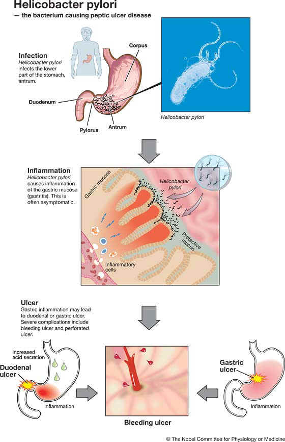
Robin Warren (born 1937), a pathologist from Perth, Australia, observed small curved bacteria colonizing the lower part of the stomach (antrum) in about 50% of patients from which biopsies had been taken. He made the crucial observation that signs of inflammation were always present in the gastric mucosa close to where the bacteria were seen.

Barry Marshall (born 1951), a young clinical fellow, became interested in Warren’s findings and together they initiated a study of biopsies from 100 patients. After several attempts, Marshall succeeded in cultivating a hitherto unknown bacterial species (later denoted Helicobacter pylori) from several of these biopsies. Together they found that the organism was present in almost all patients with gastric inflammation, duodenal ulcer or gastric ulcer. Based on these results, they proposed that Helicobacter pylori is involved in the aetiology of these diseases.

Even though peptic ulcers could be healed by inhibiting gastric acid production, they frequently relapsed, since bacteria and chronic inflammation of the stomach remained. In treatment studies, Marshall and Warren as well as others showed that patients could be cured from their peptic ulcer disease only when the bacteria were eradicated from the stomach. Thanks to the pioneering discovery by Marshall and Warren, peptic ulcer disease is no longer a chronic, frequently disabling condition, but a disease that can be cured by a short regimen of antibiotics and acid secretion inhibitors.

Peptic ulcer – an infectious disease!

This year’s Nobel Prize in Physiology or Medicine goes to Barry Marshall and Robin Warren, who with tenacity and a prepared mind challenged prevailing dogmas. By using technologies generally available (fibre endoscopy, silver staining of histological sections and culture techniques for microaerophilic bacteria), they made an irrefutable case that the bacterium Helicobacter pylori is causing disease. By culturing the bacteria they made them amenable to scientific study.

In 1982, when this bacterium was discovered by Marshall and Warren, stress and lifestyle were considered the major causes of peptic ulcer disease. It is now firmly established that Helicobacter pylori causes more than 90% of duodenal ulcers and up to 80% of gastric ulcers. The link between Helicobacter pylori infection and subsequent gastritis and peptic ulcer disease has been established through studies of human volunteers, antibiotic treatment studies and epidemiological studies.

Helicobacter pylori causes life-long infection

Helicobacter pylori is a spiral-shaped Gram-negative bacterium that colonizes the stomach in about 50% of all humans. In countries with high socio-economic standards infection is considerably less common than in developing countries where virtually everyone may be infected.

Infection is typically contracted in early childhood, frequently by transmission from mother to child, and the bacteria may remain in the stomach for the rest of the person’s life. This chronic infection is initiated in the lower part of the stomach (antrum). As first reported by Robin Warren, the presence of Helicobacter pylori is always associated with an inflammation of the underlying gastric mucosa as evidenced by an infiltration of inflammatory cells.

The infection is usually asymptomatic but can cause peptic ulcer!

The severity of this inflammation and its location in the stomach is of crucial importance for the diseases that can result from Helicobacter pylori infection. In most individuals Helicobacter pylori infection is asymptomatic. However, about 10-15% of infected individuals will some time experience peptic ulcer disease. Such ulcers are more common in the duodenum than in the stomach itself. Severe complications include bleeding and perforation.

The current view is that the chronic inflammation in the distal part of the stomach caused by Helicobacter pylori infection results in an increased acid production from the non-infected upper corpus region of the stomach. This will predispose for ulcer development in the more vulnerable duodenum.

Malignancies associated with Helicobacter pylori infection

In some individuals Helicobacter pylori also infects the corpus region of the stomach. This results in a more widespread inflammation that predisposes not only to ulcer in the corpus region, but also to stomach cancer. This cancer has decreased in incidence in many countries during the last half-century but still ranks as number two in the world in terms of cancer deaths.

Inflammation in the stomach mucosa is also a risk factor for a special type of lymphatic neoplasm in the stomach, MALT (mucosa associated lymphoid tissue) lymphoma. Since such lymphomas may regress when Helicobacter pylori is eradicated by antibiotics, the bacterium plays an important role in perpetuating this tumour.

 Disease or not – interaction between the bacterium and the human host

Helicobacter pylori is present only in humans and has adapted to the stomach environment. Only a minority of infected individuals develop stomach disease. After Marshall’s and Warren’s discovery, research has been intense. Details underlying the exact pathogenetic mechanisms are continuously being unravelled.

The bacterium itself is extremely variable, and strains differ markedly in many aspects, such as adherence to the gastric mucosa and ability to provoke inflammation. Even in a single infected individual all bacteria are not identical, and during the course of chronic infection bacteria adapt to the changing conditions in the stomach with time.

Likewise, genetic variations among humans may affect their susceptibility to Helicobacter pylori. Not until recently has an animal model been established, the Mongolian gerbil. In this animal, studies of peptic ulcer disease and malignant transformation promise to give more detailed information on disease mechanisms.

Antibiotics cure but can lead to resistance

Helicobacter pylori infection can be diagnosed by antibody tests, by identifying the organism in biopsies taken during endoscopy, or by the non-invasive breath test that identifies bacterial production of an enzyme in the stomach.

An indiscriminate use of antibiotics to eradicate Helicobacter pylori also from healthy carriers would lead to severe problems with bacterial resistance against these important drugs. Therefore, treatment against Helicobacter pylori should be used restrictively in patients without documented gastric or duodenal ulcer disease.

Microbial origin of other chronic inflammatory conditions?

Many diseases in humans such as Crohn’s disease, ulcerative colitis, rheumatoid arthritis and atherosclerosis are due to chronic inflammation. The discovery that one of the most common diseases of mankind, peptic ulcer disease, has a microbial cause, has stimulated the search for microbes as possible causes of other chronic inflammatory conditions.

Even though no definite answers are at hand, recent data clearly suggest that a dysfunction in the recognition of microbial products by the human immune system can result in disease development. The discovery of Helicobacter pylori has led to an increased understanding of the connection between chronic infection, inflammation and cancer.

Source: http://nobelprize.org/nobel_prizes/medicine/laureates/2005/press.html