Simple Plants PowerPoint Questions

Simple Seedless Nonvascular & Vascular Plants
PowerPoint Worksheet

Seedless Nonvascular plants

1. Name the 3 divisions of seedless vascular plants and a member of each division.

     a.

     b.

     c.

Division Bryophyta

2. What is the common name for mosses, liverworts, and hornworts?

3. Bryophytes lack what type of tissue?

4. Name the 2 vascular tissues lacking in bryophytes and tell their function.


5. What is the 2 stage life cycle of plants called?

6. Name the 2 life cycle stages.

7. which stage is DOMINANT in bryophytes (mosses, liverworts, & hornworts)?

8. How do bryophytes reproduce?

9. Which stage of the moss looks like a lush green carpet?

10. Name the division for moss.

11. Why are moss small plants?

12. Do moss have TRUE roots, stems, or leaves?

13. In what type of area do moss grow?  Give several examples.


14.Moss gametophytes must grow close together in moist areas. Give 2 reasons why this is so.

     a.

     b.

15. What covers the outside of a moss plant to prevent water loss?

16. What anchors moss plants?

17. Can rhizoids absorb water like true roots?

18. Where does the sporophyte generation occur on moss plants?


19. What is at  the top of the sporophyte?

20. Label the following moss plant.

21. ___________ moss is used by florist. What characteristic makes it useful?


22. Because moss will grow on bare ground, it is called a _________ plant.

23. How is peat moss used?


24. Give 4 other uses for moss.


25. Moss are capable of asexual reproduction. Name and describe 2 types of this vegetative reproduction.

     a.

     b.

26. What are gemmae?


27. How are gemmae separate from the parent plant & dispersed?


28. Which stage of the moss is haploid and which is diploid?

29. The gametophyte generation produces what 2 cells? 

30. Why do these cells have half the chromosome number?

31. ____________ have a ________ set of chromosomes and reproduce ___________.

32. the sporophyte grows attached to the top of the ______________.

33. Since sporophytes lack chlorophyll, what cellular process are they incapable of doing?

34. How does the sporophyte get its food?

35. What is the setae on a moss plant?

36. How are the moss gametes protected?


37. Name the female gametangia & tell what it produces.

38. Eggs of moss are _____________ & ___________.

39. Label the female gametangia.

40.  Name the male gametangia & tell what it produces.

41.How does the sperm cell know the direction in which to swim to the egg?


42. Label the male gametangia.

43. The moss ___________ or fertilized egg develops into the ____________.

44. Spores of the sporophyte capsule germinate into young plants called ______________.

45. Protonema develop into the _____________ stage

46. Label the protonema & developing gametophyte in this picture.

47. Label the life cycle of the moss.

Division Hepatophyta

48. ___________ are nonvascular, _________ producing bryophytes.

49. What stage is dominant in liverwort’s life cycle?

50. Describe the liverwort gametophyte.


51.Liverworts are found growing where?


52. Liverworts need lots of water for ____________.

53. How do liverworts reproduce asexually?

54. How do liverworts reproduce sexually?

Division Anthocerophyta

55._____________ are small, nonvascular ____________ with a dominant, leafy ____________ like liverworts.

56. Where are the antheridia & archegonia in hornworts?

57. Zygotes develop into ______________ sporophytes.

58. Is the horn-shaped sporophyte capable of photosynthesis?

59. Is the horn-shaped sporophyte attached to or separate from the gametophyte?

60. Label the parts of the hornwort.

Seedless Vascular Plants

61.Label these structures on the back of this fern.

62. Name and give an example of a plant in the 4 divisions of seedless vascular plants.

     a.

     b.

     c.

     d.

63. Name the vascular tissues.

64. Do seedless vascular plants go through alternation of generations?

65. Which stage is dominant?

66. How do they reproduce?

Division Psilophyta

67.Describe whisk ferns.


68. Do they have true roots, stems, or leaves?

69.How many extant genera are there?

70. Name the root like structures of whisk ferns and tell whether they can or can’t absorb water.

71. How do whisk ferns reproduce asexually?

72. How do whisk ferns reproduce sexually?

73. Make and label a sketch of an aerial branch of whisk with sporangia.


74. What is the purpose of sporangia?

Division Lycophyta

75. The division Lycophyta contains the ______________ living vascular plants.

76. Club moss are commonly called ______________ ____________.  Explain why this is true.


77.Club moss have ________ growing root like ___________.

78. Describe the habitat needed by club moss.

79. Describe the leaves of club moss.


80. Are these TRUE leaves? Explain why.


81. What is found in the axils of the leaves & what is their purpose?

82. What are strobili?

83. Some club moss are homosporous while others are heterosporous.  Explain what each of these terms means.

     a. homosporous-

     b. heteroporous-

84. Give an example of a homosporous club moss.

85. Lycopodium is used in fireworks. Explain the reason for this.


86. What do the spores of Lycopodium look like?


87.What is the purpose of each of these structures.


88. Give 3 other uses for club mosses.

     a.

     b.

     c.

Division Sphenophyta

89. How many extant species of horsetails are there?

90. Name the living genera of horsetails.

91. What is another name for horsetails?

92. Why are they called this?

93. Describe the stems of horsetails.


94. Where does photosynthesis take place in horsetails?

95. How are horsetails anchored?

96. How do horsetails reproduce?

97. Where are their spores found?

98. In prehistoric times, what was true of the size of horsetails?

99. Describe the habitat of horsetails.

100. How do horsetails prevent water loss from the parts of the plant above ground?

101. What special spore dispersing structures are found on the spores of horsetails?

102. Describe how elaters work.


103. Label the stem, node, and leaves on this horsetail.

104. Give 3 other uses for  horsetails.

     a.

     b.

     c.

104. Can animals eat horsetails? Why or why not?


Division Pterophyta

105. Ferns are in the ____________ group of extant vascular plants.

106. Describe the habitats for ferns.


107. How do ferns reproduce asexually?

108. What stage is dominant in the life cycle of the fern?

109. What is the only part of the fern plant that appears above ground?  What parts are found below ground?

110. Fern leaves are called ______________ and are attached to the plant by short stems called ______________.

111. Describe the appearance of newly forming fern fronds and tell what they are called.


112. What are sori and where are they found?

113. How are fern spores spread?

114. What forms when a fern spore lands on moist ground and germinates (starts growing)?

115. The prothallus starts what stage in the life cycle?

116. What is the shape of the gametophyte and does it live long?

117. What 2 structures grow ON the gametophyte?

118. Label the gametophyte and the male and female gametangia.

119. Label the parts of a fern.

120. Label the life cycle of the fern.

121. Give 4 uses for ferns.     a.

     b.

     c.

     d.


Sponge & Cnidarian Study Guide

Study guide for Sponge, Cnidarians, & Ctenophores

·         Know relatives of the jellyfish
·         How are sponges different from other animals
·         Know characteristics of all invertebrates
·         Know characteristics of sponges
·         What is the function of collar cells in sponges
·         What are spicules
·         Know characteristics of adult sponges
·         Be able to explain skeletal support of sponges
·         How do sponges obtain their food
·         What helps draw water into a sponge
·         What is the function of amebocytes in sponges
·         How does excess water leave a sponge
·         What is the purpose of gemmules in sponges
·         What is a hermaphrodite
·         How can sponges reproduce
·         Know animals that capture prey by using nematocysts
·         What are the 2 distinct life stages of cnidarians
·         Describe nematocysts
·         What organisms have tentacles with stinging cells
·         Know examples of cnidarians
·         Describe the life of a planula larva
·         Know the life stage that is dominant in sea anemones
·         What organisms would be anthozoans
·         Know the dominant life stage of jellyfish
·         Know the main characteristics of ctenophores

BACK


Sponges & Cnidarian

Sponges, Cnidarians, & Ctenophores

Phylum Porifera
Characteristics

  • Includes marine & freshwater sponges
  • Found in the kingdom Animalia & subkingdom Parazoa
  • Sessile as adults
  • Simplest of all animals

  • Contain specialized cells, but no tissue
  • Asymmetrical
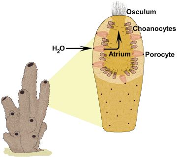
  • Bodies filled with holes or pores for water circulation
  • Marine sponges are larger & more colorful than freshwater sponges
  • Range in size from 2 centimeters to 2 meters
  • Osculum is single, large body opening at the top for water & wastes to leave
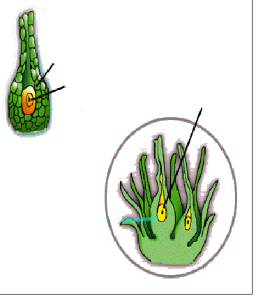
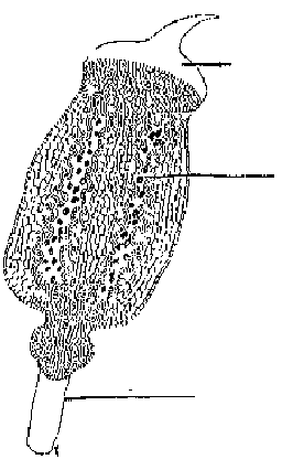
  • Spongocoel is the body cavity of sponges
  • Have only 2 cell layers (ectoderm & endoderm) separated by jellylike material
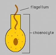
  • Flagellated cells called choanocytes or collar cells line their internal body cavity
  • Flagella of choanocytes beat & pull in water containing food which the collar traps


  • Spongin is a network of flexible, protein fibers making up the sponge’s skeleton
  • Spicules are tiny, hard particles shaped like spikes or stars in the  skeleton of some sponges
  • Spicules are made of calcium carbonate or silica

Feeding

  • Sponges are filter feeders that remove plankton (food) from the water that is brought in through pores lined with collar cells
  •  Flagella pull in bacteria, protozoans, & algae that sticks to collar of choanocytes where it is digested
  • Amebocytes are specialized cells in sponges that can roam to pick up food from choanocytes & distribute it to all other parts of the sponge
  • Amebocytes also transport carbon dioxide & wastes away from sponge cells
  • Excess water & food leaves through the excurrent osculum

Reproduction

  • Sponges can reproduce asexually by external buds that break off & form new sponges or stay attached to form sponge colonies
  • Gemmules are specialized, internal buds formed by sponges during cold or dry weather that can survive harsh conditions
  • Gemmules consist of a food-filled ball of amebocytes surrounded by a protective coat with spicules & released when adult sponge dies
  • Gemmules break open when conditions improve & the cells form new sponges

  • Sponge can also asexually regenerate missing parts or a new sponge from a small piece of sponge
  • Sponges are hermaphrodites (produce both eggs & sperm), but they exchange sperm & cross-fertilize eggs during sexual reproduction
  • Planula is the flagellated, free-swimming larva that forms from the zygote
  • Planula larva eventually settles to the bottom & attaches to develop into an adult, sessile sponge

Classes of Sponges

  • Calcarea are chalky sponges with calcium carbonate spicules
  • Hexactinella includes glass sponges & the Venus flower basket with silica spicules
  • Demospongiae include horny & bath sponges with only spongin or spongin & silica spicules
  • Sclerospongiae are coral sponges & have spongin & silica and calcium carbonate spicules

Phylum Cnidaria
Characteristics

  • Includes marine organisms such as jelllyfish, Portuguese man-of-war, coral, sea anemone, & sea fans
  • Hydra is a freshwater cnidarian

  • All carnivorous
  • Have 2 cell layers (epidermis -outer & gastrodermis-inner) with a hollow body called gastrovascular cavity
  • Contain a jelly-like layer between epidermis 7 gastrodermis  called mesoglea
  • Single opening (mouth/anus) to gastrovascular cavity where food & water enter & wastes leave; called two-way digestive system
  • Have tentacles around mouth to pull in water & capture food

  • Have a simple nerve net with to help with movement & senses
  • Sessile members include corals, sea anemones, & sea fans
  • Have radial symmetry as adults

  • Contain stinging cells called cnidocytes in their tentacles that contain coiled stingers called nematocysts that can shoot out & paralyze prey 

Body Forms

  • Have 2 basic body forms —polyp & medusa


MEDUSA

POLYP


  • Polyp forms are usually sessile with upright tentacles arranged around the mouth at the top and with a thin layer of mesoglea
  • Polyps are the asexual stage
  • Corals, hydra, & sea anemones exist in the polyp form as adults 


CORAL POLYPS

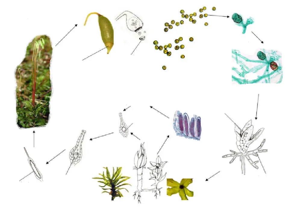
  • Medusa forms are usually free-swimming, bell-shaped animals with tentacles that hang down around the mouth and with a thick layer of mesoglea for support
  • Medusa are the sexual stage
  • Jellyfish & Portuguese man-of-war are medusa form as adults
  • Some cnidarians are dimorphic or go through both polyp & medusa stages in their life cycle

Life cycle of a jellyfish
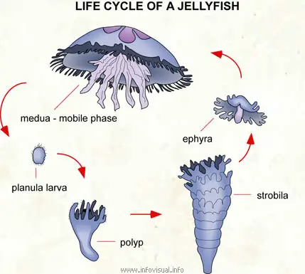
JELLYFISH LIFE CYCLE

  • Some are solitary (Hydra) others are colonial (corals)
  • Three classes include Hydrozoa (hydra), Scyphozoa (jellyfish), & Anthozoa (sea anemones & corals)

Hydrozoa

  • Includes freshwater, sessile hydra (exists only as polyps) 
  • Portuguese man-of-war (exists as colony of polyps & medusa)
  • Group of cells called basal disk produces sticky secretion for attachment & can secrete gas bubbles to unattach & let hydra float
  • Hydra also move by somersaulting (tentacles bend over to bottom as basal disk pulls free)
  • Tentacles pull food into gastrovascular cavity where enzymes digest it
  • Reproduce asexually by budding during warm weather & sexually in the fall
  • Hermaphrodites that release sperm into water to fertilize eggs of another hydra


HYDRA

Scyphozoa

  • Includes bell-shaped jellyfish
  • Medusa stage is dominant in the life cycle
  • Tentacles may be meters in length & carry poisons that cause severe pain or death
  • Have both asexual polyps & sexual medusa stages in their life cycles
  • Adult medusa stage releases eggs & sperm into water
  • Fertilization produces ciliated planula larva that settles to the bottom, attaches, & forms tentacles 
  • New medusa bud off of reproductive polyps & form adult jellyfish

jellyfish life cycle photo
JELLYFISH

Anthozoa

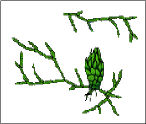
  • Include corals in a limestone case & sea anemones
  • Called “flower animals”
  • All marine
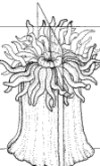
  • Sea anemone is a sessile, polyp-form that uses its tentacles to paralyze fish
  • Some anemones in the Pacific Ocean live symbiotically with the clownfish sharing food & protecting each other

#22A

  • Corals are small, colonial polyps living in limestone cases
  • Coral reefs form as polyps die & provide a home and protection for other marine animals
  • Reefs form in warm, shallow water & only the top layer has living polyps
  • Algae may live symbiotically with coral supplying them with oxygen

Phylum Ctenophora

Characteristics

  • All marine
  • Includes comb jellies

  • Have eight rows of fused cilia called “comb rows”
  • Largest animal to move by cilia
  • Move by beating cilia
  • Lack cnidocytes but have cells sticky cells called colloblasts that bind to prey
  • Colloblasts located on two ribbon-like tentacles
  • Have sensory structure called apical organ to detect direction in the water
  • Most are hermaphrodites (make eggs & sperm)
  • Produce light by bioluminescence
BACK


Squid Dissection

              Squid Dissection

Objectives:

As a result of this lesson, students will be able to:

  1. Locate and identify major external and internal features and organs of a squid.
  2. Understand and use basic dissection techniques and terms.
  3. Critically examine the functions of several squid features and organs.


Teaching Notes: This lab is a very thorough dissection of a squid and can be adapted to different grade levels. Teachers should try the lessons, considering which parts are most appropriate for their students and curriculum. The descriptions use complex dissection terminology. Be certain students understand the vocabulary of dissection prior to beginning the lab.

These lessons were tested with middle school students ages 11 to 13. They followed procedures and understood concepts well. The skills necessary to do all steps in the dissection are within the normal ability range of middle school students.


Materials:

  • squid*
  • scissors
  • toothpicks (for probes and pointers)
  • drawing paper
  • forceps
  • hand lens (5x recommended)
  • small cups (ketchup cups work well)
  • dissecting pan (or lunch trays)
  • paper towels
  • diagram of squid
  • wash bottle
  • microscope (optional)
  • dissecting scopes (optional)
  • slides (optional)
  • slide covers (optional)


*Look for squid at the local supermarket in the seafood and frozen foods sections. You may have to order it in advance. For areas that have them, you can also go to the local fish market or oriental food stores, or you can deal directly with fishermen.


Teaching Notes: Squid specimens tend to deteriorate rapidly. Keep all squid frozen until the morning before dissection. Thaw the squid in the refrigerator. If the entire dissection cannot be completed in one day, do the external activities while the specimens are still partly frozen, and the internal activities the next day after squid are thawed.

Squid may have tentacles or arms missing. Individual squid vary internally, and their relative maturity determines which organs are formed well enough to be seen clearly, and which have lost (or have yet to gain) their shape and coloration. Please advise students that they may not see everything shown in the enclosed diagram. Tissue shrinks and organs become misshapen quickly. To help maintain the freshness of the specimen, cover it with a wet paper towel as you work so it does not dry out so quickly.

Finally, this lesson is a tactile experience. You may want to explore this aspect through sensory activities, written descriptions, poetry, and/or artwork. Encourage students to experience the many textures found inside and outside the squid’s body. Moving fingertips along the suckers is suggested as well – the suckers do not scrape or hurt if you are gentle with them.


Procedure


  • Orientation:

Place the squid with the dorsal (back) side up in the dissecting pan. This means put the side with the funnel down and the fin side up. Make sure the tentacles and arms are towards you. Locate the head, eyes, beaks (mouth), arms (8), two longer feeding tentacles, fins, mantle, and skin. Use the hand lens to examine the suckers on the tentacles and arms as well as the spots on the skin, which are chromatophores.


Step 1A


What are the differences between arm and tentacle suckers? Where are the suckers located on the feeding tentacles as compared to the location of the suckers on the arms?


Step 1B


How do you account for the different locations of the suckers on the tentacles and the arms? What are chromatophores?


  1. The Mouth and Beaks: Locate the dark beaks in the center of the mouth.

    Step 2A


    Open and close the beaks, noting how the ventral beak overlaps the dorsal beak. How is this different from a parrot’s beak? Before you pull out the beaks, imagine what they will look like on the inside. With tweezers, remove the beaks and place beaks together with dark pointed parts opposite one another. Manipulate them (open and close) as if the squid were eating. What makes them work in this way?

    Step 2B Step 2B


    In order to remove the radula (a ribbon with rows of teeth on a tongue-like muscle) from inside the mouth, make small incisions in the edge of the mouth. With tweezers, locate the small, folded, plastic-like radula between beaks and remove it. It is usually very small, yellow or white in color. What is the radula’s function?   Store the radula and the beaks in water in a small cup if you are going to do a microscopic examination.
    Drawing of Squid Radula
  2. Funnel:
  3. Turn the body over, ventral side up, and locate the funnel (a deflated fleshy tube located at the base of the head). A squid swims by squirting water from the mantle through the funnel. The direction it swims depends on which way the funnel is aimed. Move the funnel and note its flexibility.

    Step 3


  4. External Anatomy:
  5. Orient the squid so that the tentacles are away from you, at the top of the dissection tray. Spread out the arms, tentacles, and fins. Draw and label the external parts of the squid: arms, tentacles (have suckers only at the tips), head, eyes, fins, mantle, funnel, tail, suckers, beaks (where each would be found on an intact squid) and mouth. If something cannot be seen, draw an arrow to show where it should be.  

    Step 4 Step 4


    If you have time, slice open an eyeball and locate the lens, pupil, retina, and iris (colored part of the eye). Look for the creamy white brain between the eyeballs. For assistance in identifying these parts, refer to the illustration below.

    Diagram of cephalopod eye


  6. Opening the Mantle:
  7. Keep the squid on its back (the side opposite the funnel). Using forceps, lift up the opening to the mantle behind the funnel (near the head) and separate the mantle from the internal organs. Close the forceps firmly so as to “pinch” the mantle flesh to keep it taut, cut along the ventral midline of the mantle, from its opening all the way to the tail. Be careful to keep the scissors lifted away from the internal organs so they are not damaged.  

    Step 5 Step 5


  8. Locating and Removing Reproductive Organs:
  9. Locate the gonad (reproductive organ) in the posterior end (refer to diagram for shape and location).

    Step 6A


    Upon opening female specimens, the large, firm, white nidamental glands are seen first. Males do not have nidamental glands. The glands lay on top of the other internal organs. These glands create the gelatinous matrix that envelops the eggs. In order to proceed further, carefully remove these glands. In females the eggs are jelly-like in a conical sac at the posterior end of the mantle. The male genital duct is a white, fluid-filled sac in the posterior end of the mantle. The sperm are stored in thin tubes in an elongated sac behind and along one gill.

    Step 6B


  10. Gills:
  11. Find the gills. These are the long, feather-shaped organs that are attached to the sides of the mantle and extend along the anterior half of the mantle. Identify the gill hearts, one on the posterior end of each gill (these are small, flat and white). Questions: Why are they white and our hearts are red or purple? The squid has a third heart (the systemic heart) that pumps blood to the rest of the body.

    Step 7


    Challenge: Why does it have separate hearts for the gills alone?
  12. Digestive Tract:

  13. Step 8A


    The long, silvery dark tube on the bottom of the liver (but appearing to be on top of the liver because of the squid’s inverted position) is the ink sac. Be careful not to break it open. Locate the stomach and caecum. These lie together as one white, silky-looking tube, like a deflated bladder and a coiled sack. The bunched up organs that look like human intestines are digestive ducts for the squid. If you are curious about the liver, wait to cut it open until the end of the dissection. It contains a lot of brown, oily liquid which may obscure other organs. If possible, open the stomach and examine its contents. Many squid will have bits of partially digested crustaceans (pink and white pieces), or tiny fish scales and bones.  

    Step 8B


  14. Removing the Ink Sac:
  15. Find and carefully remove the silvery-black ink sac that lies connected to the intestine. To do this, pinch the opening of the sac (near the back of the funnel) with forceps while gently pulling up and cutting the connective membrane along its length. After cutting about 1/3 to 1/2 of it, hold the sac with your fingers and pull the sac off the liver. Be careful not to puncture it. Squid ink stains clothing and skin. Place the sac in a small cup for later use with the gladius (pen).  

    Step 9 Step 9


  16. Removing the Gladius (Pen):
  17. The gladius is a long, clear feather-shaped structure used to support the mantle and for organ attachment. It and the cranium, or brain case, make up the “skeleton” of the squid. It feels like plastic and is made of tissue similar to a shrimp shell. There are two ways to remove it: from the tail or from inside the cut-open mantle. To remove it from inside the open mantle, grasp the head and organs firmly, and rotate them to the side with your left hand while holding on to one side of the mantle with your right hand and pulling away gently. Pulling the gladius out is like removing a splinter from your skin. You may need to cut away connective tissues that hold the gladius in place.

    Step 10A Step 10A


    The gladius is revealed, lying along the dorsal midline of the mantle.  

    Step 10B Step 10B


    Grab the forward end of the gladius and pull it carefully from its slot in the mantle. It may be helpful to have one person hold down the lower mantle while the other removes the gladius. To remove from the tail end, rotate the organs to one side, cutting connective tissues. Make sure the mantle is slit along the internal dorsal midline all the way to the tip of the tail. Pry out the tail end of the gladius and pull straight back, away from the body.

    Step 10C Step 10C


  18. Writing with the Gladius (Pen) and Squid Ink:

  19. Step 11A Step 11A


    Cut one end of the ink sac open and press it against the bottom of the cup with forceps or toothpick. You can also hold one end and push the ink out with your finger, as you would toothpaste from a tube. This will release the ink. Dip the pointed tip (the anterior end) of the gladius into the ink, filling the tip with the dark fluid. Then, using only the ink-filled tip of the gladius, write your name on your squid illustration or paper. If there is enough ink, create and write the name of your dissected squid under its picture. If the ink seems dry and pasty, add one drop of water at a time to create fluid ink. Though this is an unusual way to write, squid ink was actually used to write and draw in ancient times, and it is used today in some cultures. Unfortunately, it tends to fade over time (except from your clothes!).

    Step 11B


  20. Internal Anatomy:
  21. Draw, label, and identify the function of the following internal parts of the squid:
    • stomach
    • caecum
    • hearts (systemic and gill)
    • gills
    • reproductive organs
    • ink sac
    • liver (digestive gland)
    • gladius
    • brain
    • eyeball


  22. Microscope Slide Option:

    Step 12
    Step 12


    The following parts of the squid make excellent specimens for microscopic study:

    • eggs from the ovaries
    • suckers
    • nidamental glands
    • tips of arms and tentacles
    • spermatophores
    • connective membranes (thinly-sliced: mantle, fin, arm muscle)
    • radula
    • stomach contents
    • liver fluids
    • skin and chromatophores
    • portions of the eye
    • beak


    Teaching Note: Most of these are useful only for a dissecting microscope.

    Questions for further Investigation:

    • Identify the differences between the tentacles and the arms. Why are they different?
    • How are squid mouths and beaks like your jaw and teeth? How are they different?
    • How does the squid use the funnel and mantle for locomotion?
    • How does the squid obtain oxygen from the water?
    • How do squid reproduce?
    • Why are the chromatophores important to the squid?
    • What are the relatives of the squid?
    • What are the characteristics of cephalopods and of mollusks?
    • Why is it difficult to identify stomach contents?
    • What is the function of the fins?
    • What organ systems are the same or different from vertebrates?

    When finished, clean your area completely. Return all equipment and wash your hands. The squid odor will remain for a little while. Lemon juice will alleviate the odor if you find it offensive. To dispose of your specimen, wrap it in plastic or a zip-lock bag and throw it away. You may want to feed it to your cat, cut it up for fish bait, or even serve it as tonight’s calamari. Bon Appetite! Visit Clyde’s Kitchen on this website for tasty squid recipes!


Previous Lesson | Lesson List | Next Lesson
Site Contents | Opening Page | Home

Starfish Dissection


Starfish Dissection

Introduction:

Echinoderms are radially symmetrical animals that are only found in the sea (there are none on land or in fresh water). Echinoderms mean “spiny skin” in Greek. Many, but not all, echinoderms have spiny skin. There are over 6,000 species. Echinoderms usually have five appendages (arms or rays), but there are some exceptions.

Radial symmetry means that the body is a hub, like a bicycle wheel, and tentacles are spokes coming out of it (think of a starfish). As larvae, echinoderms are bilaterally symmetrical. As they mature, they become radially symmetrical.

Most adult echinoderms live on the bottom of the ocean floor. Many echinoderms have suckers on the ends of their feet that are used to capture and hold prey, and to hold onto rocks in a swift current.

Sea Stars
Sea Star
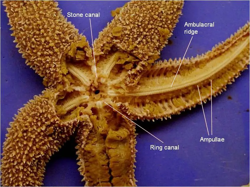
Sea stars (group name Stelleroidea) are sometimes called starfish, though they are not real fish (they lack both vertebrae and fins). There are two sub-types of sea stars:

  • Asteroideas are the true sea stars and sun stars.
  • Ophiuroideas are brittle stars and basket stars.

The differences between the two sub-types lies in how the arms connect to the central disk. Ophiuroids have arms that do not connect with each other. There is a distinct boundary between arm and central disk. Asteroids have arms that are connected to each other. Also, it is harder to tell with asteroids where the central disk ends and the arms begin.

The sea star’s top surface (or skin) looks spiny if you examine it. If you look very closely you will notice that there are different types of growths on the surface. Some bumps are used to absorb oxygen, they are called dermal branchiae. Pedicellaria are pincher-like organs used to clean the surface of the skin. Barnacle larvae could land on a sea star and start growing if it were not for these organs.

How Do Sea Stars Move?
Underside of a Sea StarEach sea star had hundreds of tiny feet on the bottom of each ray. These are tube feet, or podia. These tiny feet can be filled with sea water. The vascular system of the sea star is also filled with sea water. By moving water from the vascular system into the tiny feet, the sea star can make a foot move by expanding it. This is how sea stars move around. Muscles within the feet are used to retract them.

Each ray of a sea star has a light sensitive organ called an eyespot. Though it can not see nearly as well as we do, sea stars can detect light and its general direction. They have some idea of where they are going.

Sea Star Anatomy

Can Sea Stars Grow New Arms?
Given enough time, sea stars can grow back arms that have been damaged or removed. For a few species, the severed arm can grow back into a complete sea star! For most sea stars, however, a severed limb dies.

What Do Sea Stars Eat?
Sea stars eat many things. A sea star’s diet can include: barnacles, snails, sea urchins, clams, and mussels. A few species, such as the spiny star of the North Atlantic, eat other sea stars! Many sea stars eat mussels and clams in an interesting way. They surround the shell and use the suckers on their feet to pull the two shells (or valves) apart. The sea star has enough force in its arms to actually bend the shell! This creates an opening between the two shells that is only .01 inches wide. Using this tiny gap, the sea star puts its stomach into the clam’s shell and eats its insides. When it is done, nothing is left but an empty shell.

Materials:

Preserved starfish, dissecting pan, scissors, scalpel, forceps, T-pins, pencil, lab apron, safety glasses

Procedure:


Dorsal view of starfish showing external anatomy

Ventral view of starfish showing external anatomy

Dorsal view of a dissected starfish showing rectal cecum, anus, madreporite, pyloric stomach, pyloric duct

Dorsal view of a dissected starfish showing madreporite, stone canal, cardiac stomach, and ampullae

Dissection showing where cardiac stomach opens into the mouth

Close up of madreporite and stone canal

Dorsal view of a dissected starfish showing pyloric caecum and pyloric ducts

Dorsal view of a dissected starfish showing gonads and ampullae

Ventral view of starfish showing external anatomy