| Algal & Fungal-like Protists Kingdom Protista All Materials © Cmassengale |
 Copyright © by Holt, Rinehart and Winston |
Algal-Like Protists
Characteristics of Algae:
- Plantlike members of the kingdom Protista
- Eukaryotes
- Most unicellular, but some multicellular
- Autotrophic – contain chlorophyll & make food by photosynthesis
- Plankton = communities of organisms, mostly microscopic, that drift passively or swim weakly near the surface of oceans, ponds, and lakes
- Produce oxygen that is returned to the atmosphere
- Range in size from microscopic to seaweeds hundreds of feet in length
- Do not have true roots, stems, nor leaves
- Form gametes (eggs & sperm) in single-celled gametangia (chambers) instead of multicellular gametangia like true plants
- Found in freshwater, marine, and moist soil habitats
- Most have flagella at some time in life cycle
- Algae cells contain organelles called pyrenoids organelles that make & store starch
Structure of Algal Cells:
- The body of algae is called the thallus (1n)
- Algae may be unicellular, colonial, filamentous, or multicellular
- Unicellular algae are single-celled & make up phytoplankton (a population of photosynthetic organisms that begins many aquatic food chains)
- Phytoplankton make much world’s carbohydrates & are the major producers of oxygen
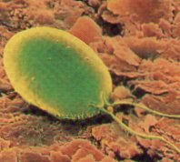
Chlamydomonas
Copyright © by Holt, Rinehart and Winston
- Colonial algae consist of groups of cells working together
- Some colonial algal cells may specialize for movement, feeding, or reproduction showing for division of labor
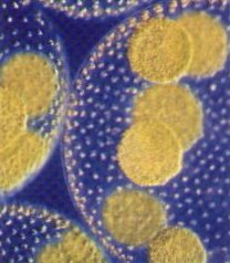
Volvox
Copyright © by Holt, Rinehart and Winston
- Filamentous algae have slender, rod-shaped thallus arranged in rows joined end-to-end
- Holdfasts are specialized structures in some filamentous algae that attaches the algae so it can grow toward sunlight at the surface
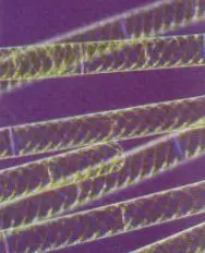
Spirogyra
Copyright © by Holt, Rinehart and Winston
- Multicellular algae often have a large, complex leaf-like thallus & may have stem-like sections and air bladders
- Macrocystis is among the largest multicellular algae
Macrocystis
Copyright © by Holt, Rinehart and Winston
Reproduction in Unicellular Algae:
Asexual Phase
- Algae absorbs its flagellum
- Haploid algal cell then divides mitotically from 2 to 3 times
- From 4 – 8 haploid flagellated cells called zoospores develop in this parent cell
- Zoospores break out of the parent cell & eventually grow to full size
Sexual Phase
- Haploid cells dividing mitotically to produce either “plus” or “minus” gametes
- A plus gamete and a minus gamete come into contact with one another, shed their cell walls, and fuse to form a diploid zygote
- This resting stage of a zygote is called a zygospore & an withstand bad environmental conditions
- When conditions are bad, the thick wall opens and the living zoospore emerges

Life Cycle of Chlamydomonas
Copyright © by Holt, Rinehart and Winston
Reproduction in Multicellular Algae:
- Oedogonium is a multicellular, filamentous green algae with specialized cells called gametangia that form gametes
- The male gametangia or antheridium makes sperm, & the female gametangia or oogonium makes eggs
- Sperm are released into the water & swim to the egg to fertilize them
- The fertilized egg or zygote is released from the oogonium & forms thick-walled zoospores
- Zoospores undergo meiosis so one cell attaches to the bottom & develops a holdfast while the other zoospores divide & form a filament

Oedogonium Life Cycle
Copyright © by Holt, Rinehart and Winston
- Spirogyra, another filamentous green algae, reproduces by conjugation
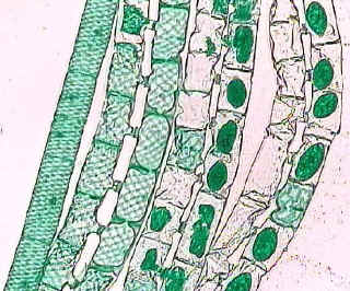
- Two filaments align side by side, their adjacent cell walls dissolve, & a conjugation tube forms between them
- Fertilization occurs when a + gamete cell moves through the tube & fuses to the – gamete cell
- Zygote forms a thick walled spore (sporangium) that breaks away from the parent & forms a new filament
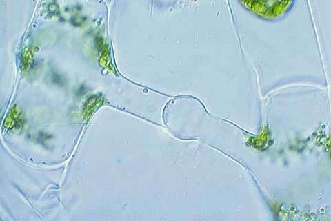
Conjugation Tube between Spirogyra
- The leaflike algae Ulva has a sexual reproductive cycle characterized by a pattern called alternation of generations
- Alternation of generations has two distinct multicellular phases- a haploid, gamete-producing phase called a gametophyte and a diploid, spore-producing phase called a sporophyte
- Alternation of Generation also occurs in more complex land plants, but the gametophyte & sporophyte do not resemble each other
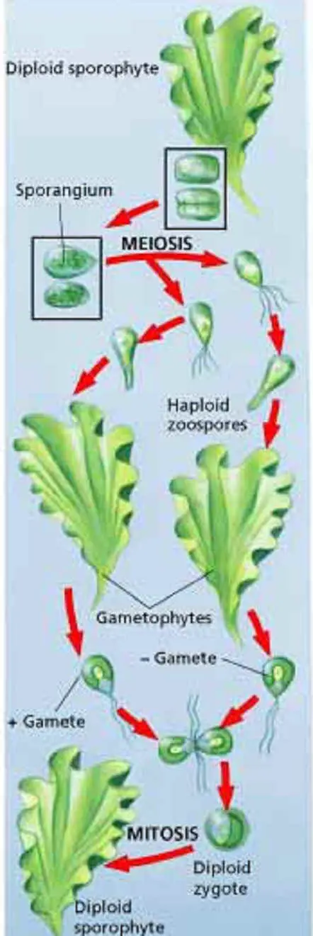
Ulva Life cycle
Copyright © by Holt, Rinehart and Winston
Classification:
- Algae are classified into 7 phyla, based on color, type of chlorophyll, form of food-storage substance, and cell wall composition
- All phyla contain chlorophyll a
- All algae live in water or moist areas (ponds, seas, moist soil, ice…)
- Act as producers making food & oxygen
- Many species of algae reproduce sexually and asexually
- Sexual reproduction in algae is often triggered by environmental stress
SEVEN PHYLA OF ALGAE |
||||
| Phylum | Structure of Thallus | Pigments | Food Storage | Cell Wall composition |
| Chlorophyta (Green Algae) |
Unicellular Colonial Filamentous Multicellular |
Chlorophyll a & b Carotenoids | Starch | Mainly Cellulose |
| Phaeophyta (Brown Algae)  |
Multicellular | Chlorophyll a & c Carotenoids Fucoxanthin Peridinin |
Laminarin | Cellulose Algin |
| Rhodophyta (Red Algae)  |
Multicellular | Chlorophyll a Phycobilins Carotenoid | Starch | Cellulose CaCO3 |
| Bacillariophyta (Diatoms) |
Unicellular Some Colonial | Chlorophyll a & c Carotenoids Xanthophyll | Starch |
Pectin SiO2 |
| Dinoflagellata (Dinoflagellates) |
Unicellular | Chlorophyll a & c Carotenoids | Starch | Cellulose |
| Chrysophyta (Golden Algae) |
Unicellular Some Colonial | Chlorophyll a & c Xanthophyll Carotenoids |
Laminarin | Cellulose |
| Euglenophyta (Euglenoids) |
Unicellular | Chlorophyll a & b Carotenoids Xanthophyll |
Paramylon |
No Cell Wall Pellicle |
Chlorophyta (green Algae):7000 species
- May be unicellular, multicellular, or colonial
- Include Spirogyra, Ulva, & Chlamydomonas
- Contain chlorophyll a & chlorophyll b and carotenoids (orange & yellow pigments) as accessory pigments
- Store food as starch
- Cell walls mainly cellulose, but some marine forms add CaCO3
- Habitat may be freshwater, moist surfaces, or marine environments
- Some have whip-like flagella for movement
- May live symbiotically as lichens
- Thought to have given rise to terrestrial plants
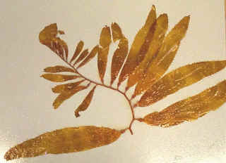
Phaeophyta (brown algae):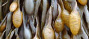 1500 species
- Contain chlorophyll a & chlorophyll c and fucoxanthin (brown pigment) as accessory pigments
- Most are multicellular growing in cooler marine habitats
- Include kelps & seaweeds
- Largest protists
- Specialized rootlike holdfasts anchor thallus to rocks
- Specialized air bladders keep leaflike blades afloat near surface to get light for photosynthesis
- Stemlike structures are called the stipe and support the blades
- Store food as a carbohydrate called laminarin
- Include Laminaria & Fucus
 |
 |
| Laminaria | Fucus |
- Macrocystis or giant kelp contains algin in its cell walls which is used in cosmetics, some drugs, ice cream, etc.
Rhodophyta (red algae):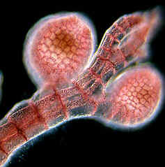 4000 species
- Multicellular algae that mainly grow deep in warm marine waters
- Some freshwater species exist
- Highly branched thallus
- Contain chlorophyll a & phycobilins (red pigments) to trap sunlight for photosynthesis
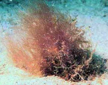
Polysiphonia (red algae)
- Store food as starch
- Cell walls contain cellulose and agar (used as a base in culture dishes to grow microbes)
- Some species contain carageenan in their cell walls used for gelatin capsules & in some cheeses
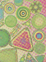
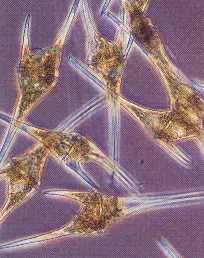
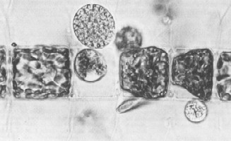
Bacillariophyta (diatoms):11,500 species
- Abundant in marine & freshwater habitats
- Called phytoplankton & start many aquatic food chains
- Contain chlorophyll a & c, carotenoids (orange pigments), & xanthophyll (yellow pigments)
- Store food as starch & contain mainly cellulose in their cell walls
- Lack cilia & flagella
- Have glass like shells or valves containing SiO2 that fit together in 2 parts
Diatoms
Copyright © by Holt, Rinehart and Winston
- Centric diatoms are marine & have circular or triangular shells
- Pennate diatoms are found in freshwater & have rectangular shells
- When diatoms die, they form a layer called diatomaceous earth that is abrasive and used in detergents, toothpaste, fertilizers, etc.
Dinoflagellata or Pyrrophyta (dinoflagellates):1100 species
- Major producers in marine habitats
- Small, unicellular organisms making up plankton
- Many are photosynthetic, but some are colorless heterotrophs
- Photosynthetic dinoflagellates are yellow to brown in color due to chlorophyll a & c and carotenoids
Copyright © by Holt, Rinehart and Winston
- Have 2 flagella that spin and move the dinoflagellate through water
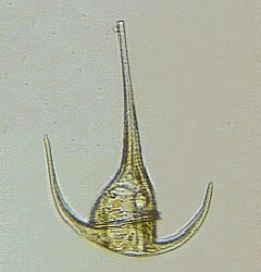
- Store food as starch
- Some dinoflagellates are covered with armor like plates & spines made of cellulose
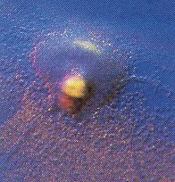
- Often undergo algal blooms where their numbers greatly increase
- Produce a toxic substance and cause poisonous red tides (water appears red due to red pigments in the dinoflagellates)

Red Tide
- Some such as Noctiluca can produce light by bioluminescence

Photograph by Robert Brons
Chrysophyta (golden algae) 850 Species:
- Most are live in freshwater habitats, but some are marine
- Unicellular algae containing chlorophyll a & c and the brown pigment fucoxanthin and carotenoids
- Many have flagella for movement
- May be naked or have cellulose cell walls or silica scales or shells
- May form highly resistant cysts to survive beneath frozen lake surfaces in winter
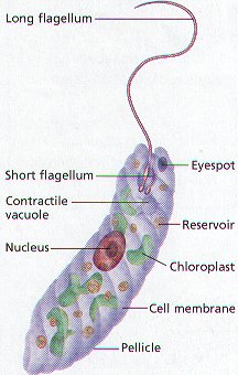
Euglenophyta 1000 Species:
- Unicellular algae that lack cell walls
- Have a flexible protein covering called the pellicle
- Called euglenoids
- Possess chlorophyll a & b and carotenoids
- Store food as paramylon (polysaccharide)
- Most live in freshwater, but some live in moist soil & the digestive tracts of certain animals
Copyright © by Holt, Rinehart and Winston
- Euglena is a common euglenoid found in freshwater
a. Elastic, transparent pellicle below cell membrane
b. Contractile vacuole to pump out excess water
c. Chloroplasts to make food by photosynthesis
d. Can be heterotrophic in the absence of light
Fungal-Like Protists
Characteristics of Fungal Protists:
- Includes cellular slime molds, plasmodial slime molds, & water molds
- Unique life cycles with two phases
- Multicellular, heterotrophic organisms
- Little tissue specialization
- Usually small & live in moist or watery habitats
- Act as decomposers breaking down dead organic matter
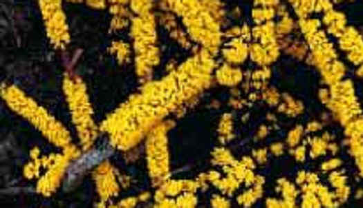
Slime molds: 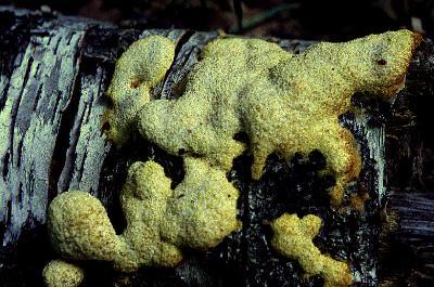
- Shiny, wet appearance
- Often brightly colored (yellow or orange)
- Have unique life cycles with 2 phases — a mobile feeding stage & a nonmotile reproductive stage
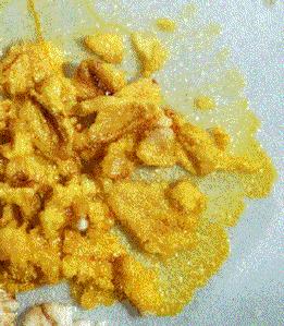
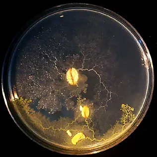
Feeding Stage of Slime Mold
Copyright © by Holt, Rinehart and Winston
- Fungal-like in nutrition (absorptive heterotrophs that break down dead organic matter)
- May be saprophytes or parasites
Saprophytic Slime Mold
- Multinucleate body mass
- May have a mobile, ameba-like feeding stage
- Make a reproductive structure or fruiting body that produces spores
- Often found on decaying wood or leaves
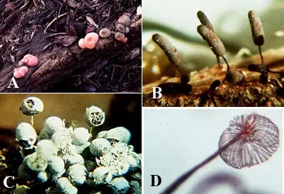
A is Lycogala epidendrum, B is Comatricha typhoides, C is Badhamia utricularia, D is Dictydium
- Two groups of slime molds exist — Cellular slime molds & Plasmodial slime molds
- Cellular Slime Molds (Phylum Acrasiomycota)
- Plasmodial Slime Molds (Phylum Myxomycota)
Copyright © by Holt, Rinehart and Winston
Acrasiomycota (Cellular Slime Molds):
- Alternate in their life cycle between amoeboid feeding stage & spore-producing fruiting body

- Live in freshwater, moist soil
- Clump together into masses called pseudoplasmodium whenever little food is available
- Cells in the pseudoplasmodium are independent but move together “slug-like”
- Pseudoplasmodium settles & forms fruiting body with spores
- Spores spread by wind to new location & form individual amoeboid feeding stage
Myxomycota (Plasmodial Slime Molds):
- Exist as a plasmodium ( a mass of cytoplasm with many nuclei)
- Plasmodium creeps along over decaying material
- Decomposes & absorbs plant material as food
- When food is scarce, the plasmodium forms stalked fruiting bodies with spores that are resistant to bad environmental conditions
- When conditions turn favorable, spores form a new plasmodium
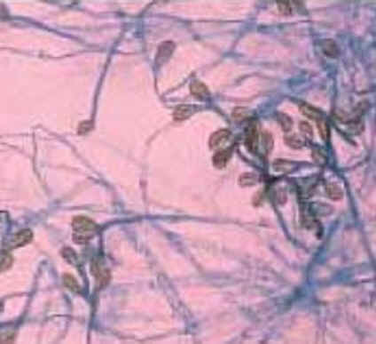
Oomycota (Water Molds):
- Fungal-like organism made of branching filaments with cell walls of cellulose

Branching Filaments of Water Mold
- Aquatic water molds are parasites on fish forming furry growths on their gills
- May act as decomposers in water of dead plants & animals
- May be pathogenic to plants
e.g. Phytophthora infestans caused blight in potatoes (Irish Potato Famine in 19th century) - Blight in plants decays & discolors stems & leaves


Blight on Leaves & Potatoes
- Water molds reproduce sexually & asexually
- Motile zoospores are asexually produced from reproductive structures called sporangium
- In sexual reproduction, cells with eggs form tubes to cells with sperm to fertilize & form new branching filaments
Chytridiomycota (Chytrids):
- Aquatic protists that form gametes & zoospores
- Most are unicellular or filamentous
- May be saprophytes (decomposers) or parasites on algae, plants, or insects
- May be a link between protists & fungi


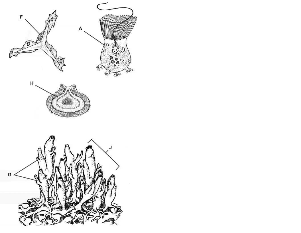
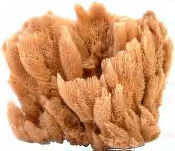 Sponges – A Coloring Worksheet
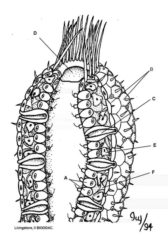


 Insulin is needed because it reduces blood glucose levels in the blood. It causes cells, especially fat and muscle cells, to absorb glucose from the blood. The glucose is needed for cellular respiration or converted into glycogen. The glycogen is stored in the liver or the muscles for future use in cellular respiration.