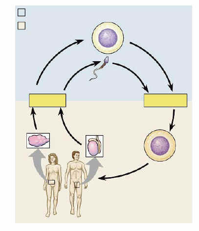
AP Lab 3 Sample 3 Mitosis


Lab 3    Mitosis and Meiosis


Introduction:


All new cells come from previously existing cells. New cells are formed by karyokinesis- the process in cell division which involves replication of the cell’s nucleus and cytokinesis-the process in cell division which involves division of the cytoplasm. Two types of nuclear division include mitosis and meiosis. Mitosis typically results in new somatic, or body, cells. Mitotic cell division is involved in the formation of an adult organism from a fertilized egg, asexual reproduction, regeneration, and maintenance or repair of body parts. Meiosis results in the formation of either gametes in animals or spores in plants. The cells formed have half the chromosome number of the parent cell.

Mitosis is best observed in cells that are growing at a rapid pace, such as in the whitefish blastula or onion root cell tips. The root tips contain a special growth region called the apical meristem where the highest percentage of cells are undergoing mitosis. The whitefish blastula is formed immediately after the egg is fertilized, a period of rapid growth and numerous cell divisions where mitosis can be observed.

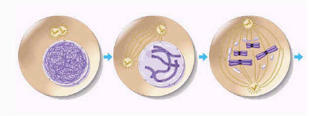
There are several stages included in before, during, and following mitosis. Interphase occurs right before a cell enters mitosis. During interphase, the cell will have a distinct nucleus with one or more nucleoli, which is filled with a fine network of threads of chromatin. During interphase, DNA replication occurs. After duplication the cell is ready to begin mitosis. Prophase is when the chromatin thickens until condensed into distinct chromosomes. The nuclear envelope dissolves and chromosomes are in the cytoplasm. The first signs of the microtubule-containing spindle also begin to appear. Next the cell begins metaphase. During this phase, the centromere of each chromosome attaches to the spindle and are moved to the center of the cell. This level position is called the metaphase plate. The chromatids separate and pull to opposite poles during the start of anaphase. Once the two chromatids are separate, each is called a chromosome. The last stage of mitosis is telophase. At this time, a new nuclear envelope is formed and the chromosomes gradually uncoil, forming the fine chromatin network seen in interphase. Cytokinesis may occur forming a cleavage furrow that will form two daughter cells when separated.

Meiosis is more complex than mitotic stages and involves two nuclear divisions called Meiosis I and Meiosis II. They result in the production of four haploid gametes and allow genetic variation because of crossing over of genetic material. Prior the process, interphase replicates the DNA. During prophase I, the first meiotic stage, homologous chromosomes move together to form a tetrad and synapsis also begins. This is where crossing over occurs, resulting in the recombination of genes. In Metaphase I, the tetrads move to the metaphase plate in the middle of the cell as on mitotic metaphase. Anaphase I brings the tetrads back to their original two stranded form and moves them to opposite poles. During Telophase I, the centriole is finished and the cell prepares for a second division. In Meiosis II, in Prophase II, centrioles move to opposite ends of the chromosome group. In Metaphase II, the chromosomes are centered within the center of each daughter cell. Anaphase II involves the centromere of the chromatids separating. Telophase II occurs when the divided chromosomes separate into different cells, known as haploid cells.

Sordaria fimicola, an ascomycete fungus, can be used to demonstrate the results of crossing over during meiosis. It spends most of its life haploid and only becomes diploid when the fusion of the mycelia of two different strains results in the fusion of two different types of haploid nuclei to form a diploid nucleus. Meiosis, followed by mitosis, in Sordaria results in the formation of eight haploid ascospores contained within a sac called an ascus. They are contained in a perithecium, a fruiting body, until mature enough to be released. The arrangement of spores directly reflects whether or not crossing over occurred. If an ascus has four tan ascospores in a row and four black ascospores in a row -4:4 arrangement, then no crossing over has taken place. If the asci has black and tan ascospores in sets of two -2:2:2:2 arrangement, or two pairs of black ascospores and four tan ascospores in the middle -2:4:2 arrangement, then crossing over has taken place.


Hypothesis:


The stages of mitosis can be examined in whitefish blastula and onion root cell tips by using a microscope. The process of crossing over and the stages of meiosis only occur during the creation of gametes and spores.


Materials:


Exercise 3A

The materials necessary for this exercise are a light microscope, prepared slides of whitefish blastula, onion root cell tips, pencil, and paper.

Exercise 3B

For this portion of the lab, materials needed are a bag of color-coded connecting beads and magnetized “centromeres,” several trays, and labels marked interphase, prophase, metaphase, anaphase, and telophase.


Methods:


Exercise 3A.1: Observing Mitosis

During this experiment, prepared slides of whitefish blastula and onion root tips should be observed under the 10X and 40X objectives of a light microscope. A cell in each stage of mitosis should be identified and sketched.

Exercise 3A.2: Time for Cell Replication

In this section of the lab, use the highest power objective on the microscope to observe and count every cell in the field of view. The cells should be counted according to the stage of mitosis they are in. At least 200 cells and 2 fields of view should be examined and counted. The percentage of cells in each stage is then recorded and the amount of time spent in each phase is calculated.

Exercise 3B.1: Simulation of Meiosis

For this portion of the experiment, a chromosome simulation kit will be used to demonstrate meiosis. Two sets of two strands with each set a different color, are connected to simulate DNA replication in both of the homologous pairs, the stage called interphase. Next, the chromosomes were entwined to represent synapsis in the stage known as prophase. Sections of beads were entwined between the pairs as in crossing over and aligned at the equator. Beads of each pair exchange places, representing metaphase. Next, anaphase was simulated by the homologous pairs being separated to opposite sides of the tray, or in terms of the “chromosomes,” the cell. Pushing the chromosomes into two separate cells, or trays, mimicked telophase.

Meiosis II was simulated as well. Prophase II is shown by the separation of the two beads, but no true change. The chromosomes again move to the equator during metaphase II, and in anaphase II, the two chromatids are separated and moved to opposite poles. Telophase II separates the chromosomes into four different cells.

Exercise 3B.2: Crossing Over during Meiosis in Sordaria

Prepared slides of Sordaria fimicola were observed under a light microscope. The asci were identified as either 4:4 or asci showing crossover. These readings were recorded. The percentage of each and map units were calculated.


Results:


Exercise 3A


Whitefish Blastula

Onion Root Cell Tips


Why is it more accurate to call mitosis “nuclear replication” rather than “cellular division”? It is more accurate to describe mitosis as “nuclear replication” because the cell does not divide in any of the mitotic steps. The entire process of mitosis is a series of steps that divides the nucleus into two separate nuclei at opposite poles. When a cell is truly split, the process is known as cytokinesis.


Explain why the whitefish blastula and onion root tips are selected for a study of mitosis. The blastula is what is formed directly following fertilization and, therefore, the cell is growing and many of the phases can be seen at this time. Onion root tip cells are also specimens that include a large amount of cell growth and a high percentage of cells experiencing mitotic activities.


Table 1: Number of Cells in Each Stage of Mitosis and Amount of Time Spent in Each Stage


Number of Cells


Field 1


Field 2

Total
 

Interphase

71 101 172 73.2% 1054.0
 

Prophase

13 15 28 12.0% 171.6
 

Metaphase

12 13 25 10.6% 153.2
 

Anaphase

1 2 3 1.3% 18.4
 

Telophase

3 4 7 3.0% 42.9
 

Total Cells Counted

235


If your observations had not been restricted to the area of the root tip that is actively dividing, how would your results differ? The majority of the cells would be in the stage of interphase and the results would be more difficult to gain and inaccurate.


Based on the data in Table 3.1, what can you infer about the relative length of time an onion root-tip cell spends in each stage of cell division? Prophase is the longest stage of mitosis (though Interphase, which occurs prior mitosis, takes up the most time of the cell’s life). Then, based on the data gained, the time spent in each stage decreases as you go further along.


Exercise 3B


List three major differences between the events of mitosis and meiosis. In mitosis, the nucleus divides once, and in meiosis, the nucleus is divided twice. Mitosis produces two identical daughter cells and meiosis produces up to four different cells. Synapsis and crossing over do not take place in mitosis, but do in meiosis.


Compare mitosis and meiosis with respect to each of the following.

Table 2: Comparing Mitosis and Meiosis


Topic Being Compared


Mitosis


Meiosis


Chromosome number of Parent Cells

Diploid (2n) Diploid (2n)
 

Number of DNA Replications

Once Once
 

Number of Divisions

One Two
 

Number of Daughter Cells

Two Four
 

Chromosome Number of Daughter Cells

Diploid (2n) Haploid (n)
 

Purpose

Growth and repair Production of gametes or spores


How are Meiosis I and Meiosis II different? Meiosis I begins with a tetrad and separates the homologous pairs. Meiosis II separates the two sister chromatids into haploids.


How do oogenesis and spermatogenesis differ? Oogenesis produces egg cells and spermatogenesis produces sperm cells.


Why is meiosis important for sexual reproduction? In meiosis the chromosome number is reduced to n so that it can be fertilized and void of any related (fertilized 2n) defects. Crossing- over occurs during meiosis, allowing for variations in the organisms created.


Table 3: The Number of Crossovers and Non-Crossovers


Number of 4:4


Number of Asci Showing Crossover


Total Asci


% Asci Showing Crossover Divided by 2


Gene to Centromere Distance (Map Units)

59 68 127 26.8% (1)


2. Draw a pair of chromosomes in MI and MII, and show how you would get a 2:4:2 arrangement of ascospores by crossing over.

Error Analysis:


Because the results gathered in the lab were based mostly on observations and sketching, chances of error are slim. However, when counting the number of cells in specific stages in Exercise 3A, mistakes could have occurred. When identifying these stages in Exercise 3A, mistakes were also possible.


Discussion and Conclusion:


The stages of mitosis were observed and timed in Exercise 3A. These stages are prophase, metaphase, anaphase, and telophase. Prophase is the most time-consuming phase, while anaphase is the least time-consuming. Mitosis is just one portion of a cell’s life. The longest time of a cell’s life (73% to be exact) is spent in interphase, a phase just prior to prophase. During this phase, DNA replication takes place. Prophase involves the first signs of cell division with a thickening of the chromatin threads until the chromatin is condensed to chromosomes. In metaphase the chromosomes move to the center of the spindle and the centromere attaches to the spindle. During anaphase the chromatids are separated and moved to opposite ends of the poles. The final stage, telophase, involves the condensation of the chromosomes and the formation of a new nuclear envelope. Following telophase, cytokinesis may occur and the cytoplasm will be divided into two cells.

During the first section of Exercise 3B, the stages of meiosis were simulated using magnetic beads and centromeres with trays serving as the “cell.” Crossing over in Sordaria was observed using a microscope in the second portion of Exercise 3B. Using the information, the map units were then determined. The distance of the gene relative to the centromere in the Sordaria was 26.8 map units.

BACK


Cell Respiration


Cell Respiration

Overview:
In this experiment, you will work with seeds that are living but dormant. A seed contains an embryo plant and a food supply surrounded by a seed coat. When the necessary conditions are met, germination occurs, and the rate of cellular respiration greatly increases. In this experiment you will measure oxygen consumption during germination. You will measure the change in gas volume in respirometers containing either germinating or non-germinating pea seeds. In addition, you will measure the rate of respiration of these peas at two different temperatures.

Objectives:
Before doing this laboratory you should understand:

  • how a respirometer works in terms of the gas laws; and
  • the general processes of metabolism in living organisms.

After doing this laboratory you should be able to:

  • calculate the rate of cell respiration from experimental data.
  • relate gas production to respiration rate; and
  • test the effect of temperature on the rate of cell respiration in ungerminated versus germinated seeds in a controlled experiment.

Introduction:
Cellular respiration is the release of energy from organic compounds by metabolic chemical oxidation in the mitochondria within each cell. Cellular respiration involves a series of enzyme-mediated reactions. The equation below shows the complete oxidation of glucose. Oxygen is required for this energy-releasing process to occur.

C6H12O6 + 6O2 —–> 6 CO2 + 6 H2O + 686 kilocalories of energy / mole of glucose oxidized

By studying the equation above, you will notice there are three ways cellular respiration could be measured. One could measure the:

1. Consumption of O2 ( How many moles of oxygen are consumed in cellular respiration?)

2. Production of CO2 ( How many moles of carbon dioxide are produced by cellular respiration?)

3. Release of energy during cellular respiration.

In this experiment, the relative volume of O2 consumed by germinating and non-germinating (dry) peas at two different temperatures will be measured.

Background Information:
A number of physical laws relating to gases are important to the understanding of how the apparatus that you will use in this exercise works. The laws are summarized in the general gas law that states:

PV = nRT

where

P is the pressure of the gas,

V is the volume of the gas,

n is the number of molecules of gas,

R is the gas constant ( its value is fixed), and

T is the temperature of the gas (in K0).

This law implies the following important concepts about gases:

1. If temperature and pressure are kept constant, then the volume of the gas is directly proportional to the number of molecules of gas.

2. If the temperature and volume remain constant, then the pressure of the gas changes in direct proportion to the number of molecules of gas present.

3. If the number of gas molecules and the temperature remain constant, then the pressure is inversely proportional to the volume.

4. If the temperature changes and the number of gas molecules is kept constant, then either pressure or volume ( or both ) will change in direct proportion to the temperature.

It is also important to remember that gases and fluids flow from regions of high pressure to regions of low pressure.

In this experiment, the CO2 produced during cellular respiration will be removed by potassium hydroxide (KOH) and will form solid potassium carbonate (K2CO3) according to the following reaction.

CO2 + 2 KOH —-> K2CO3 + H2O

Since the carbon dioxide is being removed, the change in the volume of gas in the respirometer will be directly related to the amount of oxygen consumed. In the experimental apparatus if water temperature and volume remain constant, the water will move toward the region of lower pressure. During respiration, oxygen will be consumed. Its volume will be reduced, because the carbon dioxide produced is being converted to a solid. The net result is a decrease in gas volume within the tube, and a related decrease in pressure in the tube. The vial with glass beads alone will permit detection of any changes in volume due to atmospheric pressure changes or temperature changes. The amount of oxygen consumed will be measured over a period of time. Six respirometers should be set up as follows:

Respirometer Temperature Contents
1 Room Germinating seeds
2 Room Dry Seeds and Beads
3 Room Beads
4 100C Germinating Seeds
5 100C Dry Seeds and Beans
6 100C Beads

Procedure:
 1.Prepare a room-temperature bath (approx. 25 degrees Celsius) and a cold-water bath (approx. 10 degrees Celsius).

2.Find the volume of 25 germinating peas by filling a 100mL graduated cylinder 50mL and measuring the displaced water.

3.Fill the graduated cylinder with 50mL water again and drop 25 non-germinating peas and add enough glass beads to attain an equal volume to the germinating peas.

4.Using the same procedure as in the previous two steps, find out how many glass beads are required to attain the same volume as the 25 germinating peas.

5.Repeat steps 2-4. These will go into the 10-degree bath.

6.To assemble 6 respirometers, obtain 6 vials, each with an attached stopper and pipette. Number the vials. Place a small wad of absorbent cotton in the bottom of each vial and, using a dropper, saturate the cotton with 15% KOH (potassium hydroxide). It is important that the same amount of KOH be used for each respirometer.

7.Place a small wad of dry, nonabsorbent cotton on top of the saturated cotton.

8.Place the first set of germinating peas, dry peas & beads, and glass beads in the first three vials, respectively. Place the next set of germinating peas, dry peas & beads, and glass beads in vials 4, 4, and 6, respectively. Insert the stopper with the calibrated pipette. Seal the set-up with silicone or petroleum jelly. Place a weighted collar on each end of the vial. Several washers around the pipette make good weights.

9.Make a sling of masking tape attached to each side of the water baths. This will hold the ends of the pipettes out of the water during an equilibration period of 7 minutes. Vials 1, 2, and 3 should be in the room temperature bath, and the other three should be in the 10 degree bath.

10.After 7 min., put all six set-ups entirely into the water. A little water should enter the pipettes and then stop. If the water continues to enter the pipette, check for leaks in the respirometer.

11.Allow the respirometers to equilibrate for 3 more minutes and then record the initial position of the water in each pipette to the nearest 0.01mL (time 0). Check the temperature in both baths and record. Record the water level in the six pipettes every 5 minutes for 20 minutes.

Table 5.1: Measurement of O2 Consumption by Soaked and Dry Pea Seeds at Room Temperature (250C) and 100C Using Volumetric Methods.

Temp
(oC)
Time
(min)
Beads Alone Germinating Peas

Dry Peas and Beans

Reading at time X Diff* Reading at time X Diff* Corrected Diff. ^ Reading at time X Diff* Corrected diff ^
Initial – 0
0-5
5- 10
10 -15
15-20
Initial – 0
0-5
5- 10
10 -15
15-20

* difference = ( initial reading at time 0) – ( reading at time X )

^ corrected difference = ( initial pea seed reading at time 0 – pea seed reading at time X) – ( initial bead reading at time X).

Analysis of Results:
1. In this investigation, you are investigating both the effect of germination versus non-germination and warm temperature versus cold temperature on respiration rate. Identify the hypothesis being tested in this activity.

_______________________________________________________________________

_______________________________________________________________________

2. This activity uses a number of controls. Identify at least three of the control, and describe the purpose of each control.

_______________________________________________________________________

_______________________________________________________________________

_______________________________________________________________________

_______________________________________________________________________

_______________________________________________________________________

_______________________________________________________________________

3. Graph the results from the corrected difference column for the germinating peas and dry peas at both room temperature and 100C.

a. What is the independent variable? ____________________________________________________

b. What is the dependent variable? ______________________________________________________

Graph Title: _____________________________________________________________________

Graph 5.1


4. Describe and explain the relationship between the amount of oxygen consumed and time.

_______________________________________________________________________

_______________________________________________________________________

_______________________________________________________________________

_______________________________________________________________________

5. From the slope of the four lines on the graph, determine the rate of oxygen consumption of germinating and dry peas during the experiments at room temperature and 100C. Recall that rate = delta Y/delta X.

Table 5.2

Condition Show Calculations Here Rate in ml.O2 / min
Germinating Peas/100C  


Germinating peas /Room Temperature  


Dry peas/100C  


Dry Peas /Room Temperature  


6. Why is it necessary to correct the readings from the peas with the readings from the beads?

_______________________________________________________________________

_______________________________________________________________________

_______________________________________________________________________

_______________________________________________________________________

7. Explain the effect of germination ( versus non-germination) on peas seed respiration.

_______________________________________________________________________

_______________________________________________________________________

_______________________________________________________________________

_______________________________________________________________________

8. What is the purpose of KOH in this experiment?

_______________________________________________________________________

_______________________________________________________________________

_______________________________________________________________________

_______________________________________________________________________

9. Why did the vial have to be completely sealed around the stopper?

_______________________________________________________________________

_______________________________________________________________________

_______________________________________________________________________

_______________________________________________________________________

10. If you used the same experimental design to compare the rates of respiration of a 25 g. reptile and a 25 g. mammal, at 100C, what results would you expect/ Explain your reasoning.

_______________________________________________________________________

_______________________________________________________________________

_______________________________________________________________________

_______________________________________________________________________

_______________________________________________________________________

_______________________________________________________________________

11. If respiration in a small mammal were studied at both room temperature (210C) and 100C, what results would you predict? Explain your reasoning.

_______________________________________________________________________

_______________________________________________________________________

_______________________________________________________________________

_______________________________________________________________________

12. Explain why water moved into the respirometer pipettes.

_______________________________________________________________________

_______________________________________________________________________

_______________________________________________________________________

________________________________________________________________________
AP LAB PAGE


DNA & Protein Synthesis Chapter 10 Worksheet


    DNA & Protein Synthesis


Section 10-1 DNA

1. What does DNA stand for?

2. What is DNA’s primary function?

3. What is the function of proteins?

4. What are the repeating subunits called that make up DNA?

5. Name the 3 parts of a DNA nucleotide.

6. Sketch and label a DNA nucleotide.

7. Name the 4 nitrogen bases on DNA.

8. What is the difference between a purine & a pyrimidine?

9. Name 2 purines.

10. Name 2 pyrimidines.

11.Who is responsible for determining the structure of the DNA molecule & in what year was this done?

12. The model of DNA is known as a ____________________________ because it is composed of two ___________________ chains wrapped around each other.

13. What makes up the sides of a DNA molecule?

14. What makes up the “steps” of a DNA molecule?

15. How did Rosalind Franklin contribute to determining the structure of DNA?

16. What type of bonds holds the DNA bases together? Are they strong or weak bonds?

17. What makes up the “backbone” of the DNA molecule?

18. On DNA, a ____________________ base will always pair with a __________________ base.

19. What is the most common form of DNA found in organisms?

20. How many base pairs are in a full turn or twist of a DNA molecule?

21. Name the complementary base pairs on DNA.

22. How many hydrogen bonds link cytosine & guanine? adenine & thymine?

23. How does the nucleotide sequence in one chain of DNA compare with the other chain of DNA?

24. Why must DNA be able to make copies of itself?

25. Define DNA replication.

26. What is the first step that must occur in DNA replication?

27. What acts as the template in DNA replication?

28. What is a replication fork?

29. What enzymes help separate the 2 strands of nucleotides on DNA? What bonds do they break?

30. What is the function of DNA polymerases?

31. ____________________ are joined to replicating strands of DNA by ________________ bonds.

32. If the sequence of nucleotides on the original DNA strand was A – G – G – C – T – A, what would be the nucleotide sequence on the complementary strand of DNA?

33. Does replication of DNA begin at one end and proceed to the other? Explain.

34. Why does DNA replication take place at many places on the molecule simultaneously?

35. When replication is complete, how do the 2 new DNA molecules compare to each other & the original DNA molecule?

36. Is DNA replicated (copied) before or after cell division?

37. Sketch & label DNA replication. (Figure 10-5, page 188)

38. What is the error rate in DNA replication? What helps lower this error rate to 1 in 1 billion nucleotides?

39. What is a mutation?

40. Name several things that can cause DNA mutations.


Section 10-2 RNA


41. What sugar is found on DNA?

42. What base is missing on RNA, & what other base replaces it?

43. Uracil will pair with what other on DNA?

44. Is RNA double or single stranded?

45. Name the 3 types of RNA and tell the shape of each.

46. Which type of RNA copies DNA’s instructions in the nucleus?

47. Which type of RNA is most abundant?

48. What does tRNA transport?

49. What 2 things make up ribosomes?

50. Define transcription.

51. In what part of a cell are proteins made?

52. What is RNA polymerase & tell its function.

53. What are promoters?

54. Where does RNA polymerase bind to the DNA it is transcribing?

55.What makes the beginning of a new gene on DNA in eukaryotes?

56. What do promoters mark the beginning of on prokaryotic DNA?

57. When a promoter binds to DNA, What happens to the double helix?

58. Are both strands of DNA copied during transcription?

59. As RNA polymerase moves along the DNA template strand, what is being added?

60. What bases pair with each other during transcription?

61. What is the termination signal?

62. What happens when RNA polymerase reaches the termination signal?

63. What are the products of transcription called?

64. Transcripts are actually ____________________________ molecules.

65. In transcription, ________________________’s instructions for making a protein

are copied by _______________________.

66. Which RNA molecules are involved in the synthesis (making) of a protein?

67. What happens to the newly made mRNA molecule following transcription in the nucleus?


Section 10-3 Protein Synthesis


68. What makes up proteins, what are the subunits called, & what bonds them together?

69. How many different kinds of amino acids make up proteins?

70. What determines how protein polypeptides fold into 3-dimensional structures?

71. Why does a protein need a 3-dimensional structure?

72. What is the genetic code & why is it important?

73. What is a codon & what does each codon code for?

74. How many codons exist?

75. Name the amino acid coded for by each of these codons:

a. UUA

b. AUU

c. UGU

d. AAA

e. GAG

f. UAA

76. What codon starts protein synthesis?

77. What codons stop protein synthesis?

78. Proteins are synthesized (made) at what organelle in the cytosol?

79. Sketch and label a tRNA molecule & tell its function.

80. Define translation & tell how it starts.

81. Where are amino acids found in a cell & how are they transported?

82. What is an anticodon & where is it found on tRNA?

83. What codon on mRNA would bind with these anticodons: (use table 10-1, page 194)

a. AAA

b. GGA

c. UAC

d. CGU

84. What are ribosomes made of and in what 2 places can they be found in a cell?

85. What is the difference between proteins made by free ribosomes & those made by attached, membrane proteins on the ER?

86. How many binding sites are found on the ribosomes and what does each site hold?

87. To start making a protein or _________________________________, a ribosome attaches to the ______________________________ codon on the __________________ transcript.
88. The start codon, AUG, pairs with what anticodon on a tRNA molecule?

89. What amino acid does the start codon always carry?

90. What type of bonds are the ones that attach amino acids to each other in a growing polypeptide?

91. __________________________ are linked to make proteins as a ______________________ moves along the mRNA transcript.

92. What ends translation?

93. Can more than one ribosome at a time translate an mRNA transcript? Explain.

94. What determines the primary structure of a protein?

95. What would the translation of these mRNA transcripts produce?

a. UAA CAA GGA GCA UCC

b. UGA CCC GAU UUC AGC

BACK