Osmosis Lab3 Sample


Osmosis Through the Cell Membrane of an Egg
by Brett Helms

Introduction:
An egg can be used to show the process of osmosis through the egg’s cell membrane. Osmosis is the process in which water diffuses across a cell membrane from an area of high concentration to an area of low concentration. Osmosis occurs when the cell needs to release water to the exterior of the cell membrane or it can occur when water needs to diffuse into the cell. Diffusion is the movement of molecules from an area of high concentration to an area of lower concentration. Diffusion and osmosis are both types of passive transport. A passive transport is the movement of molecules across a cell membrane without the cell having to exert any energy. Water will move into the cell when the solution is hypotonic to the cytosol. A hypotonic solution is defined as a solution whose solute concentration is lower than that inside a cell. The cell membrane is the thin outer layer of a cell. It allows certain substances to diffuse across, into and out of the cell. When a solution is hypertonic the solute concentration is higher than that inside a cell. This would result in molecule movement into a cell. When a solution is isotonic, the solute concentration equals that inside a cell. This would result into no movement of molecules across the cell membrane. Molecules that are very small but not soluble in lipids may diffuse across the membrane by moving through the pores of the cell membrane.


Hypothesis:
The water and the vinegar will diffuse through the cell membrane more easily then the syrup.


Materials:
The materials used included paper, pencil, 200ml beakers, vinegar, distilled water, syrup, 2 eggs, tray, plastic wrap, colored marker, tape, and a digital balance.


Methods:
The two eggs were weighed on a digital balance and their initial masses were recorded. The eggs were then placed in a beaker of vinegar to dissolve the calcium shell and leave the cell membrane exposed. The eggs were weighed after they were removed from the vinegar and their masses were recorded. The two eggs were then placed in a beaker of syrup.  The eggs were once again weighed on the digital balance and their masses recorded. The eggs were then placed a beaker of distilled water. After the eggs were removed and their weights and appearance recorded.


Results:


Results of Osmosis
Table for Egg 1


Solutions Egg Mass Before in Grams Observations Before Placed in Solution Egg Mass After in Grams Observations After removed from Solution
Vinegar 56.9g Hard exterior 84.3g Egg lost shell
Syrup 84.3g Shell had a firm jelly-like membrane 48.4g Outside not firm anymore, very shriveled
Water 48.5g The jelly-like membrane was soft and not firm at all 77.9g Egg firmed up and looked like the shell just came off


Results of Osmosis
Table for Egg 2


Solutions


Egg Mass Before in Grams


Observations Before placed in solution


Egg Mass After in Grams


Observations After removed from solution

Vinegar 60.5g Hard exterior 87.65g Egg Lost Shell
Syrup 87.65g Shell had a firm jelly-like membrane 54.4g Outside not firm anymore
Water 54.4g The jelly-like membrane was soft and not firm at all 79.3g Egg firmed up and looked like the shell just came off


1. When the egg was placed in the water, in which direction did the water molecules move? The water molecules moved into the egg.

2. On what evidence do you base this? The egg mass increased.

3. How do you explain the volume of liquid remaining when the egg was removed from the syrup? It was less because some had moved into the egg.

4. When the egg was placed in the water after being removed from the syrup, in which direction did the water move? The water moved into the egg.


Error Analysis:
This experiment is based upon changes in egg mass in various solutions. The balance used was not checked for accuracy with two standard masses. The changes in masses may or may not be correct.


Discussion and conclusion:
There were significant increases in egg mass when the eggs were submerged in vinegar and water. However there was a significant decreased in egg mass when the eggs were submerged in syrup causing the egg to appear shriveled. The reason for the increasing and decreasing is called diffusion, which is the movement of molecules from an area of high concentration to an area of lower concentration. That is why when the egg was submerged in syrup much of the water in the egg diffused out because of the higher amount of water in the egg compared to the smaller amount in the syrup. That is also why when the egg was placed in vinegar and in distilled water its mass increased meaning that more water molecules moved into the eggs because the solution outside the egg was lower in water than that inside the egg. These results support the hypothesis that water diffuses from an area of high to an area of low concentration across a cell membrane. This experiment demonstrated the process of osmosis through the egg cell membrane.

BACK


Monohybrid Corn Lab


Monohybrid Cross in Corn

INTRODUCTION:

A cross between individuals that involves one pair of contrasting traits is called a monohybrid cross. First we will use Punnett square diagrams to predict the results of various monohybrid crosses. We will then examine ears of corn Purple results from the dominant allele (P), and yellow from the recessive allele (p). We will be making observations and assumptions for both the genotype or genetic make-up, and the phenotype or external appearance.
Review genetics and the use of Punnett squares in a biology text before doing this experiment.

MATERIALS:

Appropriate ears of corn.
(You can purchase them from a biological supply, such as Carolina. You need a heterozygous X heterozygous 3:1, and a monohybrid test cross 1:1.)

PROCEDURE:

Theoretical: We will use a Punnett square to examine the theoretical outcome of possible monohybrid crosses.

1. The first cross is with a Homozygous dominant parent (PP), and a Homozygous recessive parent (pp).
Fill in the Punnett square. Each box represents a genotype possibility for an offspring. Place the allele donated by each parent in the corresponding box. Now list the possible genotypes and their corresponding phenotype.
Remember: The genotype is represented by the two letters for the offspring, and the phenotype is a color.
Remember: If an individual’s genotype is heterozygous, the dominant trait will be expressed in the phenotype.
Give the percent possible for the phenotypes.

2. Now look at a cross between a Homozygous dominant parent (PP), and a Heterozygous parent (Pp). Fill in as in step one.

3. Next we will examine the possibilities with a cross between a Heterozygous parent (Pp), and another Heterozygous parent (Pp). Fill-in as before.

4. Finally we will examine a Test cross. A test cross is between a Homozygous recessive parent (pp), and a Heterozygous parent (Pp). Again, fill-in as before.


Actual cross: Now we will make a count of an actual cross and compare the calculations to the phenotype percentages of the theoretical.

5. Obtain an ear of corn that is the result of Heterozygous X Heterozygous. Count and record the purple and yellow kernels, and record the numbers. Also record the total number of kernels.

TOTAL purple: _______ TOTAL yellow: _______ TOTAL all kernels: _______

6. Now find the percent of purple and yellow kernels. To find the percent of purple, divide the total purple by the total for all kernels, then multiply by 100.

Percent purple: _______ % Percent yellow: _______ %

Compare your results with the theoretical answers you obtained for the Heterozygous X Heterozygous cross.

7. Results for genetic crosses are often recorded as ratios. Calculate the ratio of purple to yellow. To do this, use their totals. The smaller total you call 1, and write it in the appropriate space. Then Divide the larger one by the smaller, and round to the nearest whole number. Record this number in the appropriate space.

Ratio of phenotypes for this cross.

PURPLE to YELLOW
____ : ____

8. Now repeat steps 5 through 7 for an ear of corn that is the result of a TEST CROSS: Homozygous recessive X Heterozygous.

TOTAL purple: _______ TOTAL yellow: _______ TOTAL all kernels: _______

Percent purple: _______ % Percent yellow: _______ %

Compare your results with the theoretical answers you obtained for the Test cross.

Ratio of phenotypes for this cross.

PURPLE to YELLOW
____ : ____


Osmosis Lab Example 2


Lab 1: Osmosis & Diffusion

Introduction:
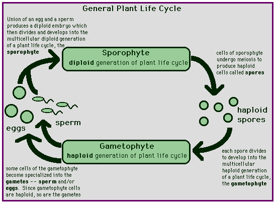
Kinetic energy, a source of energy stored in cells, causes molecules to bump into each other and move in new directions. Diffusion is the result of this contact. Diffusion is the random movement of molecules to an area of lower concentration from an area of higher concentration. Osmosis is a type of diffusion. This is the diffusion of water through a selectively permeable membrane from a region of higher water potential to a region of lower water potential. Water potential is the measure of free energy of water in a solution. A living system also contains an active transport to create movement of particles like ions that move against their concentration gradient. The energy source ATP is used during this process to move the particles across the cell membrane. This experiment takes place to measure the diffusion of small molecules through dialysis tubing. This tubing acts as a selectively permeable membrane, allowing larger molecules to pass through, but slowly. Dialysis is the movement of a solute through a selectively permeable membrane.

When the two solutions on either sides of the membrane are equal and no net movement is detected, the solutions are isotonic. This means that the solutions have the same concentration of solutes. If two solutions differ in the concentration of solutes that each has, the one with more solute is hypertonic. The solution that has less solute is hypotonic.

Water potential is predicting the movement of water into or out of plant cells. It is abbreviated by the Greek letter psi and has two components; a physical pressure component, pressure potential, and the effects of solutes, solute potential. Water always moves from an area of high to low water potential. The equation is water potential equals the sum of pressure potential and solute potential.

In a plant cell, turgor pressure is necessary. This is a pressure available to plants in a hypotonic environment. Turgor pressure gives plants their structure and strength. When a plant cell is in an isotonic solution, the turgor pressure decreases, causing wilting in the plant structure. In hypertonic solutions, plants plasma membrane shrinks away from the cell wall, an action termed plasmolysis.


Hypothesis:
Diffusion and osmosis occur between different molar solutions until the solutions are isotonic, effecting the turgor pressure of plant cells.


Materials:
Lab 1A – The materials used in conducting this experiment are as follows: one 30cm strip of dialysis tubing (presoaked), distilled water, 15%glucose/1%starch solution, 250mL beaker, Iodine Potassium Iodide solution, glucose Testape, and string.

Lab 1B – The materials used in conducting this experiment are as follows: six presoaked strips of dialysis tubing, distilled water, 0.2M, 0.4M, 0.6M, 0.8M, and 1.0M solutions of sucrose, six 250mL glass beakers, string, and an electronic balance.

Lab 1C – The materials used in conducting this experiment are as follows: six 250mL glass beakers, a potato, a core borer, a knife, distilled water, , 0.2M, 0.4M, 0.6M, 0.8M, and 1.0M solutions of sucrose, string, a ruler, and an electronic balance.

Lab 1D – The materials used in conducting this experiment are as follows: graph paper, pencil, a ruler, a calculator, and colored pencils.

Lab 1E – The materials used in conducting this experiment are as follows: a light microscope, microscope slide, cover slip, distilled water, NaCl solution, paper, pencil, and onion skin.


Procedure:
Lab 1A: Obtain a 30cm piece of dialysis tubing that has been presoaked in distilled water. Tie off one end securely. Open the other end of the dialysis tube and insert 15mL of 15%glucose/1%starch solution. Tie off the other end of the bag, leaving room for expansion. Record the color of the solution within the bag. Test the 15%glucose/1%starch solution for the presence of glucose using Testape. Fill a 250mL beaker with distilled water and add approximately 4mL of Lugol’s solution (IKI) to the distilled water. Test this solution for the presence of glucose as well with the Testape. Record the results in the data table. Immerse the bag in the beaker of solution. Let this stand for approximately 30min, or until distinct coloration is observed. Record final colors of solutions in the bag and in the beaker. Test both solutions once more for the presence of glucose with the Testape strips.

Lab 1B: Before starting this lab, wash your hands. Obtain six 30cm dialysis strips that have been presoaked in distilled water. Tie off each end securely. Pour approximately 25mL of each sucrose molar solution into its respective bags (that should be labeled, but not on the tubing itself). Tie off the other ends securely with string, careful to get any air bubbles out and leaving room for expansion. Rinse off each bag and blot off the water. Weigh and record the initial mass of the dialysis bags in the data table. Fill six 250mL glass beakers 2/3 full of distilled water and label each beaker with its respective bag’s molarity of sucrose. Immerse each bag into the distilled water. Allow this to stand for thirty minutes. Remove each bag, blot the sides to get off extra solution and weigh and record mass in grams each bag and determine the mass difference and percent change in mass. Next, compare the group percentages to the class.

Lab 1C: Pour 100mL of the assigned sucrose solutions into their 250mL beakers (pre-labeled). Obtain a large potato. Using a core borer, take 24 samples out of the potato, and measure each in centimeters so that they are all equal in length (use the knife to slice off ends). Make sure not to leave any skin with the samples. Place these cores in a covered beaker until an electronic balance can be obtained. Determine the mass of four cores at a time, placing the four in their sucrose solutions. Record this data for each of the six beakers. Allow these potato samples to sit immersed in the solutions overnight, covered. Remove the cores, blot off excess solution, and weigh the samples, recording the mass in the data table. Determine the mass difference, the percent change in mass and the class average percent change in mass. Graph the increase and decrease in mass of the potato cores according to the molarity of the solutions they were placed in on graph 1.2.

Lab 1D: Using paper, a pencil, and a calculator determine the solute potential of the sucrose solution, the pressure potential, and the water potential. Also, obtain graph paper and graph the values given for the zucchini percent change in mass and molarity of sucrose solutions in the graph 1.3.

Lab 1E: Prepare a wet mount slide of onion skin. Observe under a light microscope and sketch what you see. Add a few drops of the NaCl solution, observe, and sketch what you see there as well.


Data:
Table 1.1 The presence of glucose in beaker and bag solutions


Initial Contents


Initial Solution Color


Final Solution Color


Initial Presence of Glucose


Final Presence of Glucose


Bag

15%glucose/1%starch solution Clear Midnight blue + +
 

Beaker

Water and IKI indicator Amber Amber +

Table 1.2 Dialysis Bag Results: Individual Data


Contents in Bag


Initial Mass


Final Mass


Mass Difference


Percent Change in Mass


Distilled water

28 g 28g 0g 0%
 

0.2M sucrose

26.9g 28.5g 1.6g 5.95%
 

0.4M sucrose

27.0g 29.4g 2.4g 8.89%
 

0.6M sucrose

28.4g 32.6g 4.2g 14.79%
 

0.8M sucrose

28.2g 32.0g 3.8g 13.48%
 

1.0M sucrose

29.9g 34.8g 4.9g 16.39%

Bags immersed @ 12:01pm, removed at 12:31pm.

Table 1.3 Dialysis Bag Results: Class Data


Group 1


Group 2


Group 3


Group 4


TOTAL


Class Average


Distilled water

0% 0% .71% .38% 1.09% 0.27%
 

0.2M sucrose

5.95% 4.01% 5.41% 1.75% 17.12% 4.28%
 

0.4M sucrose

8.89% 8.63% 8.89% 8.42% 34.83% 8.71%
 

0.6M sucrose

14.79% -8.06% 10.69% 9.61% 27.03% 6.76%
 

0.8M sucrose

13.48% 15.19% 12.33% 15.70% 56.70% 14.10%
 

1.0M sucrose

16.39% 7.29% 15.13% 12.50% 51.31% 12.83%

Table 1.4: Potato Core: Individual Data


Contents in Beaker


Initial Mass


Final Mass


Mass Difference


Percent Change in Mass


Class Average % Change


Distilled water

3.4g 4.1g .7g 20.59% 15.93%
 

0.2M sucrose

3.4g 3.5g .1g 2.94% -2.77%
 

0.4M sucrose

6.2g 5.3g 0.9g -14.52% -16.55%
 

0.6M sucrose

6.3g 4.8g 1.5g -23.81% -22.83%
 

0.8M sucrose

6.2g 4.4g 1.8g -29.03% -25.72%
 

1.0M sucrose

6.0g 4.3g 1.7g -28.33% -28.17%

Table 1.5: Potato Core Results: Class Data


Group 1


Group 2


Group 3


Group 4


TOTAL


Class Average


Distilled water

20.59% 13.33% 14.81% 15.00% 63.73% 15.93%
 

0.2M sucrose

2.94% -3.39% -7.69% -2.94% -11.08% -2.77%
 

0.4M sucrose

-14.52% -18.64% -15.38% -17.65% -66.19% -16.55%
 

0.6M sucrose

-23.81% -26.23% -23.08% -18.18% -91.30% -22.83%
 

0.8M sucrose

-29.03% -26.32% -26.92% -20.59% -102.86% -25.72%
 

1.0M sucrose

-28.33% -29.31% -30.78% -24.24% -112.66% -28.17%

Questions:

Which substance(s) are entering the bag and which are leaving the bag? What experimental evidence supports your answer? Iodine Potassium Iodide is entering the bag because the indicator’s color is concentrated in the bag when the IKI started only in the beaker. Glucose and water left the bag, the evidence was the Testape color.


Explain the results you obtained. Include the concentration differences and membrane pore size in your discussion. The results simply state that the water, glucose, and IKI were small enough molecules to pass through the selectively permeable membrane. The starch didn’t leave the beaker because its color was amber and the starch molecule was much too large to pass through the selectively permeable membrane.


Quantitative data uses numbers to measure observed changes. How could this experiment be modified so that quantitative data could be collected to show that water diffused into the dialysis bag? Quantitative data would include the initial and final percent concentrations of the glucose, therefore showing the concentration of water by the change of percent in glucose. The movement would be noticeable in the concentration because it is known that water moves from high water potential to a low water potential.


Based on your observations, rank the following by relative size, beginning with the smallest: glucose molecules, water, IKI, membrane pores, and starch molecules. The smallest substance was IKI, followed by water, glucose, the membrane pores, then the starch molecules.


What results would you expect if the experiment started with a glucose and IKI solution inside the bag and only starch and water outside? Why? Based on the size of the molecules, the glucose and IKI would move out of the bag, the water in, and the starch left in the beaker again.


Explain the relationship between the change in mass and the molarity of sucrose within the dialysis bags. These two things are directly proportional. As the mass increases, so does the molarity.


Predict what would happen to the mass of each bag in this experiment if all the bags were placed in a 0.4M sucrose solution instead of distilled water. Explain your response. These are inversely proportional because whenever the sucrose molarity inside the bag is more concentrated, it will become more dilute and vise versa. The solutions will reach equilibrium somewhere between the two concentrations.


Why did you calculate the percent change in mass rather than simply using the change in mass? The differences in mass don’t deal with the proportional aspect of the solutions, making the real results less accurate. The percent was calculated to give the exact difference, along with considering the quantities of solution.


A dialysis bag is filled with distilled water and then placed in a sucrose solution. The bag’s initial mass is 20g, and its final mass is 18g. Calculate the percent change of mass, showing your calculations in the space below. 18g(final mass) – 20g(initial mass)=-2/20g(initial mass) x 100, which gives you a 10% change of mass.


If a potato is allowed to dehydrate by sitting in the open air, would the water potential of the potato cells decrease or increase? Why? The water potential of the potato would decrease because water moves from a high water potential region to a low potential region, and a dehydrated potato cell is hypertonic in comparison with the environment, forcing water to come into the cell. The moving in part shows that the potato cell had a low water potential.


If a plant cell has a lower water potential than its surrounding environment, and if pressure is equal to zero, is the cell hypertonic or hypotonic to its environment? Will the cell gain water or lose water? Explain your response. If the plant cell has lower water potential, that means the water will come into the cell, the cell is hypertonic to its environment. This cell will gain water because water follows its concentration gradient.


In figure 1.5, the beaker is open to the atmosphere. What is the pressure potential of the system? The pressure potential in this figure is equal to zero.


In figure 1.5, where is the greatest water potential? The greatest water potential is within the dialysis bag.


Water will diffuse__the bag. Why? Water will diffuse out of the bag because the highest water potential is inside the bag, forcing the water out.


Calculate solute potential of the sucrose solution in which the mass of the zucchini cores does not change. Show work. Y s =iCRT therefore Y s=(1)(1.0mole/liter)(0.0831 liter bar/mole degrees K)(295 degrees K) Y s=-24.51 bars.


Calculate the water potential of the solutes within the zucchini cores. Show work. Y =Y s+Y p so Y =0+-24.51 , Y =-24.51bars


What effect does adding solute have on the solute potential component of that solution? Why? Adding solute to a solution would increase the solute potential and decrease the water potential.


Consider what would happen to a red blood cell placed in distilled water:
Which would have the higher concentration of water molecules?The distilled water would have the higher concentration of water molecules.


Which would have the higher water potential? The red blood cell would have the higher water potential.


What would happen to the red blood cell? Why? The red blood cell would take in a lot of water and might lyse due to pressure inside. This is a possibility because animal cells have no tolerance under hypotonic situations.


Describe the appearance of the onion cells. The onion cells appear to have great turgor pressure, spread out, thick and bright in the inside. The cell walls were very defined and it was clear where one cell ended and another began.


Describe the appearance of the onion cells after the NaCl was added. The plasma membrane shriveled from the cell wall, causing plasmolysis. The cells looked wrinkly or weak. The turgor pressure dropped tremendously.


Remove the cover slip and flood the onion with fresh water. Observe and describe what happened. The onion cells were again hypertonic to their environment, and gathered water, increasing in turgor pressure and restoring themselves to the normal state of being.


What is plasmolysis? Plasmolysis is the separation of the plasma membrane from the cell wall in a plant cell.


Why did the onion cell plasmolyze? The environment became hypertonic to the cell and the water left the cell running with its concentration gradient due to the NaCl. With all the water leaving the cell, it shrank, leaving behind its cell wall.


In the winter, grass often dies near roads that have been salted to remove ice. What causes this to happen. The salt causes the grass’s environment to become hypertonic, and the water leaves the plant cells, causes withering and eventually death of the plant.


Error Analysis:
Lab 1A: One possible source of error could be the tightness of the string that tied off the dialysis tubing. If there was a leak or a break in the dialysis tubing, all of the data would be off.

Lab 1B: A possible source of error in this lab could have been in the first step. If the handler of the dialysis tubing did not wash their hands and accidentally touched the sac part of the tubing, the oils from their hands could have blocked some of the pores on the tubing, distorting the data.

Lab 1C: A piece of potato skin could have been left in the beakers along with the potato. This causes problems in the data tables. Another possible source of error could be that the students did not pat dry the potato sample well enough causing drops to be left on the electronic balance, tarring it incorrectly, causing all other data to be off slightly.

Lab 1D: Simple mathematical errors always occur, so there is always room for simple algebraic mistakes in this section of the lab.

Lab 1E: If the wet sample was not prepared correctly, or the salt solution added to fast not giving the cells time to react, this lab would have different results. The sources of error also include the possible concept that the onion cells might have dried out by the time the observer got around to sketching. This could cause error in observances, and data in conclusion.


Discussion and Conclusion:
During Lab 1A, the data suggests what molecules can and cannot diffuse across a selectively permeable membrane. The coloration showed that the Iodine Potassium Iodide was small enough to pass through the pores of the membrane because the color of this indicator moved from within the beaker to in the bag. Water and glucose moved out because water is small enough to pass through the membrane and the glucose tested positive with the Testape inside the beaker. The glucose at the beginning was only in the bag, so it obviously moved out.

Lab 1B proved that water moves across the selectively permeable membrane of the dialysis tubing much easier than sucrose sugar does. The water moved to reach equilibrium between the solutions. Sucrose must be too large a molecule to pass through the membrane quickly.

Lab 1C showed that the potato samples took in water when immersed in a distilled water solution. Potatoes must contain sucrose molecules due to the conclusion of this lab because the potatoes take in water in the distilled water beaker. Potatoes had a lower water potential and higher solute potential than the distilled water. It is just the opposite inside the beaker.


Lab 1D’s calculations made it evident that all of the results could be determined and proved correct with simple algebra equations and formulas. This gives the lab much better illustration and a stable understanding of diffusion and osmosis.

Lab 1E showed the plasmolysis clearly and allowed the student to see exactly what goes on in this action. This particular part of the lab illustrated the shrinking of the plasma membrane from the cell wall in a plant cell. It shows how plant cells react in a hypertonic environment. The turgor pressure decreases a lot, and the cells become very weak when the water leaves the cell.

BACK


Moss & Fern

Mosses & Ferns
fern gametophyte
Kingdom Plantae
All Materials © Cmassengale   

Seedless Nonvascular Plants

  • Includes mosses, liverworts, and hornworts
  • Lack vascular tissue (xylem & phloem) to carry water & food
  • Have a Sporophyte & Gametophyte stage known as alternation of generations
  • Gametophyte is dominant stage
  • Reproduce by spores

Division  Bryophyta

 Mosses:

  • Small, nonvascular land plants
  • No true roots, stems, or leaves
  • Class Musci
  • Most common bryophyte
  • Grow on moist areas (brick walls, as thick mats on forest floors, and on the shaded side of trees)
  • Some can survive periodic dry spells & revive when H2O becomes available
  • Must grow close together and must have H2O to complete their life cycle 
  • Sperm swims to egg through drops of water during fertilization
  • H2O moves cell-to-cell by osmosis
  • Sphagnum moss is known for its moisture holding capacity, absorbing up to 20 times its dry weight with water.


MOSS SPOROPHYTES & FERN GAMETOPHYTES

LIFE CYCLE OF MOSSES:

  • Mosses alternate between a haploid (n) gametophyte stage & a diploid (2n) sporophyte stage 
  • Gametophyte is the dominant generation


Moss Gametophyte Moss Sporophyte
Polytrichum formosum with moss flowers Tortula muralis?


  • Called alternation of generations

  • The haploid gametophyte stage contains half the chromosome number & produces gametes (egg & sperm) 
  • Gametophyte stage is dominant in the moss’s life cycle
  • Gametophytes are photosynthetic & have root-like rhizoids
  • The diploid sporophyte has a complete set of chromosomes & produces spores by meiosis
  • Sporophyte of a moss is smaller than, & attached to the Gametophyte
  • Sporophytes lack chlorophyll & depend on the photosynthetic gametophyte for food
  • Sporophyte has a long, slender stalk topped with a capsule
  • Capsule forms haploid (n) spores 


Moss Capsules

Sexual Reproduction in Moss:

  • Mosses produce 2 kinds of gametes (egg & sperm)
  • Gametes of Bryophytes are surrounded by a jacket of sterile cells that keep the cells from drying out
  • Female gametes or eggs are larger with more cytoplasm & are immobile
  • Flagellated sperm must swim to the egg through water droplets for fertilization
  • Moss gametes form in separate reproductive structures on the Gametophyte — Archegonium & Antheridium


Archegonium Antheridium
moss archegonial head X 40.jpg (102370 bytes) Mnium antheridial head 40X.jpg (660893 bytes)


  • Each Archegonium forms one egg, but each Antheridium forms many sperm
  • Fertilization can occur only after rain when the Gametophyte is covered with water
  • Sperms swim to the egg by following a chemical trail released by the egg 
  • A zygote (fertilized egg) forms that undergoes mitosis and becomes a Sporophyte
  • Cells inside mature Sporophyte capsule undergoes meiosis and form haploid spores
  • Haploid spores germinate into juvenile plants called protonema
  • Protonema begin the Gametophyte generation

Protonema of Funaria hygrometrica
Protonema

  • Spores are carried by wind & sprout on moist soil forming a new Gametophyte

Asexual reproduction in Mosses:

  • Asexual reproduction in moss may occur by fragmentation or gemmae
  • Pieces of a Gametophyte can break off & form new moss plants (fragmentation)
  • Gemmae are tiny, cup shaped structures on the Gametophytes 
  • Raindrops separate gemmae from the parent plant so they can spread & form new Gametophytes


Gemmae cups


Uses for Moss:

  • Help decomposer dead logs
  • Serve as pioneer plants on bare rock or ground
  • Help prevent erosion
  • Provide shelter for insects & small animals
  • Used as nesting materials by birds & mammals
  • Sphagnum or peat moss forms peat bogs (wet ecosystem)
  • Peat is burned as fuel in some areas

Division  Hepatophyta  

Liverworts:

  • Nonvascular
  • Undergo alternation of generations with Sporophyte attached to Gametophyte
  • Gametophytes are green & leafy and the dominant generation


Liverwort

  • Need abundant water for fertilization
  • Reproduce by spores
  • Grow on moist rocks or soil
  • Reproduce asexually by gemmae and by growing new branches

Division  Anthocerophyta

Hornworts:

  • Small, nonvascular bryophytes
  • Gametophyte leafy like liverworts
  • Archegonia & antheridia form inside the plant
  • After fertilization, zygotes develop into long, horn-shaped Sporophytes
  • Horn-shaped Sporophytes capable of photosynthesis so not completely dependent on Gametophyte


Hornwort

Seedless Vascular Plants

  • Includes club mosses, whisk ferns, horsetails, & ferns
  • Have specialized vascular tissues (xylem & phloem) to transport H2O, food, etc.
  • Have a Sporophyte & Gametophyte stage known as alternation of generations
  • Sporophyte is the dominant stage
  • Reproduce by spores

Division  Psilophyta

Whisk Ferns:

  • Photosynthetic, aerial stem forks repeatedly to form a small twiggy bush
  • No true roots, stems, or leaves
  • Have horizontal, underground stems called rhizomes
  • Root-like structures called rhizoids anchor plant
  • Reproduce by spores & vegetatively from rhizomes
  • Only 2 living genera


Whisk Fern

Division  Lycophyta

Club Mosses:

  • Low growing plants resembling pine trees
  • Have a club-shaped spore producing structure


Club Moss

  • Some like Lycopodium contain chemicals that burn quickly
  • Resurrection moss is green (after rains) when moist and brown when dry.


Resurrection Plant
resurrection plant


Division  Sphenophyta

Horsetails:

  • Equisetum called scouring rush is the only living species
  • Photosynthetic aerial stems & underground rhizomes
  • Stems contain silica & were once used to scrub pots
  • Reproduce by means of spores made in small cones at the tip of branches
  • In prehistoric times, some plants of this family grew to be large trees
  • Found in wetlands


Horsetail

Division  Pterophyta

Fern Gametophyte:

  • Largest group of living seedless vascular plants
  • Live in moist habitats
  • Alternates between dominant Sporophyte stage & Gametophyte stage
  • Sporophyte stage has true roots, stems, & leaves
  • Produce spores on the underside of leaves 

fern sporangia.jpg (47544 bytes)

  • Leaves are called fronds & are attached by a stem-like petiole


FERNS

Fern Life Cycle:

  • Spores produced on underside of fronds in clusters of sporangia called sori
  • Spores undergo meiosis, are spread by wind, & germinate on moist soil to form prothallus
  • Prothallus begins the Gametophyte stage
  • Mature Gametophytes are small, heart-shaped structures that live only a short time
  • Male antheridia & female archegonia grow on the prothalli
  • Sperm must swim to the egg to fertilize it & developing embryo becomes the Sporophyte generation
  • Newly forming fronds are called fiddleheads & uncurl

Uses for Ferns:

  • Prevent erosion
  • Fiddleheads serve as food
  • Ornamental plants
  • Formed coal million of years ago
BACK


Osmosis Lab Report Sample 4 PreAP

Osmosis Through a Cell membrane of an Egg

Joe Lockwood

Introduction:

When a cell membrane is said to be selectively permeable, it means that the cell membrane controls what substances pass in and out through the membrane.  This characteristic of cell membranes plays a great role in passive transport.  Passive transport is the movement of substances across the cell membrane without any input of energy by the cell.  The energy for passive transport comes entirely from kinetic energy that the molecules have. The simplest type of passive transport is diffusion, which is the movement of molecules from an area of high concentration to an area of lower concentration.  Diffusion moves down the concentration gradient, which is the difference in the concentration of molecules across a space.  Osmosis is a type of diffusion in which water molecules move down the concentration gradient.

When the concentration of solute molecules outside the cell is lower than the concentration of solute in the cytosol , the solution outside is hypotonic to the cytosol.  If the concentration of solute molecules is higher outside of the cell, the solution outside is said to be hypertonic.  The solution outside is isotonic if the concentration is equal on both sides of the cell membrane.

The egg shell is made of calcium carbonate and vinegar contains acetic acid.  These two can react to produce calcium acetate and carbonic acid which then decompose into water and carbon dioxide as shown in the two chemical equations:

Text Box:

Text Box:

 Hypothesis:

The eggs will increase in mass in all three solutions, showing that diffusion and osmosis occur when the concentration of two solutions is different, so that equilibrium can be established. 

Materials:

To conduct this experiment, these materials will be needed:  1-2 fresh hen eggs in their shells, masking tape and marker, distilled water, clear sugar syrup, vinegar, clear jar with lid, tongs, electronic balance, paper towels, paper, and a pencil.

 Methods:

On Day 1, the first step should be to label the jar with your lab group and the word “vinegar”.  Next, the group will mass the egg with the electronic balance and record the results in the data table.  After this, the group will carefully place the raw egg into the jar and cover the egg with vinegar.  Finally, the group is to loosely recap the jar and allow the jar to sit for 24 to 48 hours until the outer calcium shell is removed.

On Day 2, the day should begin with the group opening the jar and pouring off the vinegar.  Next, they will use tongs to carefully remove the egg to a paper towel and pat it dry.  When this is done, the group should mass the egg on an electric balance and record the size, mass, and appearance of the egg.  After this, they will clean and re-label the jar with their lab group and the word “distilled water”.  They will carefully place the egg into the jar and cover the egg with distilled water.  Finally, they will loosely re-cap the jar and allow it to sit for 24 hours.

On Day 3, the first step is to open the jar and clean out the distilled water.  Then tongs should be used to carefully remove the egg to a paper towel and pat it dry. The size is to be recorded and so should the appearance of the egg on the table.  Next, the group will mass the egg on an electric balance and record the results.  After this, the jar should be cleaned and re-labeled with the name of the group and the word “syrup”.  Finally, the group should place the egg into the jar cover it with clear syrup, loosely re-cap the jar and allow it to sit for 24 hours.

On Day 4, the day should begin by the group opening the jar and pouring off the syrup.  Next, the group will use tongs to very carefully remove the egg, rinse off the excess syrup under slow running water, and pat the egg dry on a paper towel.  After this, the size and appearance of the egg should be recorded in the data table.  Then, the mass of the egg should be taken on an electronic balance and recorded.  Finally, the work area should be cleaned and all the lab equipment should be put away.

 Results:         

Questions:

1. Vinegar is made of acetic acid and water.  Explain how it was able to remove the calcium shell.  The reaction of the acetic acid and calcium carbonate of the egg shell produces calcium acetate and carbonic acid, which then decomposes into water and carbon dioxide.


2.(a) What happened to the size of the egg after remaining in vinegar? The egg got bigger.

(b) Was there more or less liquid left in the jar?  There was less liquid left in the jar.

(c) Did water move into or out of the egg? Why?  Water moved into the egg because there was a lower concentration of solute molecules in the vinegar than there was inside the egg.

3.(a) What happened to the size of the egg after remaining in distilled water? The egg got a little bit bigger, but not by very much.

(b) Was there more or less liquid left in the jar?  There was a little bit less liquid left in the jar, but the change was very small.

(c) Did water move into or out of the egg? Why?  A small amount of water moved into the egg because the distilled water had a slightly lower concentration of solute molecules than inside the egg.

4. (a) What happened to the size of the egg after remaining in syrup? The egg became smaller.

(b) Was there more or less liquid left in the jar?  There was more liquid left in the jar.
(c) Did water move into or out of the egg? Why?  Water moved out of the cell because the syrup molecules were hypotonic to the solute molecules inside the egg.

5.  Was the egg larger after remaining in water or vinegar? Why?  The egg was larger after remaining in water because the water has the lower concentration of solute molecules than the vinegar so more water would diffuse to an area of higher concentration of solute particles.

6.  Why are fresh vegetables sprinkled with water at markets?  They do this so that water will diffuse into the vegetables and keep them plump and allow them to keep their look of freshness.

7.  Roads are sometimes salted to melt ice.  What does this salting do to the plants along roadside and why?  This salting dehydrates the plants because the higher salt concentration causes the water to diffuse out of the plant to even up the concentration.


Error Analysis:

A few errors may have happened over the course of this experiment. The washing of the egg could have affected the mass.  Also, the jars might not have been thoroughly cleaned out before putting in the next substance.  This could have affected the rate of diffusion because it would have changed the concentration of the solute particles.  These errors and a few others may have occurred.

 Discussion and Conclusion:

The hypothesis was not correct.  While two of the solutions caused the eggs to increase in mass, syrup caused the egg to lose mass.  This shows that the syrup was hypertonic to the solution inside of the egg, causing water to diffuse out of the egg to try and establish equilibrium. The egg’s mass increased in the distilled water and vinegar because they were hypotonic to the solution in the egg, causing water to diffuse into the cell.  The shell on the egg dissolved because    the egg shell is made of calcium carbonate and vinegar contains acetic acid.  These two can react to produce calcium acetate and carbonic acid which then decompose into water and carbon dioxide.


BACK