 Charles Robert Darwin |
Darwin and Evolution All Materials © Cmassengale |
History of Evolution:
- Plato & Aristotle believed species were fixed & could be arranged according to their complexity
- In the mid eighteenth century, Carolus Linnaeus developed a system of classification that called binomial nomenclature
- George Cuvier, in the eighteenth century, explained changes in the fossil record by proposing that a whole series of catastrophes (extinctions) and re-populations from other regions had occurred giving the appearance of change over time
- Prior to Darwin, it was thought that the world was young & species did not change
- Lamarck (1744-1829) was first to state that descent with modification occurs and that organisms become adapted to their environments
- Inheritance of acquired characteristics was the Lamarckian belief that organisms become adapted to their environment during their lifetime and pass on these adaptations to their offspring
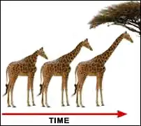
- Lamarck believed that the long necks of giraffes evolved as generations of giraffes reached for ever higher leaves; known as the Law of Use & Disuse
- Because it is supported by so many lines of evidence, evolution is no longer considered a hypothesis
- Evolution is one of the great unifying theories of biology
Darwin’s Background & Voyage:
- His nature was too sensitive to become a doctor like his father so he studied divinity
- He attended biology and geology lectures and was tutored by the Reverend John Henslow who arranged his trip on the HMS Beagle
- In 1831, at the age of 22, Charles Darwin accepted a naturalist position aboard the ship HMS Beagle & began a five-year voyage around the world

- He read Principles of Geology by Charles Lyell that stated that the observed massive geological changes were caused by slow, continuous processes (erosion, uplifting…)
- Darwin carried this book with him on his voyage as he witnessed Argentina coast earthquakes raising the earth several feet, & marine shells occurring far inland and at great heights in the Andes
- Darwin’s many observations led him to the idea that species slowly change over time
- Darwin’s comparison of the animals of South America and the Galapagos Islands caused him to conclude that adaptation to the environment can cause diversification, including origin of new species
Examples: Patagonian hares replaced rabbits in the South American grasslands
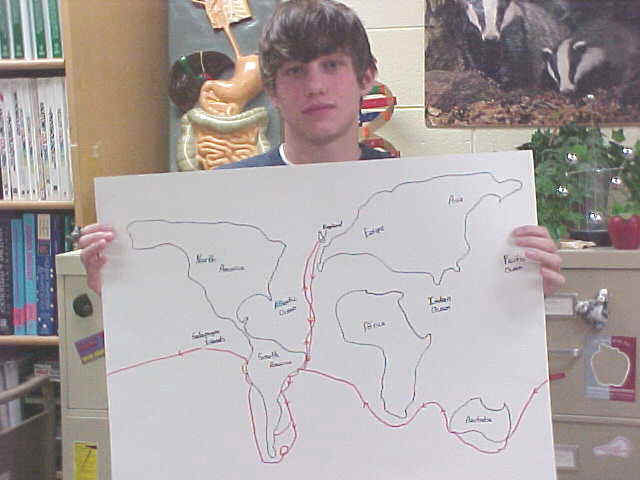
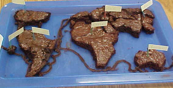
The Galapagos Islands:
- Volcanic islands off the South American coast
- Island species varied from the mainland species, and from island-to-island
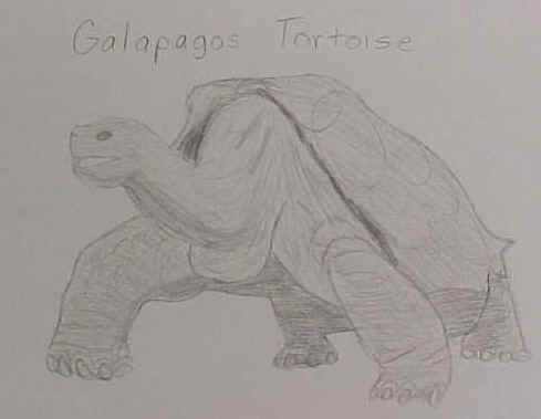
- Each island had either long or short necked tortoises depending on the island’s vegetation
- Finches on the Galapagos Islands resembled a mainland finch, but there were more types
- Bill shapes are adaptations to different means of gathering food.

- Galapagos finch species varied by nesting site, beak size, and eating habits

Darwin’s Theory of Evolution:
- An adaptation is a trait that helps an organism be more suited to its environment
- Darwin decided adaptations develop over time
- Natural selection was proposed by both Alfred Russell Wallace and Darwin as a driving mechanism of evolution
- Darwin and Wallace both read an essay by Thomas Malthus that proposed that human populations outgrow resources so there is a constant struggle for existence
- Fitness is a measure of an organism’s reproductive success
- Organisms most fit to reproduce are selected by environment which results in adaptation of the population
- Natural selection is also called “survival of the fittest”
- Conditions for natural selection include:
a. Variations exist among members of a population
b. Many more individuals are produced each generation than will survive
c. Some individuals are better adapted so they survive & reproduce
d. Members of a population compete for food, space, mates… - Variations that make adaptation possible are those that are passed on generation to generation
- Extinction occurs when previous adaptations are no longer suitable to a changed environment
On the Origin of Species by Darwin:
- After the HMS Beagle returned to England in 1836, Darwin waited over 20 years to publish
- Darwin was forced to publish Origin of Species after reading a similar hypothesis by Alfred Russell Wallace
- Both men concluded that life forms arose by descent from a common ancestor, and that natural selection is the mechanism by which species change and new species arise
Fossil Evidence:
- Fossils are relics or impressions of ancient organisms
- Most fossils are found in layers (strata) of sedimentary rock
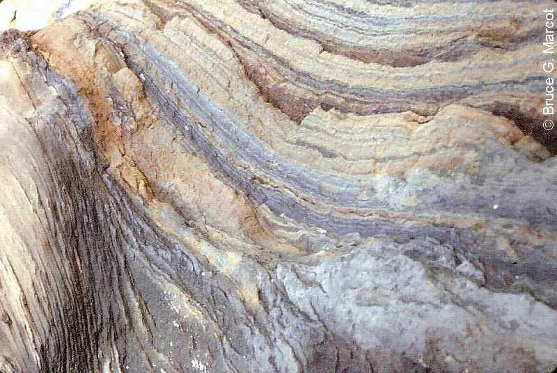
- The fossil record traces history of life and allows us to study history of particular organisms
- Through radioactive dating, geologists estimate the age of the earth at about 4.6 billion years
| ERA | PERIOD | EPOCH | DATES MYA |
AGE of | Notes | |
|---|---|---|---|---|---|---|
| Cenozoic | Quaternary | Holocene | 0-2 | Mammals | Humans | |
| Pleistocene | Other Mammal Species | |||||
| Tertiary | Pliocene | 2-5 | ||||
| Miocene | 5-24 | |||||
| Oligocene | 24-37 | |||||
| Eocene | 37-58 | |||||
| Paleocene | 58-66 | Extinction of dinosaurs | ||||
| Mesozoic | Cretaceous | 66-144 | Reptiles | Flowering plants | ||
| Jurassic | 144-208 | 1st birds & mammals | ||||
| Triassic | 208-245 | First Dinosaurs | ||||
| Paleozoic | Permian | 245-286 | Amphibians | End of trilobites | ||
| Carboniferous | Pennsylvanian | 286-320 | First reptiles | |||
| Mississippian | 320-360 | Large primitive trees | ||||
| Devonian | 360-408 | Fishes | First amphibians | |||
| Silurian | 408-438 | First land plant fossils | ||||
| Ordovician | 438-505 | Invertebrates | First Fish | |||
| Cambrian | 505-570 | 1st shells, trilobites dominant | ||||
Precambrian |
570-2,500 | 1st Multi-celled organisms | ||||
| 2,500-3,800 | 1st one-celled organisms | |||||
| 3,800-4,600 | ||||||
- Fossils are at least 10,000 years old and include skeletons, shells, seeds, insects trapped in amber, imprints of organisms, organisms frozen in ice (wooly mammoth), or trapped in tar pits (saber-toothed tiger)
- Transitional forms reveal links between groups (Example: Therapsids were mammal-like reptiles and Pterosaurs were bird like reptiles)
PTEROSAURS
Biogeographical Evidence:
- Biogeography is the study of the geographic distribution of life forms on earth
- Physical factors, such as the location of continents, determine where a population can spread
- Example: Placental mammals arose after Australia separated from the other continents, so only marsupials diversified in Australia
| KOALA | KANGAROO |
Anatomical Evidence:
- Organisms have anatomical similarities when they are closely related because of common descent
- Homologous structures in different organisms are inherited from a common ancestor have have similar structures
- Example : Vertebrate forelimbs contain the same sets of bones organized in similar ways, despite their dissimilar functions

- Analogous structures are inherited from different ancestors and have come to resemble each other because they serve a similar function
- Example: Bird wing & bat wing are both for flight but they are structurally different
- Vestigial Structures are remains of a structure that is no longer functional but show common ancestry
- Example: Humans have a tailbone but no tail
Embryological Evidence:
- During development, all vertebrates have a post-anal tail and paired pharyngeal pouches
- Organisms that show similarities in their embryonic development may have a common ancestry

Biochemical Evidence:
- Almost all living organisms use the same basic biochemical molecules, e.g., DNA, ATP, enzymes …
- Similarities in amino acid sequences, DNA codes, etc. can be explained by descent from a common ancestor
Examples of Evolution in Modern Times:
- Peppered moth — light colored vs. dark colored (industrialization influence) Manchester, England
- Insect resistance to insecticides
- Bacterial resistance to antibiotics