
Use this clam dissection guide to recognize the shell, mantle, gills, foot, adductor muscles, and other structures of a bivalve. Labeled diagrams and existing specimen photographs support identification, while the observation sequence connects anatomy with water flow, feeding, and movement.
Student resource: Clam Dissection Questions. Read the teacher notes below before assigning the legacy questions.
Learning goals and materials
- Identify a bivalve’s main external and internal structures.
- Explain how the mantle, gills, foot, and adductors support different functions.
- Distinguish the movement of water through the mantle cavity from the movement of food through the gut.
Prerequisites: animal tissues, organ systems, and classification terminology. Clams belong to Animalia, Mollusca, and Bivalvia; “Pelecypoda” is an older name seen in legacy resources. Clams include marine and freshwater forms. Unlike many other mollusks, bivalves do not have a radula.
Materials: a commercially preserved clam, tray, appropriate PPE, blunt probe, forceps, and teacher-approved instruments. A teacher-prepared opened specimen avoids asking students to force tightly closed valves apart. A diagram/photo route can meet the identification and explanation goals without cutting.
Teacher preparation and safety
This guide is for commercially preserved specimens under teacher supervision. Review the specimen label, supplier’s safety data sheet (SDS), school risk assessment, and disposal rules. Demonstrate tools before students begin. Use the required splash goggles, preservative-compatible gloves, protective clothing, and ventilation. Keep hands out of the cutting path and use a tray; no food or drink.
Offer a diagram, model, or teacher-demonstration route when needed. Keep specimens and preservative out of sinks and ordinary waste unless the school’s approved procedure permits it. Clean equipment and surfaces as directed and wash hands. Begin with the biology lab safety expectations. See NSTA’s dissection safety guidance and the supplier’s dissection FAQs; product-specific instructions take priority over a general web guide.
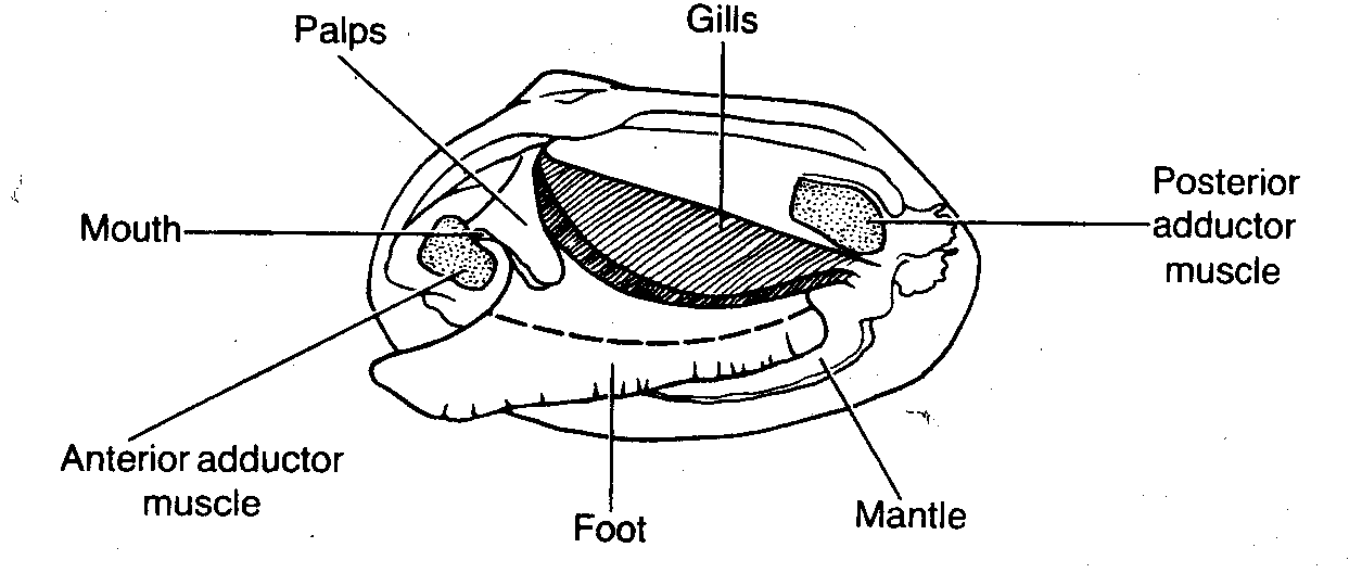
Labeled internal anatomy of a clam
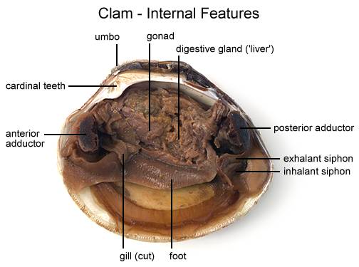
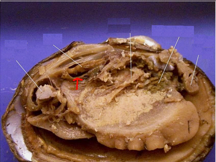
Use the specimen’s hinge and siphon end to orient the photo. An opened shell can be rotated in a tray, so “left on the page” is not a reliable anatomical direction. Colors vary with species, preservation, and reproductive condition.
External shell, umbo, and orientation
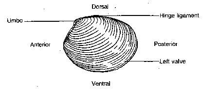

The two shell halves are valves. Find the umbo, the early-formed region near the hinge, and inspect the growth lines. Lines record episodes of shell growth; do not assume every visible ring equals one year without a species-specific method. The hinge ligament helps open the valves when the adductor muscles relax; the adductors pull them closed.
A step-by-step observation sequence
- Observe the unopened specimen: sketch the valves, hinge, umbo, and growth lines. Mark dorsal and ventral, then use the teacher’s specimen reference to establish anterior and posterior.
- Have the teacher prepare access: follow the supplier’s opening method. Do not pry a shell in your hand or push a screwdriver or blade toward fingers. A prepared opened clam is suitable for the remaining steps.
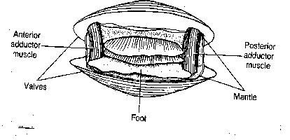
- Locate attachments: on an opened specimen, identify adductor muscles or their scars. If detaching a valve is part of the approved procedure, the teacher demonstrates how to separate the attachment near the shell without slicing through the organs.
- Find the mantle: observe the thin tissue lining the valves. Lift only the edge indicated by the teacher to expose the mantle cavity and gills; distinguish that space from the visceral mass.
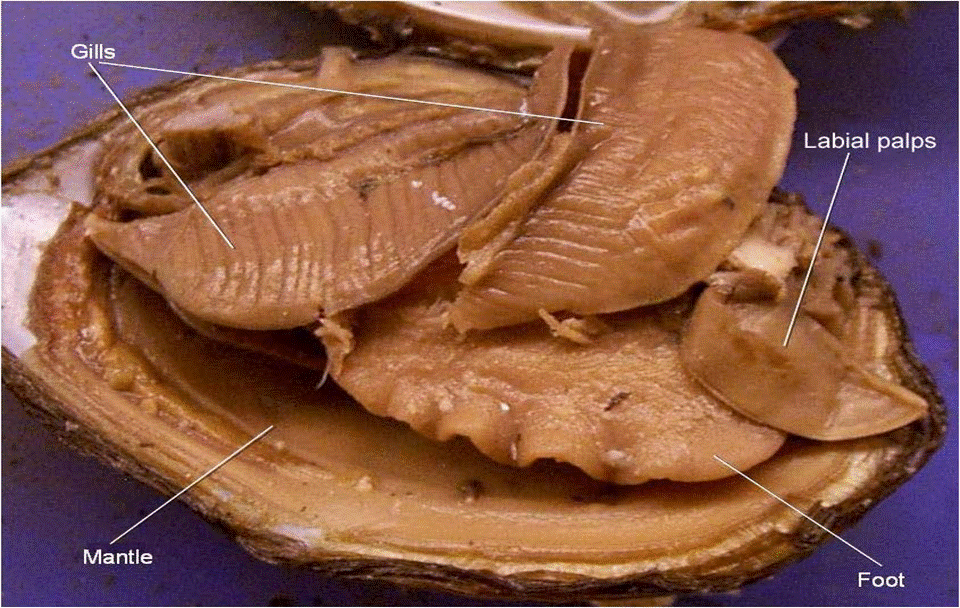
- Locate the foot, gills, and palps: compare their shapes and connections with the labeled photographs. Work with a blunt probe rather than cutting away the foot just to find labels.
- Study the visceral mass: locate the region containing digestive and reproductive organs. Deeper exploration of the stomach, intestine, gonad, or dorsal heart region is an optional teacher-directed extension, not a requirement to destroy the specimen.
- Record and finish: label a sketch, trace water and food with different arrows, note structures not visible, and follow the class cleanup and disposal plan.
Structures and functions
| Structure | What it does |
|---|---|
| Mantle and mantle cavity | The mantle covers the soft body and secretes shell material. The mantle cavity is the space associated with the mantle that contains the gills; it is not the name for every internal body space. |
| Adductor muscles and hinge ligament | Adductors close the valves. The elastic ligament helps separate them when the muscles relax. |
| Gills | Support gas exchange; in typical suspension-feeding clams, cilia and mucus also help capture and move food particles. |
| Labial palps and mouth | Palps help sort/direct particles toward the mouth. Food enters the digestive tract through the mouth, not through the gill tissue. |
| Muscular foot | Used for burrowing in many clams. Bivalves vary in mobility; not every adult is permanently fixed in one place. |
| Visceral mass and digestive gland | The visceral mass contains many internal organs. The digestive gland assists digestion and nutrient processing. |
| Heart and circulatory spaces | The heart lies dorsally in a pericardial region near the hinge. Clams have an open circulatory system; the hemocoel and mantle cavity are different spaces. |


Water flow and food flow are different pathways
Water: enters at the incurrent opening, passes through the mantle-cavity/gill system, and leaves at the excurrent opening. The gills exchange gases with the moving water.
Food: particles captured from the water are moved toward the palps and mouth, then pass through the esophagus, stomach, intestine, and anus. Material leaving the anus enters the outgoing water route. Draw separate arrows for the two pathways: water is not simply sent through the stomach as if the digestive tract were a pipe between siphons.
For a comparison with a vertebrate food pathway, use Frog Dissection. Similar functions do not mean identical anatomy.
Before assigning the legacy lab questions
- The prompt “What muscles open and close the clam?” needs discussion: adductors close it, while elastic action of the ligament helps opening.
- A blank asking for “the body cavity” is ambiguous. Ask specifically about mantle cavity, hemocoel, or reduced coelom instead of marking one unspecified answer as universally correct.
- Do not use growth rings alone as an exact age test. Larval names and mobility also depend on which bivalve is being discussed.
- The existing lab questions and unlabeled figure remain available. This guide adds clarification, not a newly verified key for every legacy item.
Original check: A student identifies the mantle and gill as the same structure. What evidence could distinguish them? Follow attachments: the mantle lines the valve and covers the body, while gills form folded surfaces within the mantle cavity. Original check: Does an unchanging preserved specimen show cilia moving water? No. Function is inferred using anatomy and a reliable reference; it is not directly observed in that specimen.
Additional original images
Open the existing shell and specimen image gallery




Related resources and further reading
- Animal Dissections directory — return to the existing collection.
- University of Hawaiʻi: molluscan body plans.
- OpenStax Biology 2e: mollusks and annelids.
- CUNY general biology lab manual: invertebrates.
- SDPB Dissection 101 resources — choose the clam section; external access and classroom-network restrictions may vary.
Use these sources for additional instruction; their assessments and illustrations have not been copied into this page.