Lab 4 Plant Pigments

Lab 4 Plant Pigments & Photosynthesis

Introduction:
The purpose of this lab experiment was to separate plant pigments using paper chromatography, and to measure the rate of photosynthesis in isolated chloroplasts. Because of capillary action the solvent moves up the paper causing the pigments to become visible at certain distances.

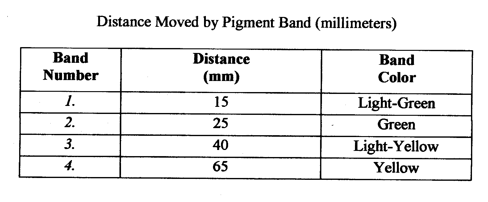
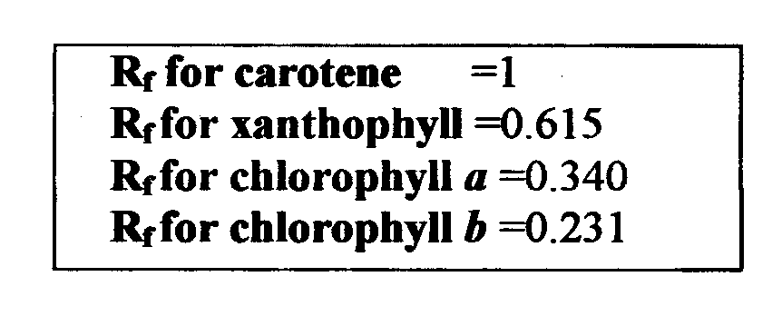
The substances visible on the paper are called pigments. Chlorophyll a is the main pigment that makes up about 75% of the pigmentation in plants. Chlorophyll b makes up about 25% of the pigmentation. And carotenes and xanthophylls are accessory pigments that make up the rest of the pigmentation. Carotene is the most soluble of the pigments and as a result will be carried the farthest by the solvent. The paper will display a spectrum of the pigments found in the spinach leaves. Using the formula Rf one can determine the relationship between the distance the solvent traveled to the distance the pigment traveled.


Rf=distance l2igment migrated (mm) distance solvent front migrated

Light is a part of a continuum of radiation or energy waves. The energy from visible light is used in the photosynthetic process. Light is absorbed in the leaf pigments, electrons within each photosystem are boosted to a higher energy level to produce ATP and to reduce NADP and NADPH. The ATP is then used in carbon fixation. This is the incorporation of CO2 into organic molecules.
To measure light transmittance in chloroplasts a spectrophotometer will be used. The reason behind measuring the light transmittance is to calculate the rate of photosynthesis in the chloroplasts. A solution called DPIP will be used in place of NADP to judge the color change of the chloroplast solutions. This technique is known as dye- reduction and it tests the hypothesis that light and chloroplasts are required for light reactions to occur.

Hypothesis:
In this experiment it is hypothesized that the cuvette with boiled chloroplasts and the cuvette kept in the dark containing unboiled chloroplasts will have very slight changes in light transmittance, whereas the cuvette containing unboiled chloroplasts that have been exposed to light will have an increasingly higher % transmittance over the course of time.

Materials and Methods:
Lab 4A:
The materials used in this section of the lab were: filter paper, glass vial, small amount of solvent, a quarter, and spinach leaves. The first step was to cut a point on one end of the filter paper and draw a pencil line 1.5cm from the tip of this point. Next a spinach leave was placed on the strip of paper and rolled over with a quarter on top of the pencil line. This gives a green line across the paper, which contains the pigments of the leave. Then the strip of paper was placed into the vial with the point down in the bottom. When the pigment reached the point 1 cm from the top of the vial then it was removed. The solvent front was then quickly marked with a pencil and then each pigment front was marked as well. From the distance the pigment traveled and the distance the solvent traveled the Rf value was calculated.

Lab 4B:
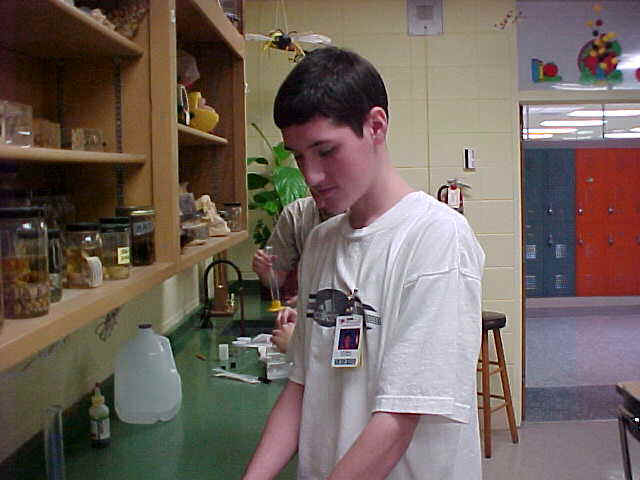
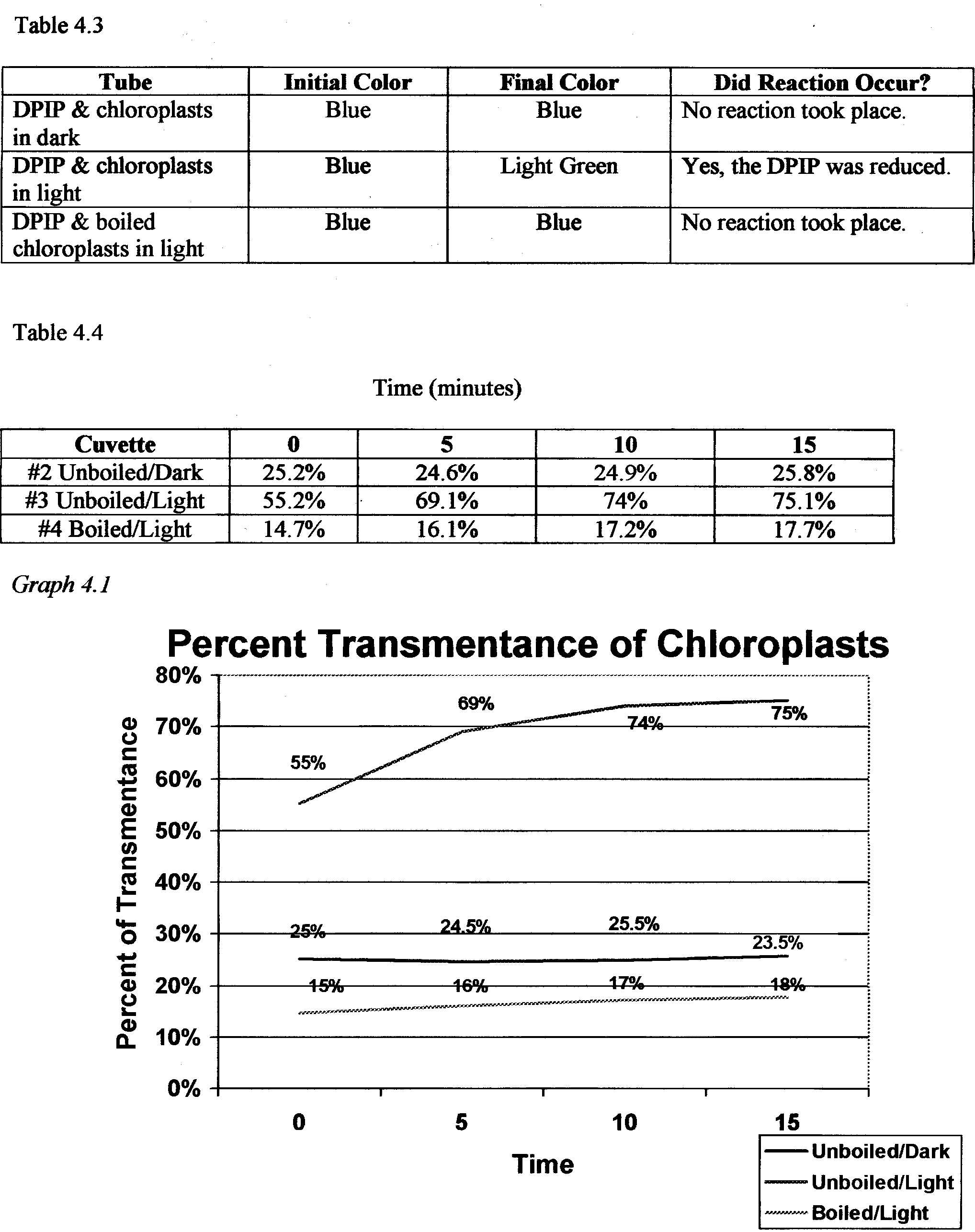
The materials used in this lab were: a spectrophotometer, 4 cuvettes, phosphate buffer, distilled water, boiled chloroplasts, unboiled chloroplasts, and DPIP .First the cuvettes were labeled 1-4 and cleaned with lens paper because even the oil from your hands can affect the transmittance of light through the cuvette. Cuvette 2 was then wrapped with foil to keep the contents in the dark. Next 1 ml of phosphate buffer was added to all four cuvettes, 4ml of distilled water was added to cuvette 1, 3rn1 of distilled water was added to cuvettes 2,3, and 4, and Iml of DPIP was added to cuvettes 2,3, and 4. Then 3 drops of unboiled chloroplasts were added to cuvette 1, it was covered with parafilm, placed into the spectrophotometer, and set to 100% transmittance. This cuvette was used to recalibrate between readings as well. Three drops of unboiled chloroplasts were placed in cuvette 2 and 3, and three drops of boiled chloroplasts were placed in cuvette 4. The cuvettes were then covered with parafilm. Each was placed in the spectrophotometer and the % transmittance of each, every five minutes for 15 minutes, was recorded.

Data:
Table 4.1

Questions:

1. Which pigment migrated the furthest and why? Carotene, it was the most soluble and didn’t form bonds with the filter paper.
2. Which of the 2 types of chlorophyll is more soluble? Chlorophyll a
3. Why do leaves change color in autumn? The chlorophyll production in the leaves slows down.
4. What is the function of the chlorophyll in photosynthesis? They absorb red and blue light rays.
5 .What are the accessory pigments and what are their functions? Carotene and xanthophylls both absorb different wavelengths of light than chlorophyll does.
6. What are some other ways chromatography is used to separate plant pigments? There are three types: Column, Paper, and Thin Layer chromatography.
7 .What does the & value represent? The distance the pigment traveled and the distance the solvent traveled expressed as one value.
8. What factors involved in the separation of the pigments? In this test it was solubility.
9 .Would you expect the Rf value of a pigment to be same if a different solvent were used? Explain. No, for a different solute there would be a different solvent rate.
10. What kind of chlorophyll does the reaction center contain? What are the roles of the other pigments? Chlorophyll a” the other pigments catch different wavelengths of light.

Error Analysis:

Fingerprints on either the filter paper or the cuvettes may have affected the experiment because the oil from your hands can get on these things and affect the results. The spectrophotometer may have not been calibrated correctly because this was the first time this particular one had been used. Other than these there were few places for error in this lab.

Conclusion:

From lab 4a we discovered that the many pigments found in chloroplasts are all involved in gathering energy from sunlight. The spectrum of color displayed on the filter paper showed the pigments and the solubility of each. In lab 4b the spectrophotometer measured the light transmittance through the various cuvettes and the chloroplast solutions in each. The actual purpose of this was to observe the DPIP go from a blue color to a clear color. This indicated that photosynthesis was occurring and at what rate it was occurring. The cuvette with the unboiled chloroplasts that had been exposed to light showed the biggest change in % transmittance, which indicates that the amount of light available has a very big effect on the rate at which the light reactions of photosynthesis occur .


BACK

Lab & AP Sample 2


Plant Pigments and Photosynthesis

Introduction:
Photosynthesis has two main parts, which are the light dependent and the light –independent. In the light-dependent reactions pigments trap energy from light, and this energy is used to split water molecules (photolysis). The light-independent reactions or dark phase of photosynthesis involve the fixing of carbon dioxide. It makes glucose and fructose chains and also releases oxygen , which passes through the stomata of the plant.

Organisms that carry out photosynthesis making their own organic molecules are called autotrophic. Some autotrophic organisms include plants, algae, and blue-green bacteria. Plants have many varieties of pigments, all of which absorb different colors of light. Chlorophyll a is the primary plant pigment and makes up about three-fourths of all the plant pigments. It absorbs red and blue light and is not found in photosynthetic bacteria.

Chlorophyll b is another plant pigment. It absorbs blue-green and orange-red light. Carotenoids are a type of accessory pigment that absorb blue and blue-green light. These pigments are fat soluble and usually masked by chlorophyll a. Anthocyanin is another accessory pigment that absorbs bright red colors. There is also chlorophyll c and d that sometimes take the place of chlorophyll b.

Chromatography is a process used to separate mixtures that can separate plant pigments. This lab uses paper chromatography where a piece of paper is used to wick solvent up to the pigments and separate them according to solubilities. The rate of migration on a chromatogram is the Rf value.


Hypothesis


Plants contain several different pigments, and the rate of photosynthesis in plant cells is directly related to light and temperature.


Materials


Exercise 4A: Plant Pigment Chromatography

This exercise required 1 50-mL graduated cylinder, a small amount of a solvent, a stopper, filter paper, scissors, a pencil, spinach leaves, and a quarter.

Exercise 4B: Photosynthesis/The Light Reaction

The materials needed for this part of the lab were a spectrophotometer, a light, a water flask, a test tube rack, ice, 5 labeled cuvettes, lens tissue, foil, and parafilm. The substances put in the cuvettes were 5 mL of phosphate buffer, approximately 16 mL of distilled water, 9 drops of unboiled chloroplasts, and 3 drops of boiled chloroplasts.


Methods


Exercise 4A: Plant Pigment Chromatography

A 50-mL graduated cylinder was filled with about 1 cm of solvent and then tightly stoppered. The filter paper was then cut to a point on one end, and a line was drawn 1.5 cm above the point. Using the ribbed edge of a quarter, spinach cells were extracted onto the pencil line. This procedure was repeated 8-10 times using a new portion of the leaf each time. The filter paper was then placed in the cylinder with the tip barely touching the solvent and none of the edges touching the sides. When the solvent reached 1 cm below the top of the paper, it was removed from the cylinder. The solvent location was immediately marked, and then the bottom of each pigment band was also marked.

Exercise 4B: Photosynthesis/The Light Reaction

The spectrophotometer was set to 605 nm and allowed to warm up. The chloroplast suspensions were prepared the previous day, part of which were boiled, and stored on ice until they were ready for use. An incubation area was prepared with a flood light, water flask, and test tube rack, by using the flask as a heat sink between the light and the rack. Five cuvettes were numbered respectively and then wiped with lens tissue. The walls and bottom of cuvette 2 were covered with foil and a foil cap was made for the top.

To each cuvette 1 mL of phosphate buffer was added. Then, to cuvette 1 4 mL of distilled water was added, but to cuvettes 2, 3, and 4 3 mL of distilled water was added. Next, 1 mL of DPIP was added to cuvettes 2, 3,and 4. To cuvette 5, 3 mL plus 3 drops of distilled water were added and 1 mL of DPIP. To cuvette 1, 3 drops of unboiled chloroplasts were added.

The spectrophotometer was brought back to zero and the contents of cuvette 1 were mixed by inverting and placed in the sample holder. Cuvette 1 was used periodically through this experiment to recalibrate the spectrophotometer. Three drops of unboiled chloroplasts were added to cuvette 2. After removing the foil sleeve, it was placed in the sample holder and the transmittance was recorded. Additional readings were also taken at 5, 10, and 15 minutes. Next, three drops of unboiled chloroplasts were transferred to cuvette 3. The percent transmittance was recorded at 0, 5, 10, and 15 minutes. Three drops of boiled chloroplasts were added to cuvette 4, and the transmittances were recorded at the same times. Finally, cuvette 5 was mixed and placed in the sample holder. The transmittance readings were recorded.


Results

Table 4.1 Distance Moved by Pigment Band


Band Number


Distance (mm)


Band Color

1. 0 mm Yellow-brown
2. 5 mm Light green
3. 30 mm Green
4. 48 mm Yellow


Distance Solvent Front Moved 60 mm.

Table 4.2 Rf Values


0.8 = Rf for xanthophyll (yellow)

0.5 = Rf for chlorophyll a (bright green to blue green)

0 = Rf for chlorophyll b (yellow green to olive green)


Table 4.4 Transmittance (%)


Cuvette


0


5


10


15


2 Unboiled/Dark

41% 43% 44% 43%
 

3 Unboiled/Light

35% 38% 39% 37%
 

4 Boiled/Light

51% 52% 53% 54%
 

5 No Chloroplasts

57% 57% 56% 55%


Lab 4B Color Chart


Cuvette


Initial Color


Final Color


1

Clear Clear
 

2

Light clear blue Blue/green
 

3

Light clear blue Dark clear blue
 

4

Light clear blue Light clear blue
 

5

Light clear blue Dark clear blue


Questions:
Exercise 4A: Plant Pigment Chromatography


What factors are involved in the separation of the pigments?


The solubility, size of particles, and their attractiveness to the paper are all involved in the separation.


Would you expect the Rf value of the pigment to be the same if a different solvent were used? Explain.


No, the different solubilities of the pigments would change the Rf values. For example chlorophyll b is only soluble to fat solutions.


What type of chlorophyll does the reaction center contain? What are the roles of the other pigments?


The reaction center contains chlorophyll a. The other pigments collect different light waves and transfer the energy to chlorophyll a.


Exercise 4B: Photosynthesis/The Light Reaction


What is the purpose of DPIP in this experiment?


DPIP is the electron acceptor in this experiment.


What molecule found in chloroplasts does DPIP “replace” in this experiment?


DPIP substitutes for the NADP molecules.


What is the source of the electrons that will reduce DPIP?


The electrons come from the photolysis of water.


What was measured with the spectrophotometer in this experiment?


The spectrophotometer measures the percentage of light transmittance through the cuvette due to DPIP reduction.


What is the effect of darkness on the reduction of DPIP? Explain.


The effect of darkness is that no reaction will occur.


What is the effect of boiling the chloroplasts on the subsequent reduction of DPIP? Explain.


Boiling denatures the protein molecules and stops the reduction.


What reasons can you give for the difference in the percent transmittance between the live chloroplasts that were incubated in the light and those that were kept in the dark?


In the dark cuvette, there was no light energy available, so there was no flow of electrons and no photolysis of water, while in the lighted cuvette these processes were allowed to continue.


Error Analysis
In Lab 4A, several mistakes could have been made with the chromatography paper. Too much handling, bending, or allowing the paper to touch the sides of the cylinder could all have affected the outcome of this experiment. The different pigment bands were also difficult to distinguish.

In Lab 4B, the procedure was very complicated and required a lot of pre-lab planning and reading. Incorrect usage of the spectrophotometer or neglecting to recalibrate often enough could have caused errors in this portion of the lab.


Discussion and Conclusion

Lab 4A demonstrated the different plant pigments by chromatography and showed how to calculate Rf values and explained their importance. There are 4-5 main pigments present in plants ranging from green to yellow in color. Lab 4B proves that light and chloroplasts are required for the light reactions of photosynthesis to occur. It showed the effects of boiling, denaturing, which did not allow photosynthesis to occur.

BACK

Lab 3 Sample Ap Mitosis & Meiosis


Mitosis and Meiosis


Introduction
There are two types of nuclear division, mitosis and meiosis. Mitosis is usually used for the growth and replacement of somatic cells, while meiosis produces the gametes or spores used in an organism’s reproduction.

Mitosis is the first of these studied in this lab. It is easily observed in cells that are growing at a rapid pace such as whitefish blastula or onion root tips, which are used in this lab. The root tips contain an area called the apical meristem that has the highest percentage of cells undergoing mitosis. The whitefish blastula is formed directly after the egg is fertilized. This is a period of rapid growth and numerous cellular divisions where mitosis can be observed.

Just before mitosis the cell is in interphase. In this part of the cell cycle the cell will have a distinct nucleus and nucleoli where the thin threads of chromatin are duplicated. After duplication the cell is ready to begin mitosis and its starts with a step called prophase. In prophase, the chromatin thicken into distinct chromosomes and the nuclear envelope breaks open releasing them into the cytoplasm. The first signs of the spindle begin to appear. Next the cell begins metaphase, where the spindle attaches to the centromere of each chromosome and moves them to the same level in the middle of the cell. This level position is called the metaphase plate. Anaphase begins when the chromatids are separated and pulled to opposite poles. Then, the final stage is telophase. The nuclear envelope is reformed and the chromosomes gradually uncoil. Cytokinesis may occur, in which case, a cleavage furrow will form and the two daughter cells will separate.

Meiosis is more complex and involves two nuclear divisions. The two divisions are called Meiosis I and Meiosis II and they result in the production of four haploid gametes. This process allows increased genetic variation due to crossing over where genes can be exchanged. The process, like mitosis, depends on interphase to replicate the DNA. Meiosis begins with Prophase I. In this stage, homologous chromosomes move together to form a tetrad and synapsis begins. This is where crossing over occurs resulting in the recombination of genes. Metaphase I moves the tetrads to the metaphase plate in the middle of the cell, and Anaphase I reduces the tetrads to their original two stranded form and moves them to opposite poles. Telophase I then prepares the cell for its second division. Meiosis II generally resembles mitosis except that the daughter cells are haploid instead of diploid. DNA replication does not occur in Interphase II, and prophase, metaphase, anaphase, and telophase occur as usual. The only change is the number of chromosomes.

The process of crossing over can be easily studied in Sordaria fimicola, an ascomycete fungus. Sordaria form a set of eight ascospores called an ascus. They are contained in a perithecium until they are mature and ready for release. Crossing over can be observed in the arrangement and color of these asci. If an ascus has four tan ascospores in a row and four black ascospores in a row (4:4 arrangement), then no crossing over had taken place. However, if the asci has black and tan ascospores in sets of two (2:2:2:2 arrangement) or two pairs of black ascospores and four tan ascospores in the middle (2:4:2 arrangement), then crossing over had taken place.


Hypothesis
Mitosis occurs in whitefish blastula and onion root tip, and it is easily observable. Meiosis and crossing over occurs in the production of gametes and spores.

Materials
This lab requires prepared slides of whitefish blastula, onion root tips, and Sordaria, pencil, paper, a light microscope, and a chromosome simulation kit.


Methods
Exercise 3A.1: Observing Mitosis

Prepared slides of whitefish blastula and onion root tips were observed under the 10X and 40X objectives. A cell in each stage of mitosis were identified, and then sketched.

Exercise 3A.2: Time for Cell Replication

Using a high power objective, every cell in a field of view was observed. Each cell was counted as being in one of the stages of mitosis and recorded. At least 200 cells and 3 fields of vision were counted and recorded. Next, the percentage of cells in each stage was recorded and the amount of time spent in each phase was calculated.

Exercise 3B.1: Simulation of Meiosis

In this part of the lab, a chromosome simulation kit was used to demonstrate meiosis. Two strands of the same color were connected to simulate DNA replication in both of the homologous pairs. Next, the chromosomes were entwined to represent synapsis. Sections of beads were switched between the pairs as in crossing over and were aligned at the equator. Next, anaphase was simulated as the homologous pairs were separated and then telophase was simulated by pushing the chromosomes into two separate cells (circles).

Meiosis II was simulated as well. The DNA is not replicated in Interphase II. The chromosomes again move to the equator and in Anaphase II the two chromatids were separated and moved to opposite poles. Telophase II separates them into four different cells.

Exercise 3B.2: Crossing Over during Meiosis in Sordaria

Prepared slides of Sordaria fimicola were observed under a light microscope. Over 100 asci were identified as either 4:4 or asci showing crossover and recorded. The percentage of each and the map units were calculated.

Results

Whitefish Blastula

Onion Root Tip

Table 3.1: Time for Cell Replication


Number of Cells

Field 1 Field 2 Field 3 Total
 

Interphase

42 36 47 125 61.27% 14 hours 42 minutes
 

Prophase

10 13 18 41 20.10% 4 hours 49 minutes
 

Metaphase

6 5 4 15 7.35% 1 hour 46 minutes
 

Anaphase

2 3 2 7 3.43% 49 minutes
 

Telophase

7 5 4 16 7.84% 1 hour 59 minutes

204


Table 3.2: Compare Mitosis and Meiosis


Mitosis


Meiosis


Chromosome number of parent cells

Diploid (2n) Diploid (2n)
 

Number of DNA replications

Once Once
 

Number of divisions

One Two
 

Number of daughter cells produced

Two Four
 

Chromosome number of daughter cells

Diploid (2n) Haploid (n)
 

Purpose

Growth and repair Production of gametes or spores


Simulation of the Meiosis I

Table 3.3: Sordaria


Number of 4:4


Number of Asci Showing Crossover


Total Asci


% Asci Showing Crossover Divided by 2


Gene to Centromere Distance (Map Units)

53 64 117 27.35% 27.35


Meiosis with Crossing Over – 2:4:2 Arrangement

Questions:

Why is it more accurate to call mitosis “nuclear replication” rather than “cellular division”?

It is more accurate to say “nuclear replication” to describe mitosis because the actual cell splitting occurs in cytokinesis. The whole process of mitosis is a series of steps that split the nucleus into two separate nuclei at opposite poles.

Explain why the whitefish blastula and onion root tips are selected for a study of mitosis.

The blastula is a hollow ball of cells that forms from the fertilization of an egg. Rapid growth occurs and numerous cellular divisions making mitosis in various stages easy to observe. Onion root tips are also a region of high percentage of cells going through mitosis because this is where most of the root growth takes place.

If your observations had not been restricted to the area of the root tip that is actively dividing, how would your results differ?

There would be virtually no cells undergoing division, so many more of the cells observed would have been in interphase where they elongate an differentiate.

Based on the data in Table 3.1, what can you infer about the relative length of time an onion root-tip cell spends in each stage of cell division?

Prophase is the longest stage of mitosis and then going in sequential order each decreases in the length of time it takes to complete.

List three major differences between the events of mitosis and meiosis.

In mitosis, the nucleus is only divided once, while in meiosis the nucleus is divided twice. Another difference is that mitosis produces two identical daughter cells, but meiosis produces up to four different daughter cells. Also, synapsis and crossing over do not take place in mitosis, but do take place in meiosis.

How are Meiosis I and Meiosis II different?

Meiosis I begins with a tetrad and separates the homologous pairs. Meiosis II separates the two sister chromatids.

How do oogenesis and spermatogenesis differ?

Oogenesis produces an egg cell, while spermatogenesis produces sperm cells.

Why is meiosis important for sexual reproduction?

In meiosis the chromosome number is reduced to n so that it can be fertilized. Also, meiosis allows for crossing over, which results in variations in organisms.

Error Analysis
There was little chance for error in this lab. It was mostly observation and sketching. However in Exercise 3A.2, the numbers for telophase were off. The calculations obtained for its time were too high; it should have been the shortest stage of meiosis. This may be caused by misidentifying the stages or counting the daughter cells as two different cells. Misidentification could have caused errors in the other parts of this lab as well.


Discussion and Conclusion
Mitosis was observed and timed in Lab 3A. The stages of mitosis are prophase, metaphase, anaphase, and telophase, prophase being the longest and telophase the shortest. Meiosis was simulated in Lab 3B and then crossing over was observed in Sordaria and the map units were determined. The gene to centromere distance in the Sordaria was 27.35 map units.

BACK


Lab & Ap Sample 2 Mitosis & Meiosis

Mitosis & Meiosis -AP lab 3

Introduction
Cells come from preexisting cells. New cells are formed during cell division which involves both replication of the cell’s nucleus, karyokinesis, and division of the cytoplasm, cytokinesis. The two kinds of cellular division are mitosis and meiosis. Mitosis usually makes body cells, somatic cells. Making an adult organism from an egg, asexual reproduction, regeneration, and the maintenance and repair of body parts are performed during mitotic cell division. This process called meiosis makes gametes, in animals, and spores, in plants. Gamete or spore cells have half the chromosomes that the parent cell has.

In plants mitosis takes place in the meristems which are normally found at the tips of stems or roots. However, in animal cells cell division takes place every where as new cells are formed and old ones are replaced. Studying mitosis can be accomplished by looking at tissues where there are many cells in a process of meiosis. Two examples are an onion root tip, or developing embryos, in animals such as whitefish blastula. A blastula is formed after an egg is fertilized and the egg begins to divide. There are several phases of the mitotic cell cycle. A precursor to mitosis is interphase. The actual steps of the mitotic cell cycle are prophase, metaphase, anaphase, and telophase. Interphase is a stage in the cell cycle in which the cell is not dividing. The nucleus contains a nucleolus and also contains chromatin. During interphase DNA replication occurs. The first phase of mitotic cell division is prophase. During prophase the chromatin begins to thicken until noticeable chromosomes are formed. Each chromosome has two chromatids that are joined at the centromere. During the later part of prophase, the nuclear envelope and nucleolus disappear. Mitotic spindle fibers, composed of microtubules, also become apparent. Following prophase is metaphase. By the time the cell has reached metaphase the chromosomes have moved to the center of the mitotic spindle. The centromere of the chromosome attaches to the spindle. The centromeres of each chromosome line up on an area called the metaphase plate. Metaphase is followed by anaphase. In the beginning of anaphase, the centromeres of each pair of chromatids separate and moved by the spindle fibers to the opposite ends of the cell. When the daughter chromosomes reach the ends of the cell the form a clump at each spindle pole. The final phase of mitosis is telophase. Telophase is identified by a recognizable condensation of the chromosomes, which is followed by the formation of a new nuclear envelope. The chromosomes slowly uncoil into chromatin once again and the nucleoli and nuclear envelope reform. It is then possible for cytokinesis, the division of the cytoplasm into two cells, to occur. In an animal cells a cleavage furrow forms and the cell pinches off into two new daughter cells.

The process of meiosis involves two nuclear divisions that result in the formation of four haploid cells. Meiosis I, a reduction division, is the first division to reduce the chromosome number from diploid to haploid and separates the homologous pairs. Meiosis II separates the sister chromatids resulting in four haploid gametes. Unlike mitosis meiosis increases genetic variation. In meiosis I each pair of homologous chromosomes come together which is known as a synapse. Chromatids of homologous chromosomes may exchange parts which is called crossing over. The distance between two genes on a chromosome may be estimated by calculating the percentage of crossing over that takes place between them. Meiosis I is preceded by interphase. During interphase DNA synthesis occurs and each chromosome is made of two chromatids joined at the centromeres. The first step of meiosis I is prophase I. During prophase I homologous chromosomes come together and synapse. A tetrad consisting of four chromatids is also formed. Prophase I is followed by metaphase I. In metaphase I the crossed over tetrads line up in the center of the cell. In anaphase I the homologous chromosomes separate and are moved to opposite ends of the cell. The final phase of meiosis I is telophase I. During telophase I centriole duplication is completed. Most of the time cytokinesis and formation of the nuclear envelope occur in order two make to cells. Meiosis II a second mitotic cell division then takes place in order to separate the chromatids in the two daughter cells made in meiosis I. This reduces the amount of DNA to one strand per chromosome. This is the only difference between meiosis I and II. Before meiosis II there is period called interphase or interkenesis. DNA replication does not take place in interphase II. Interphase II is followed by prophase II, No DNA replication occurs in prophase II and replicated centrioles separate and move to opposite sides of the chromosome groups. During metaphase II the chromosomes are centered in the middle of each daughter cell. During anaphase II the centromere regions of the chromatids are separate. The last stage of meiosis II is telophase II. In telophase II the chromosomes are at opposite ends of the cell and a nuclear envelope forms, and sometimes the cytoplasm divides.

Sordaria fimicola is fungus that may be used to show the results of crossing over during meiosis. Sordaria throughout most of its life is haploid, but becomes diploid after the fusion of two different types of nuclei, which forms a diploid nucleus. In Sordaria meiosis results in the making of eight haploid ascospores found in a sac called an ascus. Most asci are found in a perithecium. The life cycle of Sordaria fimicola is as follows: a spore is discharged through an ascus. The ascospore then undergoes mitosis, which forms a filament. The filament then undergoes mitosis, which forms a mycelium. Mycelial fusion and fertilization then takes place. This forms a diploid zygote. The zygote undergoes mitosis to form four haploid nuclei. The nuclei also undergo mitosis and form eight haploid nuclei, which then form eight ascospores. When mycelia of a mutant strain of Sordaria and a wild type of Sordaria undergo meiosis four black and four tan ascospores form. The arrangement of the ascospores reflects whether crossing over has occurred or not. Gametes, egg and sperm, are made during meiosis. Each egg and sperm cell contains half the total chromosomes a normal cell of that species would have. When the egg and sperm unite during fertilization the total chromosome number is restored.

Exercise 3A.1 –  Hypothesis
While looking at prepared slides of onion root tip cells and whitefish blastula cells under a microscope I will be able to identify and draw the stages of mitosis in these cells.

Materials
The materials used in this experiment were a light microscope and prepared slides of onion root tip cells and whitefish blastula cells.

Methods
Using the microscope examine the slides of onion root tip cells and whitefish blastula cells. Begin by locating the merismatic region of the onion or the blastula using the 10 X objective. Then use the 40 X objective to study individual cells. Identify one cell that clearly represents each phase. Sketch and label the cell on a separate piece of paper.

Exercise 3A.2  –  Hypothesis
When undergoing mitosis most of the cells in an onion root tip will be in interphase. More cells will be in the stage of prophase than metaphase. More cells will be in metaphase than anaphase and more cells will be in anaphase than telophase.

Materials
The materials used in this experiment were a prepared slide of an onion root tip and a light microscope.

Methods
Obtain a prepared slide of an onion root tip and observe every cell in one high power field of view and determine which phase of the cell cycle it is in. Make sure to do this in pairs so one person can observe the cells and the other person can record which phase the cell is in. Make sure to count three full fields of view and at least 200 cells. Then, record your data in table 3.1. Next, calculate the percentage of cells in each phase by using the equation; percentage of cells in stage X 1,440 minutes =_________ minutes of cell cycle spent in stage.

Exercise 3B.1  –  Hypothesis
Using beads it will be possible to show the stages of meiosis I and meiosis II.

Materials
The materials used in this exercise were chromosome simulation kits containing four strands of beads. Two strands will be one color and the other two strands should be another color.

Methods
To show the process of interphase place one strand of each color near the center of your work area. Next, simulate DNA replication by bringing the magnetic centromere region of one strand in contact with the centromere region of the other of the same color. Do the same with its homolog. Next, to show crossing over in prophase I pop the beads apart on one chromatid at the fifth bead. Do the same with the other chromatid. Then reconnect the beads to those of the other color. Proceed through prophase I of meiosis and note how crossing over results in recombination of genetic information. Then to show metaphase I place the chromosomes near the middle of the cell. During anaphase I, the homologous chromosomes separate and are “pulled” to opposite ends of the cell. Next, to show telophase I place each chromosome at opposite sides of the cell.

In prophase II of meiosis II replicated centrioles separate and move to opposite sides of the chromosome groups. Next, to show metaphase II arrange the chromosomes so they are centered in the middle of each daughter cell. Then, separate the chromatids of the chromosomes and pull the daughter chromosomes toward the opposite sides of the daughter cell in order to show anaphase II. Finally in order to show telophase II, place the chromosomes at opposite sides of the dividing cell.

Exercise 3B.2  –  Hypothesis
There will be more asci that maintain a 4:4 relationship of not crossing over than asci that do cross over.

Materials
The materials used in this exercise were a prepared slide of Sordaria fimicola and a light microscope.

Methods
Begin by obtaining a prepared slide that contain asci of Sordaria fimicola. Then, using the 10 X objective, view the slide and locate a group of hybrid asci. Make sure to count at least 50 hybrid asci and enter your data in table 3.3.

Results
Exercise 3A.1


Exercise 3A.2

Table 3.1


Number of Cells

Field 1 Field 2 Field 3 Total
Interphase 42 36 47 125 61.27 14 hours 42 min
Prophase 10 14 18 32 20.10 4 hours 49 min
Metaphase 6 5 4 15 7.35 1 hour 45 min
Anaphase 2 3 2 7 3.43 49 min
Telophase 7 5 4 16 7.84 1 hour 52 min


Exercise 3B.2

Table 3.3


Number of 4:4 Number of Asci showing crossover Total Asci % Asci showing crossover divided by 2 Gene to centromere distance (map units)
60 45 105 21.4 % 21.4 map units


Questions:
Exercise 3A.1

1.Why is it more accurate to call mitosis “nuclear replication” than “cellular division”?

In mitosis two new nuclei are being formed. Also cytokinesis is actually a part of mitosis.

2. Explain why the whitefish blastula and onion root tip are selected for a study of mitosis?

A blastula is a hollow ball of cells that forms when an egg divides quickly; a large amount of mitosis is taking place here. The onion root tip is the place where growth occurs in the onion so a large amount of mitosis is taking place here.

Exercise 3A.1

1. If your observations had not been restricted to the area of the root tip that is actively dividing, how would your results have been different?

The tips are located in the meristem. Cells that are not in the meristem do not divide as quickly but they are elongating and differentiating. None of the phases would have been visible.

2. Based on the data in table 3.1 what can you infer about the relative length of time an onion root-tip cell spends in each stage of cell division?

Prophase is the longest stage and telophase is the shortest.

Exercise 3B.1

1. List three major differences between the events of mitosis and meiosis?

Mitosis has one nuclear division and meiosis has two nuclear divisions. Mitosis makes two identical daughter cells. Meiosis makes four daughter cells that half the number of chromosomes that their parent cells had. Crossing over and the exchange of genes occurs in meiosis but not in mitosis.

2.Compare mitosis and meiosis with respect to the following:


Mitosis Meiosis
Chromosome number of parent cells 2n 2n
Number of DNA replications 1 1
Number of divisions 1 2
Number of daughter cells produced 2 4
Chromosome number of daughter cells 2n n
Purpose Growth and repair Gamete and spore production


3. How are meiosis I and meiosis II different?

Meiosis I starts with a tetrad and separates the homologs. In meiosis the strands separate into 4.

4. How do oogenesis and spermatogenesis differ?

In oogenesis an egg is formed and three cells called polar bodies die. In spermatogenesis sperm are formed.

5. Why is meiosis important for sexual reproduction?

In meiosis the chromosome number is reduced by half. When fertlization occurs chromosome number is restored. Gene exchange causes variation.

6.Using your data in table 3.3 determine the distance between the gene for spore color and the centromere. Record your results in table 3.3.

Table 3.3


Number of 4:4 Number of Asci showing crossover Total Asci % Asci showing crossover divided by 2 Gene to centromere distance (map units)
60 45 105 21.4 % 21.4 map units


7. Draw a pair of chromosomes in MI and MII and show how you would get a 2:4:2 arrangement of ascospres by crossing over.

Error Analysis
In exercise 3A.2 inaccurate results were received. There should have been fewer cells in telophase than any of the other phases and cells should have spent less time in telophase than in any of the other phases. The results received are inaccurate because the number of cells that were in telophase were improperly counted. We received results that more cells were in telophase and spent more time in telophase than anaphase. In exercise 3B.2 improperly identifying some of the asci as crossover or non-crossover might have caused the results that were received to be inaccurate.

Conclusions:

From these experiments one can conclude that it is possible to look at mitotic stages of onion root tip cells and whitefish blastula through a microscope and draw them. Also, from these experiments one can conclude that most of the cell cycle in an onion root tip is spent in interphase. Prophase is after interphase in time spent in each cycle. Metaphase is after prophase. Anaphase is after metaphase. The least amount of time is spent in telophase. Also, a person can simulate the chromosomes in meiosis I and meiosis II using a chromosome simulation kit. Finally, one can conclude form the results of the experiments that more asci do not cross over in Sordaria fimicola than the number of asci that do cross over.

BACK


AP Lab 2 Report 2001


Enzyme Catalysis


Introduction
Enzymes are proteins produced by living cells that act as catalysts, which affect the rate of a biochemical reaction. They allow these complex biochemical reactions to occur at a relatively low temperature and with less energy usage.

In enzyme-catalyzed reactions, a substrate, the substance to be acted upon, binds to the active site on an enzyme to form the desired product. Each active site on the enzyme is unique to the substrate it will bind with causing each to have an individual three-dimensional structure. This reaction is reversible and is shown as following:

E + S—-ES—- E + P

Enzymes are recyclable and unchanged during the reaction. The active site is the only part of the enzyme that reacts with the substrate. However, its unique protein structure under certain circumstances can easily be denatured. Some of the factors that affect enzyme reactions are salt concentration, pH, temperature, substrate and product concentration, and activators and inhibitors.

Enzymes require a very specific environment to be affective. Salt concentration must be in an intermediate concentration. If the salt concentration is too low, the enzyme side chains will attract each other and form an inactive precipitate. Likewise, if the salt concentration is too high, the enzyme reaction is blocked by the salt ions. The optimum pH for an enzyme-catalyzed reaction is neutral (7 on the pH scale). If the pH rises and becomes basic, the enzyme begins losing its H+ ions, and if it becomes too acidic, the enzyme gains H+ ions. Both of these conditions denature the enzyme and cause its active site to change shape.

Enzymes also have a temperature optimum, which is obtained when the enzyme is working at its fastest, and if raised any further, the enzyme would denature. For substrate and product concentrations, enzymes follow the law of mass action, which says that the direction of a reaction is directly dependent on the concentration. Activators make active sites better fit a substrate causing the reaction rate to increase. Inhibitors bind with the enzymes’ active site and block the substrate from bonding causing the reaction to subside.

The enzyme in this lab is catalase, which produced by living organisms to prevent the accumulation of toxic hydrogen peroxide. Hydrogen peroxide decomposes to form water and oxygen as in the following equation:

2H2O2 ® 2H2O + O2

This reaction occurs spontaneously without catalase, but the enzyme speeds the reaction considerably. This lab’s purpose is to prove that catalase does speed the decomposition of hydrogen peroxide and to determine the rate of this reaction.


Hypothesis
The enzyme catalase, under optimum conditions, effectively speeds the decomposition of hydrogen peroxide.


Materials
Exercise 2A: Test of Catalase Activity

In Part 1, the materials used were 10mL of 1.5% H2O2, 50-mL glass beaker, 1 mL catalase, and 2 10-mL pipettes and pipette pumps. In Part 2, the materials used were 5 mL of catalase, a boiling water bath, 1 test tube, a test tube rack, 10 mL of 1.5% H2O2, 50-mL beaker, and 2 10-mL pipettes and pipette pumps. In Part 3, the materials used were 10 mL of 1.5% H2O2, 50-mL beaker, liver, and a syringe.

Exercise 2B: The Baseline Assay

This part of the lab required 10 mL of 1.5% H2O2, 1 mL distilled H2O, 10 mL of H2SO4, 2 50-mL beakers, a sheet of white paper, 5 mL KMnO4, 2 5-mL syringes, and 2 10-mL pipettes and pumps.

Exercise 2C: The Uncatalyzed Rate of H2O2 Decomposition

The materials used for this section were 15 mL of 1.5% H2O2, 1 mL distilled H2O, 10 mL H2SO4, 2 50-mL beakers, a sheet of white paper, 5 mL KMnO4, 2 5-mL syringes, and 2 10-mL pipettes and pumps.

Exercise 2D: An Enzyme-Catalyzed Rate of H2O2 Decomposition

The materials required for Exercise 2D were 70 mL of 1.5% H2O2, 70 mL of H2SO4, 6 mL of catalase solution, 13 plastic, labeled cups, 3 100-mL beakers, 1 50-mL beaker, 1 10-mL syringe, 1 5-mL syringe, 1 60-mL syringe, a sheet of white paper, a timer, and 30 mL of KMnO4.


Method
Exercise 2A: Test of Catalase Activity

In Part 1, 10 mL of 1.5% H2O2 were transferred into a 50-mL beaker. Then, 1 mL of fresh catalase solution was added and the reaction was observed and recorded. In Part 2, 5 mL of catalase was placed in a test tube and put in a boiling water bath for five minutes. 10 mL of 1.5% H2O2 were transferred to a 50-mL beaker and 1 mL of the boiled catalase was added. The reaction was observed and recorded. In Part 3, 10mL of 1.5% H2O2 were transferred to a 50 mL beaker. 1 cm3 of liver was added to the beaker and the reaction was observed and recorded.

Exercise 2B: The Baseline Assay

10 mL of 1.5% H2O2 were transferred to a 50-mL beaker. 1 mL of H2O was added instead of catalase, and then, 10 mL of H2SO4 were added. After mixing well, a 5 mL sample was removed and placed over a white sheet of paper. A 5-mL syringe was used to add KMnO4, 1 drop at a time until a persistent brown or pink color was obtained. The solution was swirled after every drop, and the results were observed and recorded. The baseline assay was calculated.

Exercise 2C: The Uncatalyzed Rate of H2O2 Decomposition

A small quantity of H2O2 was placed in a beaker and stored uncovered for approximately 24 hours. To determine the amount of H2O2 remaining, 10 mL of 1.5% H2O2 were transferred to a 50-mL beaker. 1 mL of H2O was added instead of catalase, and then, 10 mL of H2SO4 were added. After mixing well, a 5 mL sample was removed and placed over a white sheet of paper. A 5-mL syringe was used to add KMnO4, 1 drop at a time until a persistent brown or pink color was obtained. The solution was swirled after every drop, and the results were observed and recorded. The percent of the spontaneously decomposed H2O2 was calculated.

Exercise 2D: An Enzyme-Catalyzed Rate of H2O2 Decomposition


The baseline assay was reestablished following the directions of Exercise 2B. Before starting the actual experiment a lot of preparation was required. Six labeled cups were set out according to their times and 10 mL of H2O2 were added to each cup. 6 mL of catalase were placed in a 10-mL syringe, and 60 mL of H2SO4 were placed in a 60-mL syringe. To start the actual lab, 1 mL of catalase was added to each of the cups, while simultaneously, the timer was started. Each of the cups were swirled. At 10 seconds, 10 mL of H2SO4 were added to stop the reaction. The same steps were repeated for the 30, 60, 120, 180, and 360 second cups, respectively.

Afterwards, a five 5 mL sample of each of the larger cups were moved to the corresponding labeled smaller cups. Each sample was assayed separately by placing each over a white sheet of paper. A 5-mL syringe was used to add KMnO4, 1 drop at a time until a persistent brown or pink color was obtained. The solution was swirled after every drop, and the results were observed and recorded.


Results

Table 1
Enzyme Activity


Activity


Observations

Enzyme activity The solution only bubbled slightly and slowly.
Effect of Extreme temperature


The catalase had no reaction with the H2O2; there were no bubbles
Presence of catalase The solution foamed up immediately


Table 2
Establishing a Baseline


Volume


Initial reading


5.0 mL


Final reading


0.8 mL


Baseline ( final volume – initial volume)


4.2 mL


Table 3
Rate of Hydrogen Peroxide Spontaneous Decomposition


Volume


Initial KMnO4


5.0 mL


Final KMnO4


1.2 mL


Amount of KMnO4 used after 24 hours


3.8 mL


Amount of H2O2 spontaneously decomposed
( ml baseline – ml after 24 hours)


0.4 mL


Percent of H2O2 spontaneously decomposed
( ml baseline – ml after 24 hours/ baseline)


9.52%


Table 4
Rate of Hydrogen Peroxide Decomposition by Catalase


Time ( Seconds)
10 30 60 120 180 360
 

Baseline KMnO4


4.0 mL


4.0 mL


4.0 mL


4.0 mL


4.0 mL


4.0 mL


Initial volume KMnO4


5.0 mL


5.0 mL


5.0 mL


5.0 mL


5.0 mL


5.0 mL


Final volume KMnO4


2.2 mL


1.4 mL


2.0 mL


1.7 mL


2.4 mL


2.3 mL


Amount KMnO4 used
(baseline – final)


2.8 mL


3.6 mL


3.0 mL


3.3 mL


2.6 mL


2.7 mL


Amount H2O2 used
(KMnO4 – initial)


1.2 mL


0.4 mL


1.0 mL


0.7 mL


1.4 mL


1.3 mL


Amount of Hydrogen Peroxide Decomposed by Catalase

Exercise 2A: Test of Catalase Activity

1. Observing the reaction of catalase on hydrogen peroxide:

a. What is the enzyme in this reaction?  catalase

b. What is the substrate in this reaction? Hydrogen peroxide

c. What is the product in this reaction? Oxygen & water

d. How could you show that the gas evolved is O2? The gas could be shown to be O2 if the gas were collected in a tube, and a glowing splint was held in the tube. If the splint glowed, it would prove the gas was oxygen.

2. Demonstrating the effect of boiling on enzyme action:

a. How does the reaction compare to the one using the unboiled catalase? Explain the reason for this difference. While the unboiled catalase caused bubbles to form in the solution, the boiled catalase did not react at all because boiling an enzyme causes the protein to unfold and therefore denatures it.

3. Demonstrating the presence of catalase in living tissue:

a. What do you think would happen if the potato or liver was boiled before being added to the H2O2? The catalase in the liver would have been denatured by the boiling and would not have reacted with the H2O2.

Analysis of Results

1. Determine the initial rate of the reaction and the rates between each of the time points.


Time Intervals (Seconds)


Initial 0 to 10


10 to 30


30 to 60


60 to 120


120 to 180


180 to 360


Rates


0.12 mL/sec


-0.04 mL/sec


0.02 mL/sec


-0.005 mL/sec


0.01167 mL/sec


-0.00083

mL/sec


2. When is the rate the highest? Explain why.


The rate is the highest in the initial ten seconds because the concentration of catalase is at its highest. As more of the product is formed, it blocks the reaction between the catalase and the hydrogen peroxide.

3. When is the rate the lowest? For what reasons is the rate low?

The rate is lowest during the 180-360 seconds time period because of the law of mass action. This law says that when there is a high concentration of product as in this period, the enzymes will be blocked by the product (water) from reaching and reacting with the substrate (H2O2).


4. Explain the inhibiting effect of sulfuric acid on the function of catalase. Relate this to enzyme structure and chemistry


Sulfuric acid has an inhibiting effect on catalase function because it causes the pH level in the solution to lower considerably. Acidic solutions cause the protein structure of the enzyme to gain H+ ions causing it to denature.


5. Predict the effect lowering the temperature would have on the rate of enzyme activity. Explain your prediction.


Lowering the temperature of the catalase would slow the rate of reaction until it finally caused the enzyme to denature, and it would no longer react with the substrate. Most enzymes are only affective in a temperature range between 40° – 50° C.

6. Design a controlled experiment to test the effect of varying pH, temperature, or enzyme concentration.

Part 1: Enzyme Activity at Room Temperature

Add 10 mL of 1.5% H2O2 to a 50-mL beaker, and add 1 mL of room temperature catalase. Mix well and add 10 mL of H2SO4. Watch the reaction and record the results.

Part 2: The Effect of Excessive Heat on Enzyme Activity

Put 5 mL of catalase into a test tube and heat thoroughly over a Bunsen burner. Add 1 mL of the heated catalase to 10 mL of 1.5% H2O2 in a 50-mL beaker. Add 10 mL of H2SO4. Watch the reaction and record the results.

Part 3: The Effect of Excessive Cooling on Enzyme Activity

Put 5 mL of catalase in a freezer until completely frozen. Add 1 mL of the frozen catalase to 10 mL of 1.5% H2O2 in a 50-mL beaker. Add 10 mL of H2SO4. Watch the reaction and record the results.


Error Analysis
Any number of factors in this lab could have affected the results of this experiment. To get the desired results all of the measurements had to be precisely accurate and fully planned before hand. In Exercise D especially, the factor of planning became increasingly essential. The first attempt at 2D was unsuccessful due to several reasons. First of all, the measurements, which were taken, could have possibly been inaccurate and the 60-mL syringe containing H2SO4 also dripped into one of the cups early which did not allow the reaction to fully take place. There was also some confusion on the operation of the timer and precise planning in its use. The second attempt at 2D contained errors as well. The measurements were still not as accurate as they should have been, and the solution did not appear entirely uniform. In one cup, for example, the first drop of KMnO4 left a persistent pink color, and then after over a minute, it returned back to being clear. It then took several milliliters more to get it back to a pink color.


Discussion and Conclusion
This lab showed how catalase increased the rate of decomposition of hydrogen peroxide. In 2A, it was shown that catalase causes a visual reaction with H2O2, that when boiled catalase is no longer reactive, and that catalase is present in living tissue. Lab 2C shows that the natural decomposition of H2O2 is much slower than the enzymatic reaction. Lab 2D showed the decomposition of H2O2 over just a period of six minutes, and it had already decomposed more than the uncatalyzed H2O2 had done in 24 hours.

BACK