Mitosis & Meiosis -AP lab 3
Introduction
Cells come from preexisting cells. New cells are formed during cell division which involves both replication of the cell’s nucleus, karyokinesis, and division of the cytoplasm, cytokinesis. The two kinds of cellular division are mitosis and meiosis. Mitosis usually makes body cells, somatic cells. Making an adult organism from an egg, asexual reproduction, regeneration, and the maintenance and repair of body parts are performed during mitotic cell division. This process called meiosis makes gametes, in animals, and spores, in plants. Gamete or spore cells have half the chromosomes that the parent cell has.
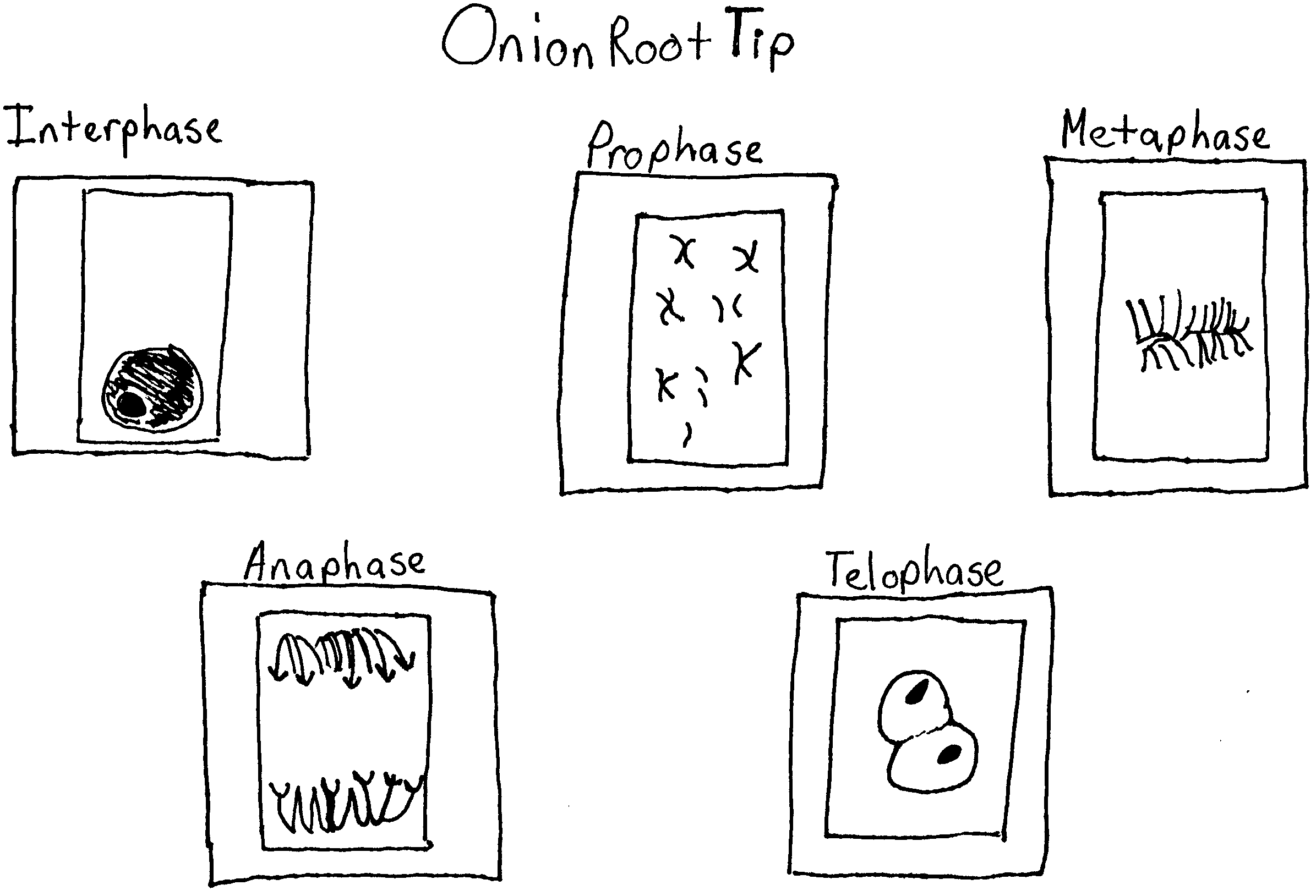
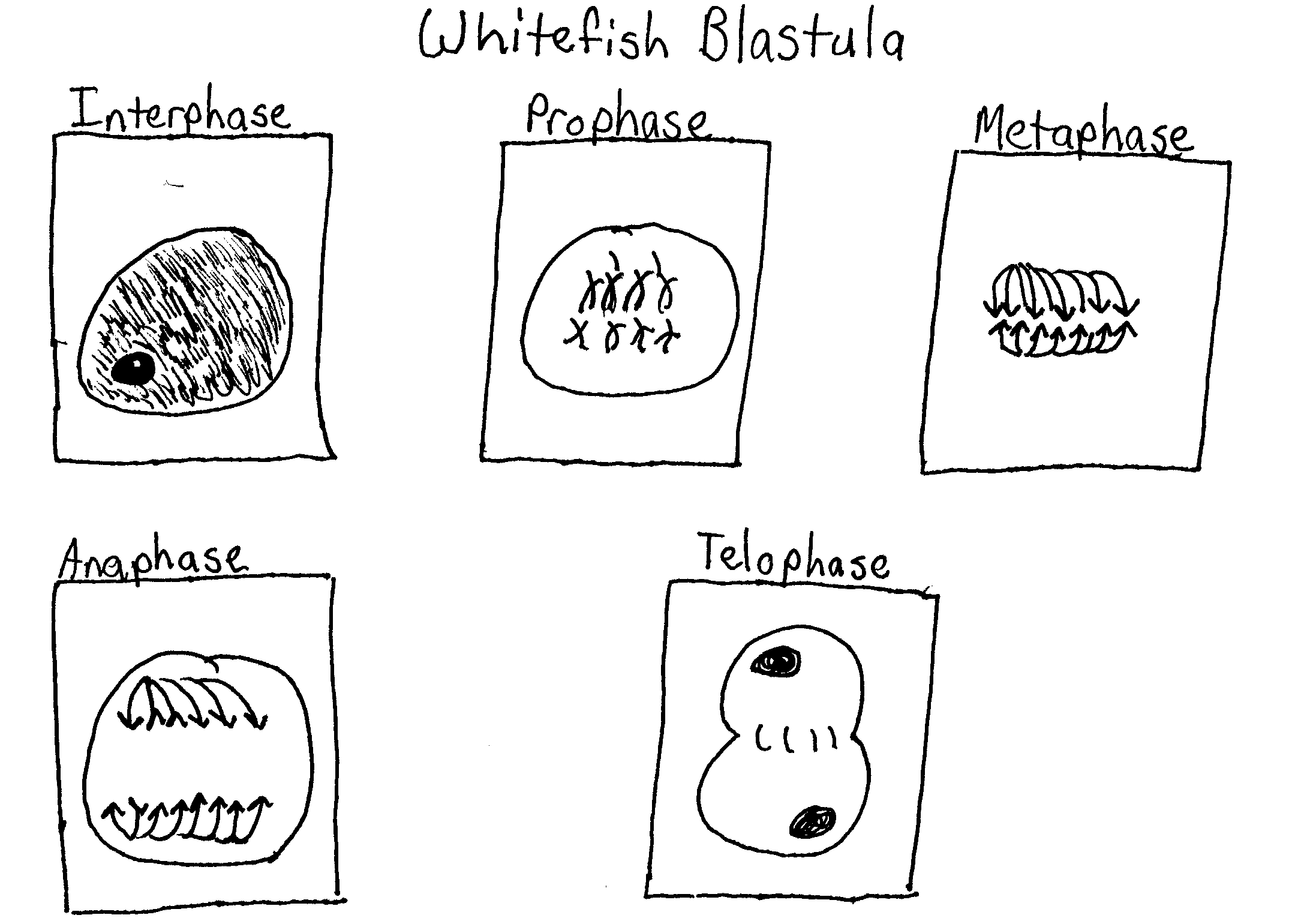
In plants mitosis takes place in the meristems which are normally found at the tips of stems or roots. However, in animal cells cell division takes place every where as new cells are formed and old ones are replaced. Studying mitosis can be accomplished by looking at tissues where there are many cells in a process of meiosis. Two examples are an onion root tip, or developing embryos, in animals such as whitefish blastula. A blastula is formed after an egg is fertilized and the egg begins to divide. There are several phases of the mitotic cell cycle. A precursor to mitosis is interphase. The actual steps of the mitotic cell cycle are prophase, metaphase, anaphase, and telophase. Interphase is a stage in the cell cycle in which the cell is not dividing. The nucleus contains a nucleolus and also contains chromatin. During interphase DNA replication occurs. The first phase of mitotic cell division is prophase. During prophase the chromatin begins to thicken until noticeable chromosomes are formed. Each chromosome has two chromatids that are joined at the centromere. During the later part of prophase, the nuclear envelope and nucleolus disappear. Mitotic spindle fibers, composed of microtubules, also become apparent. Following prophase is metaphase. By the time the cell has reached metaphase the chromosomes have moved to the center of the mitotic spindle. The centromere of the chromosome attaches to the spindle. The centromeres of each chromosome line up on an area called the metaphase plate. Metaphase is followed by anaphase. In the beginning of anaphase, the centromeres of each pair of chromatids separate and moved by the spindle fibers to the opposite ends of the cell. When the daughter chromosomes reach the ends of the cell the form a clump at each spindle pole. The final phase of mitosis is telophase. Telophase is identified by a recognizable condensation of the chromosomes, which is followed by the formation of a new nuclear envelope. The chromosomes slowly uncoil into chromatin once again and the nucleoli and nuclear envelope reform. It is then possible for cytokinesis, the division of the cytoplasm into two cells, to occur. In an animal cells a cleavage furrow forms and the cell pinches off into two new daughter cells.
The process of meiosis involves two nuclear divisions that result in the formation of four haploid cells. Meiosis I, a reduction division, is the first division to reduce the chromosome number from diploid to haploid and separates the homologous pairs. Meiosis II separates the sister chromatids resulting in four haploid gametes. Unlike mitosis meiosis increases genetic variation. In meiosis I each pair of homologous chromosomes come together which is known as a synapse. Chromatids of homologous chromosomes may exchange parts which is called crossing over. The distance between two genes on a chromosome may be estimated by calculating the percentage of crossing over that takes place between them. Meiosis I is preceded by interphase. During interphase DNA synthesis occurs and each chromosome is made of two chromatids joined at the centromeres. The first step of meiosis I is prophase I. During prophase I homologous chromosomes come together and synapse. A tetrad consisting of four chromatids is also formed. Prophase I is followed by metaphase I. In metaphase I the crossed over tetrads line up in the center of the cell. In anaphase I the homologous chromosomes separate and are moved to opposite ends of the cell. The final phase of meiosis I is telophase I. During telophase I centriole duplication is completed. Most of the time cytokinesis and formation of the nuclear envelope occur in order two make to cells. Meiosis II a second mitotic cell division then takes place in order to separate the chromatids in the two daughter cells made in meiosis I. This reduces the amount of DNA to one strand per chromosome. This is the only difference between meiosis I and II. Before meiosis II there is period called interphase or interkenesis. DNA replication does not take place in interphase II. Interphase II is followed by prophase II, No DNA replication occurs in prophase II and replicated centrioles separate and move to opposite sides of the chromosome groups. During metaphase II the chromosomes are centered in the middle of each daughter cell. During anaphase II the centromere regions of the chromatids are separate. The last stage of meiosis II is telophase II. In telophase II the chromosomes are at opposite ends of the cell and a nuclear envelope forms, and sometimes the cytoplasm divides.
Sordaria fimicola is fungus that may be used to show the results of crossing over during meiosis. Sordaria throughout most of its life is haploid, but becomes diploid after the fusion of two different types of nuclei, which forms a diploid nucleus. In Sordaria meiosis results in the making of eight haploid ascospores found in a sac called an ascus. Most asci are found in a perithecium. The life cycle of Sordaria fimicola is as follows: a spore is discharged through an ascus. The ascospore then undergoes mitosis, which forms a filament. The filament then undergoes mitosis, which forms a mycelium. Mycelial fusion and fertilization then takes place. This forms a diploid zygote. The zygote undergoes mitosis to form four haploid nuclei. The nuclei also undergo mitosis and form eight haploid nuclei, which then form eight ascospores. When mycelia of a mutant strain of Sordaria and a wild type of Sordaria undergo meiosis four black and four tan ascospores form. The arrangement of the ascospores reflects whether crossing over has occurred or not. Gametes, egg and sperm, are made during meiosis. Each egg and sperm cell contains half the total chromosomes a normal cell of that species would have. When the egg and sperm unite during fertilization the total chromosome number is restored.
Exercise 3A.1 – Hypothesis
While looking at prepared slides of onion root tip cells and whitefish blastula cells under a microscope I will be able to identify and draw the stages of mitosis in these cells.
Materials
The materials used in this experiment were a light microscope and prepared slides of onion root tip cells and whitefish blastula cells.
Methods
Using the microscope examine the slides of onion root tip cells and whitefish blastula cells. Begin by locating the merismatic region of the onion or the blastula using the 10 X objective. Then use the 40 X objective to study individual cells. Identify one cell that clearly represents each phase. Sketch and label the cell on a separate piece of paper.
Exercise 3A.2 – Hypothesis
When undergoing mitosis most of the cells in an onion root tip will be in interphase. More cells will be in the stage of prophase than metaphase. More cells will be in metaphase than anaphase and more cells will be in anaphase than telophase.
Materials
The materials used in this experiment were a prepared slide of an onion root tip and a light microscope.
Methods
Obtain a prepared slide of an onion root tip and observe every cell in one high power field of view and determine which phase of the cell cycle it is in. Make sure to do this in pairs so one person can observe the cells and the other person can record which phase the cell is in. Make sure to count three full fields of view and at least 200 cells. Then, record your data in table 3.1. Next, calculate the percentage of cells in each phase by using the equation; percentage of cells in stage X 1,440 minutes =_________ minutes of cell cycle spent in stage.
Exercise 3B.1 – Hypothesis
Using beads it will be possible to show the stages of meiosis I and meiosis II.
Materials
The materials used in this exercise were chromosome simulation kits containing four strands of beads. Two strands will be one color and the other two strands should be another color.
Methods
To show the process of interphase place one strand of each color near the center of your work area. Next, simulate DNA replication by bringing the magnetic centromere region of one strand in contact with the centromere region of the other of the same color. Do the same with its homolog. Next, to show crossing over in prophase I pop the beads apart on one chromatid at the fifth bead. Do the same with the other chromatid. Then reconnect the beads to those of the other color. Proceed through prophase I of meiosis and note how crossing over results in recombination of genetic information. Then to show metaphase I place the chromosomes near the middle of the cell. During anaphase I, the homologous chromosomes separate and are “pulled” to opposite ends of the cell. Next, to show telophase I place each chromosome at opposite sides of the cell.
In prophase II of meiosis II replicated centrioles separate and move to opposite sides of the chromosome groups. Next, to show metaphase II arrange the chromosomes so they are centered in the middle of each daughter cell. Then, separate the chromatids of the chromosomes and pull the daughter chromosomes toward the opposite sides of the daughter cell in order to show anaphase II. Finally in order to show telophase II, place the chromosomes at opposite sides of the dividing cell.
Exercise 3B.2 – Hypothesis
There will be more asci that maintain a 4:4 relationship of not crossing over than asci that do cross over.
Materials
The materials used in this exercise were a prepared slide of Sordaria fimicola and a light microscope.
Methods
Begin by obtaining a prepared slide that contain asci of Sordaria fimicola. Then, using the 10 X objective, view the slide and locate a group of hybrid asci. Make sure to count at least 50 hybrid asci and enter your data in table 3.3.
Results
Exercise 3A.1
Exercise 3A.2
Table 3.1
Number of Cells | ||||||
| Field 1 | Field 2 | Field 3 | Total | |||
| Interphase | 42 | 36 | 47 | 125 | 61.27 | 14 hours 42 min |
| Prophase | 10 | 14 | 18 | 32 | 20.10 | 4 hours 49 min |
| Metaphase | 6 | 5 | 4 | 15 | 7.35 | 1 hour 45 min |
| Anaphase | 2 | 3 | 2 | 7 | 3.43 | 49 min |
| Telophase | 7 | 5 | 4 | 16 | 7.84 | 1 hour 52 min |
Exercise 3B.2
Table 3.3
| Number of 4:4 | Number of Asci showing crossover | Total Asci | % Asci showing crossover divided by 2 | Gene to centromere distance (map units) |
| 60 | 45 | 105 | 21.4 % | 21.4 map units |
Questions:
Exercise 3A.1
1.Why is it more accurate to call mitosis “nuclear replication” than “cellular division”?
In mitosis two new nuclei are being formed. Also cytokinesis is actually a part of mitosis.
2. Explain why the whitefish blastula and onion root tip are selected for a study of mitosis?
A blastula is a hollow ball of cells that forms when an egg divides quickly; a large amount of mitosis is taking place here. The onion root tip is the place where growth occurs in the onion so a large amount of mitosis is taking place here.
Exercise 3A.1
1. If your observations had not been restricted to the area of the root tip that is actively dividing, how would your results have been different?
The tips are located in the meristem. Cells that are not in the meristem do not divide as quickly but they are elongating and differentiating. None of the phases would have been visible.
2. Based on the data in table 3.1 what can you infer about the relative length of time an onion root-tip cell spends in each stage of cell division?
Prophase is the longest stage and telophase is the shortest.
Exercise 3B.1
1. List three major differences between the events of mitosis and meiosis?
Mitosis has one nuclear division and meiosis has two nuclear divisions. Mitosis makes two identical daughter cells. Meiosis makes four daughter cells that half the number of chromosomes that their parent cells had. Crossing over and the exchange of genes occurs in meiosis but not in mitosis.
2.Compare mitosis and meiosis with respect to the following:
| Mitosis | Meiosis | |
| Chromosome number of parent cells | 2n | 2n |
| Number of DNA replications | 1 | 1 |
| Number of divisions | 1 | 2 |
| Number of daughter cells produced | 2 | 4 |
| Chromosome number of daughter cells | 2n | n |
| Purpose | Growth and repair | Gamete and spore production |
3. How are meiosis I and meiosis II different?
Meiosis I starts with a tetrad and separates the homologs. In meiosis the strands separate into 4.
4. How do oogenesis and spermatogenesis differ?
In oogenesis an egg is formed and three cells called polar bodies die. In spermatogenesis sperm are formed.
5. Why is meiosis important for sexual reproduction?
In meiosis the chromosome number is reduced by half. When fertlization occurs chromosome number is restored. Gene exchange causes variation.
6.Using your data in table 3.3 determine the distance between the gene for spore color and the centromere. Record your results in table 3.3.
Table 3.3
| Number of 4:4 | Number of Asci showing crossover | Total Asci | % Asci showing crossover divided by 2 | Gene to centromere distance (map units) |
| 60 | 45 | 105 | 21.4 % | 21.4 map units |
7. Draw a pair of chromosomes in MI and MII and show how you would get a 2:4:2 arrangement of ascospres by crossing over.
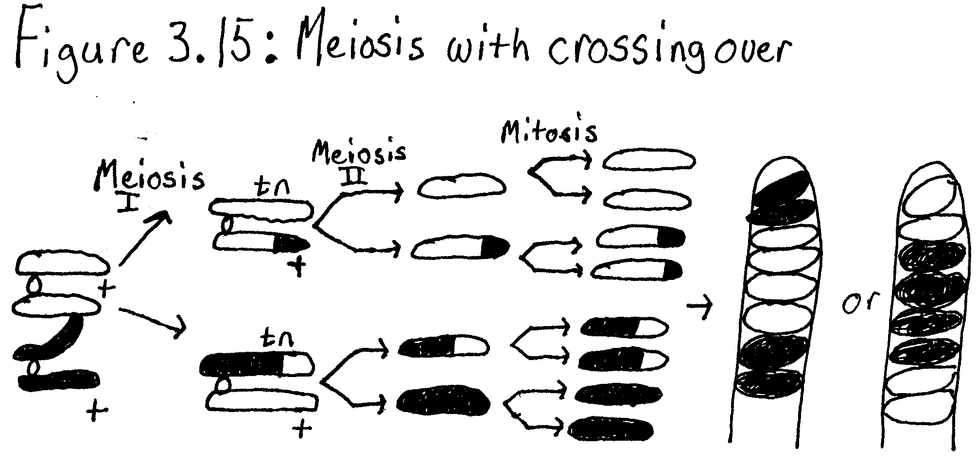
Error Analysis
In exercise 3A.2 inaccurate results were received. There should have been fewer cells in telophase than any of the other phases and cells should have spent less time in telophase than in any of the other phases. The results received are inaccurate because the number of cells that were in telophase were improperly counted. We received results that more cells were in telophase and spent more time in telophase than anaphase. In exercise 3B.2 improperly identifying some of the asci as crossover or non-crossover might have caused the results that were received to be inaccurate.
Conclusions:
From these experiments one can conclude that it is possible to look at mitotic stages of onion root tip cells and whitefish blastula through a microscope and draw them. Also, from these experiments one can conclude that most of the cell cycle in an onion root tip is spent in interphase. Prophase is after interphase in time spent in each cycle. Metaphase is after prophase. Anaphase is after metaphase. The least amount of time is spent in telophase. Also, a person can simulate the chromosomes in meiosis I and meiosis II using a chromosome simulation kit. Finally, one can conclude form the results of the experiments that more asci do not cross over in Sordaria fimicola than the number of asci that do cross over.
