Lab 3 Sample Ap Mitosis & Meiosis


Mitosis and Meiosis


Introduction
There are two types of nuclear division, mitosis and meiosis. Mitosis is usually used for the growth and replacement of somatic cells, while meiosis produces the gametes or spores used in an organism’s reproduction.

Mitosis is the first of these studied in this lab. It is easily observed in cells that are growing at a rapid pace such as whitefish blastula or onion root tips, which are used in this lab. The root tips contain an area called the apical meristem that has the highest percentage of cells undergoing mitosis. The whitefish blastula is formed directly after the egg is fertilized. This is a period of rapid growth and numerous cellular divisions where mitosis can be observed.

Just before mitosis the cell is in interphase. In this part of the cell cycle the cell will have a distinct nucleus and nucleoli where the thin threads of chromatin are duplicated. After duplication the cell is ready to begin mitosis and its starts with a step called prophase. In prophase, the chromatin thicken into distinct chromosomes and the nuclear envelope breaks open releasing them into the cytoplasm. The first signs of the spindle begin to appear. Next the cell begins metaphase, where the spindle attaches to the centromere of each chromosome and moves them to the same level in the middle of the cell. This level position is called the metaphase plate. Anaphase begins when the chromatids are separated and pulled to opposite poles. Then, the final stage is telophase. The nuclear envelope is reformed and the chromosomes gradually uncoil. Cytokinesis may occur, in which case, a cleavage furrow will form and the two daughter cells will separate.

Meiosis is more complex and involves two nuclear divisions. The two divisions are called Meiosis I and Meiosis II and they result in the production of four haploid gametes. This process allows increased genetic variation due to crossing over where genes can be exchanged. The process, like mitosis, depends on interphase to replicate the DNA. Meiosis begins with Prophase I. In this stage, homologous chromosomes move together to form a tetrad and synapsis begins. This is where crossing over occurs resulting in the recombination of genes. Metaphase I moves the tetrads to the metaphase plate in the middle of the cell, and Anaphase I reduces the tetrads to their original two stranded form and moves them to opposite poles. Telophase I then prepares the cell for its second division. Meiosis II generally resembles mitosis except that the daughter cells are haploid instead of diploid. DNA replication does not occur in Interphase II, and prophase, metaphase, anaphase, and telophase occur as usual. The only change is the number of chromosomes.

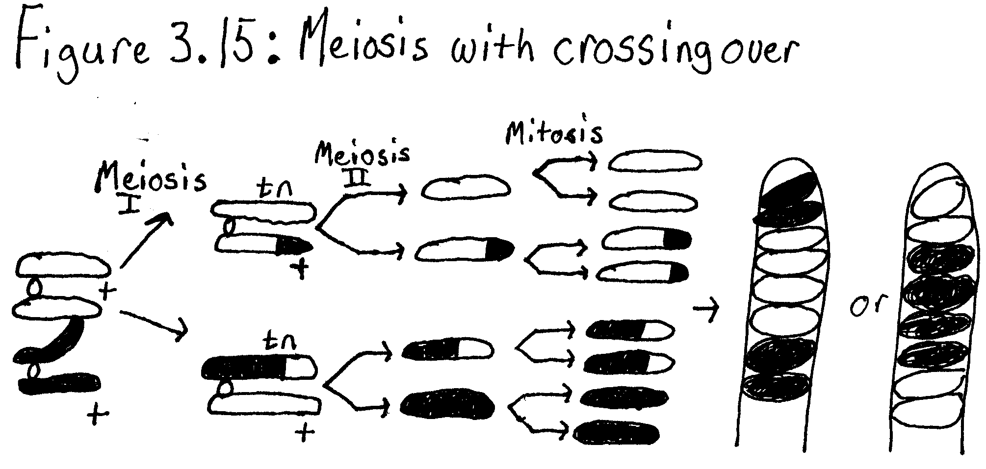
The process of crossing over can be easily studied in Sordaria fimicola, an ascomycete fungus. Sordaria form a set of eight ascospores called an ascus. They are contained in a perithecium until they are mature and ready for release. Crossing over can be observed in the arrangement and color of these asci. If an ascus has four tan ascospores in a row and four black ascospores in a row (4:4 arrangement), then no crossing over had taken place. However, if the asci has black and tan ascospores in sets of two (2:2:2:2 arrangement) or two pairs of black ascospores and four tan ascospores in the middle (2:4:2 arrangement), then crossing over had taken place.


Hypothesis
Mitosis occurs in whitefish blastula and onion root tip, and it is easily observable. Meiosis and crossing over occurs in the production of gametes and spores.

Materials
This lab requires prepared slides of whitefish blastula, onion root tips, and Sordaria, pencil, paper, a light microscope, and a chromosome simulation kit.


Methods
Exercise 3A.1: Observing Mitosis

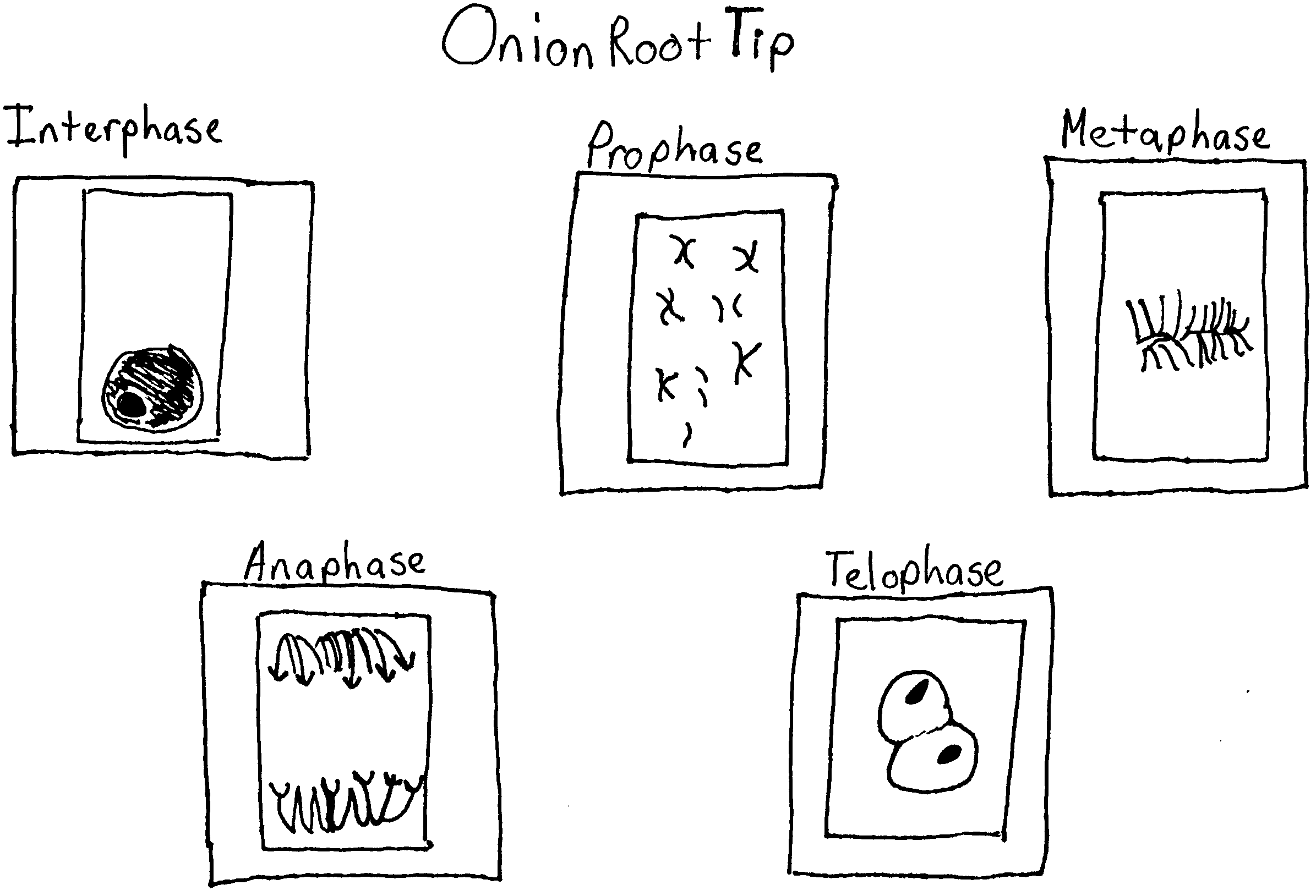
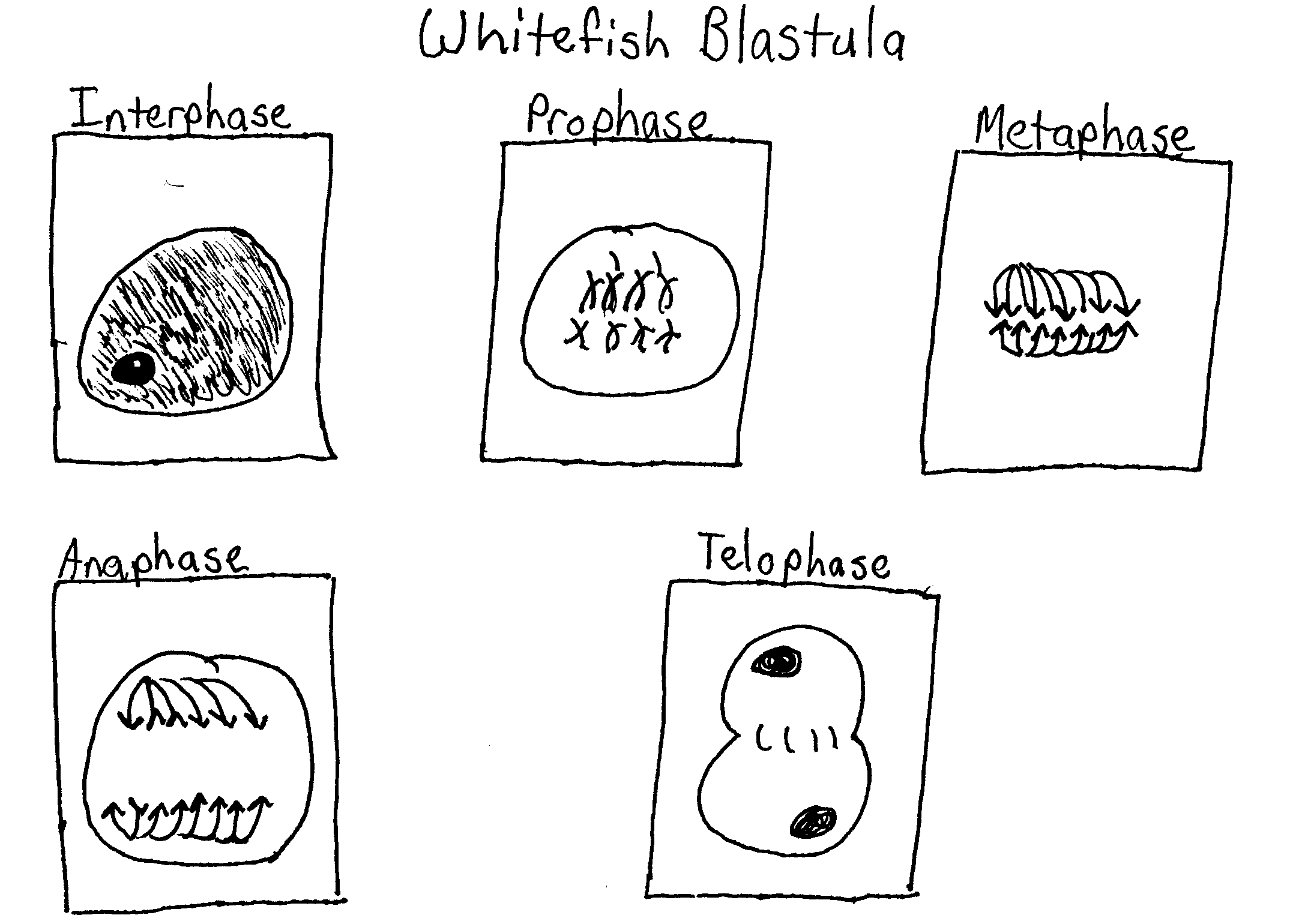
Prepared slides of whitefish blastula and onion root tips were observed under the 10X and 40X objectives. A cell in each stage of mitosis were identified, and then sketched.

Exercise 3A.2: Time for Cell Replication

Using a high power objective, every cell in a field of view was observed. Each cell was counted as being in one of the stages of mitosis and recorded. At least 200 cells and 3 fields of vision were counted and recorded. Next, the percentage of cells in each stage was recorded and the amount of time spent in each phase was calculated.

Exercise 3B.1: Simulation of Meiosis

In this part of the lab, a chromosome simulation kit was used to demonstrate meiosis. Two strands of the same color were connected to simulate DNA replication in both of the homologous pairs. Next, the chromosomes were entwined to represent synapsis. Sections of beads were switched between the pairs as in crossing over and were aligned at the equator. Next, anaphase was simulated as the homologous pairs were separated and then telophase was simulated by pushing the chromosomes into two separate cells (circles).

Meiosis II was simulated as well. The DNA is not replicated in Interphase II. The chromosomes again move to the equator and in Anaphase II the two chromatids were separated and moved to opposite poles. Telophase II separates them into four different cells.

Exercise 3B.2: Crossing Over during Meiosis in Sordaria

Prepared slides of Sordaria fimicola were observed under a light microscope. Over 100 asci were identified as either 4:4 or asci showing crossover and recorded. The percentage of each and the map units were calculated.

Results

Whitefish Blastula

Onion Root Tip

Table 3.1: Time for Cell Replication


Number of Cells

Field 1 Field 2 Field 3 Total
 

Interphase

42 36 47 125 61.27% 14 hours 42 minutes
 

Prophase

10 13 18 41 20.10% 4 hours 49 minutes
 

Metaphase

6 5 4 15 7.35% 1 hour 46 minutes
 

Anaphase

2 3 2 7 3.43% 49 minutes
 

Telophase

7 5 4 16 7.84% 1 hour 59 minutes

204


Table 3.2: Compare Mitosis and Meiosis


Mitosis


Meiosis


Chromosome number of parent cells

Diploid (2n) Diploid (2n)
 

Number of DNA replications

Once Once
 

Number of divisions

One Two
 

Number of daughter cells produced

Two Four
 

Chromosome number of daughter cells

Diploid (2n) Haploid (n)
 

Purpose

Growth and repair Production of gametes or spores


Simulation of the Meiosis I

Table 3.3: Sordaria


Number of 4:4


Number of Asci Showing Crossover


Total Asci


% Asci Showing Crossover Divided by 2


Gene to Centromere Distance (Map Units)

53 64 117 27.35% 27.35


Meiosis with Crossing Over – 2:4:2 Arrangement

Questions:

Why is it more accurate to call mitosis “nuclear replication” rather than “cellular division”?

It is more accurate to say “nuclear replication” to describe mitosis because the actual cell splitting occurs in cytokinesis. The whole process of mitosis is a series of steps that split the nucleus into two separate nuclei at opposite poles.

Explain why the whitefish blastula and onion root tips are selected for a study of mitosis.

The blastula is a hollow ball of cells that forms from the fertilization of an egg. Rapid growth occurs and numerous cellular divisions making mitosis in various stages easy to observe. Onion root tips are also a region of high percentage of cells going through mitosis because this is where most of the root growth takes place.

If your observations had not been restricted to the area of the root tip that is actively dividing, how would your results differ?

There would be virtually no cells undergoing division, so many more of the cells observed would have been in interphase where they elongate an differentiate.

Based on the data in Table 3.1, what can you infer about the relative length of time an onion root-tip cell spends in each stage of cell division?

Prophase is the longest stage of mitosis and then going in sequential order each decreases in the length of time it takes to complete.

List three major differences between the events of mitosis and meiosis.

In mitosis, the nucleus is only divided once, while in meiosis the nucleus is divided twice. Another difference is that mitosis produces two identical daughter cells, but meiosis produces up to four different daughter cells. Also, synapsis and crossing over do not take place in mitosis, but do take place in meiosis.

How are Meiosis I and Meiosis II different?

Meiosis I begins with a tetrad and separates the homologous pairs. Meiosis II separates the two sister chromatids.

How do oogenesis and spermatogenesis differ?

Oogenesis produces an egg cell, while spermatogenesis produces sperm cells.

Why is meiosis important for sexual reproduction?

In meiosis the chromosome number is reduced to n so that it can be fertilized. Also, meiosis allows for crossing over, which results in variations in organisms.

Error Analysis
There was little chance for error in this lab. It was mostly observation and sketching. However in Exercise 3A.2, the numbers for telophase were off. The calculations obtained for its time were too high; it should have been the shortest stage of meiosis. This may be caused by misidentifying the stages or counting the daughter cells as two different cells. Misidentification could have caused errors in the other parts of this lab as well.


Discussion and Conclusion
Mitosis was observed and timed in Lab 3A. The stages of mitosis are prophase, metaphase, anaphase, and telophase, prophase being the longest and telophase the shortest. Meiosis was simulated in Lab 3B and then crossing over was observed in Sordaria and the map units were determined. The gene to centromere distance in the Sordaria was 27.35 map units.

BACK


Lab 7 Sample 3 Fruitflies

Drosophila Genetics

Introduction

Drosophila Melanogaster, the fruit fly, is a great organism for genetic use because it has simple food requirements, occupies little space, is hardy, completes its life cycle in 12 days, makes a large number of offspring, can be knocked out easily, and it has many types of hereditary variations that can be seen with low power microscopes. Drosophila has a small number of chromosomes, four pairs. They are easily located in the large salivary glands. The Drosophila can be obtained from many places. Research of Drosophilae has led to a lot of knowledge about many of its genes.

Many factors combine to affect the length of the Drosophila life cycle. Temperature affects the life cycle the most. At room temperature the average life cycle of the Drosophila is about 12 days. Eggs of the Drosophila are small, oval shaped, and have two filaments at one end. They are usually laid on the surface of the culture medium, and with practice, can be seen with the naked eye. After one day the eggs hatch into the larva.

The larval stage of the Drosophila eats all the time. Larvae tunnel into the culture medium when they eat. The larva will shed its skin as it increases in size. In the last of the three larval stages, the cells of the salivary glands contain giant chromosomes that can be seen under low power in a microscope.

The pupal stage. Before a larva becomes a pupa it climbs the side of the container. The last larval covering then becomes harder and darker, forming the pupal case. Through this case the later stages of metamorphosis to an adult fly can be seen. In particular, the eyes, the wings, and the legs become visible.

The adult stage. When metamorphosis is over, the adult fly emerges form the pupal case. They are fragile and light in color and their wings are not fully expanded. They get darker in about an hour. They live about a month and then die. A female refrains from mating for about 12 days after she emerges from the pupal case. After she mates her receptacles contain large amounts of sperm and she lays her eggs. Make sure that the first flies you use are virgins.

The experiment will take several weeks. You will be assigned Drosophila with well-defined mutant traits by your teacher. You will keep a close record of what happens as each of these flies mate and pass there traits off to their offspring over a few generations.

There are three types of crosses that are studied in this lab. In monohybrid crosses the mode of inheritance is determined when a single contrasting pair of characteristics is involved. In a dihybrid cross the mode of inheritance is determined when the two pairs of contrasting of characteristics are considered simultaneously. In a sex-linked cross the mode of inheritance is determined when the mutant characteristic is associated with the X chromosome.

Hypothesis

In the sex linked cross of Drosophila Melanogaster, a phenotypic ratio of 1:1 will be obtained.

Materials

The materials used in this lab are as follows: a vile of Drosophilia with c designated trait, vials containing a medium, a refrigerator, ice packs, Petri dishes, a light microscope, a vial of wild type flies, an incubator, a pencil and paper.

Methods

Begin by obtaining a vial of wild type flies. Practice immobilizing and sexing these flies. Make sure to examine the flies and determine the characteristics of their eyes, wings, bristles, and antennae. Next, these are the steps for immobilizing the flies. Hold the vial containing the flies at an angle and place it in a refrigerator for several minutes. When the flies are immobilized, place them into a small plastic Petri dish. Then place the Petri dish on top of the icepack in order to maintain the cool temperature necessary to keep flies immobilized. Use the dissecting microscope to view the flies. Make sure to top the petri dish on when viewing the flies.

You can easily distinguish male flies from females by looking for the following characteristics: males are usually smaller than the females, males have dark blunt abdomens and females have lighter pointed abdomens. The males have sex combs, which are black bristles on the uppermost joint of the forelegs. Next, get a vial containing experimental flies. Make sure to write down the number of the vial that you have. The flies you now have are the P1 generation. The females should have laid eggs. The eggs and larvae are the F1 generation. Then after there are eggs present knock out remove the adult flies from the vial. Sex the adult flies and write down any mutations. Place the flies in the morgue that contains alcohol. Make sure to label the vial with the symbols for the mating.

After about another week has passed knock out and record characteristics of the remaining F1 flies and record the results in table 7.1. Then place the six pairs of these flies in a new vial and place the remaining flies in the morgue. Label the new vial F1, and tell the cross, date and your name.

After another week has passed, remove the F1 flies and put them in the morgue. The F2 generation are the eggs and larvae in the vial. Place the vial back into the incubator. Once again, after another week has passed remove the F2 flies and record their sex and characteristics and place the results in Table 7.2. Recording a greater number of F2 flies will make your results more accurate. Try to collect at least 200 flies. In order to analyze your data you will first have to be able to be able to complete Chi-Square Analysis.

Results

Table 7.1 F1 Generation


Phenotype females males
Red eyes 33 0
White eyes 0 31
 

Table 7.2 F2 Generation

Phenotype Male Female
Red eyes 50 52
White eyes 56 67


1. Describe the observed mutations? In the F1 generation the males had white eyes and the females had red eyes. In the F2 generation the males and females could have had either red or white eyes.

2. Write a hypothesis which describes the mode of inheritance of the trait you studied. This is your null hypothesis ( as described in the Statistical Analysis Section). For a sex linked cross there will always be a one to one ratio of the phenotypes. In the F1 generation there will be a one to one ratio of red eyed females to the number of white eyed males. In the F2 generation there will be a one to one ratio of red eyed females to white eyed females. There will also be a one to one ratio of red eyed males to white eyed males.

3. Refer to a textbook and review Punnett squares. In the space below construct two Punnett squares to predict the expected results of both the parental and F1 crosses from your null hypothesis.

Parental cross


Y Xr
Xr YXr Xr Xr
Xr YXr Xr Xr


F1 cross


Y Xr Y XR
XR YXR XR Xr YXR XRXR
XR YXR XR Xr YXr XR Xr
Xr YXr XrXr YXr X RXr
Xr YXr XrXr YXr XRXr


4. Refer to the Punnett squares above. Record the expected ratios for the genotypes and phenotypes of the F1 and F2 in the experiment below.


Expected Genotypic Ratio Expected Phenotypic ratio
F1 1:1 1:1
F2 1:1 1:1


        5. Do the actual results deviate from what was expected? If so, explain how.
No my results do no deviate much from what was expected. However in the F2 generation there were 67 white females and 52 red females.

6. For the results describe your cross? My cross is a sex linked cross.


7. Are the deviations for the phenotypic ratio of the F2 generation within the limits expected by chance?
To answer this question, statistically analyze the data using the Chi-Square-Analysis. Calculate the Chi-Square for the F2 generation in the chart below. Refer to the critical values of the Chi Square distribution table to determine the P value that is associated with your statistic.


Observed Phenotypes (o) Expected (e) (o-e) (o-e)2 (o-e)2

e

67 Xr Xr 56 11 121 2.16
52 XR XR 56 -4 16 .28
50 YXR 56 -6 36 .64
56YXr 56 0 0 0
3.02


(a) Calculate the Chi-Square value for these data.

1. How many degrees of freedom are there? 3 degrees of freedom

2. Chi Square=2.52

3. Referring to the critical values chart what is the probability value for these data? Greater than .05 probabilities that the null hypothesis is right.

(b) According to the probability value, can you accept or reject your null hypothesis? Explain why. I can accept the null hypothesis because my Chi-Square answer is less than the critical value form the table. I have 3 degrees of freedom and my Chi- Square answer was3.0 which is less than 7.82.

        1. Why was it necessary for the females of the parental generation to be virgins? The females store sperm in their receptacles and if they were not virgins we would not be able to tell who the fathers were.

2. Why was it not necessary to isolate virgin females for the F1 cross? The females store sperm in their receptacles and if they were not virgins we would not be able to tell who the fathers were

3. Why the adult flies were removed from the vials at weeks 2 and 4? So, they are not be able to mate with the next generation.

Chi-Square Analysis

Introduction

Statistics can be used to determine if differences among groups are significant, or simply the result of predictable error. The statistical test most frequently used to determine whether data obtained experimentally provide a good fit, or approximation to the expected or theoretical data is the Chi-square test. This test can be used to determine if deviations from the expected values are due to chance alone or to comeother circumstance.

To determine if the observed data fall with in acceptable limits, a Chi-Square analysis is performed to test the validity of a null hypothesis; that there is no statistically significant difference between the observed and expected data. If the Chi-Square analysis indicates that the data vary too much from the expected 3: 1 an alternative hypothesis is accepted.

Methods

The formula for Chi-square is:

X2=E(o-e)2
E

O= observed number of individuals

e= expected number of individuals

E= the sum of the values

The (df) are determined by taking the number of possible phenotypes and subtracting one from it. If the Chi- Square answer is greater than the number from the critical values chart then the null hypothesis is incorrect. The results are said to be significant at .05. This means that only 5 % of the time you would expect to see similar data if the null hypothesis were correct. The probability can also be rejected at .001. This time it means that less than 1 % of the time would you expect to see similar data.

Results

Critical Values Chart


Degrees of Freedom (df)

1 2 3 4 5
.05 3.84 5.99 7.82 9.49 11.1
.01 6.64 9.21 11.3 13.2 15.1
.001 10.8 13.8 16.3 18.5 20.5


Practice Problem

An investigator observes that when pure-breeding long winged Drosophila are mated with pure breeding short wing flies the F1 have an intermediate wing length. When several intermediate wing length flies are allowed to interbreed the following results are obtained. 230 long wings. 510 intermediate length wings. 260 short wings.

a. What is the genotype of the F intermediate wing length flies? The genotype is Ll.

b. Write a hypothesis describing the mode of inheritance of wing length in Drosophila. There will be 333 long winged flies. 666 intermediate winged flies. There will be 33 short wing flies.

c. Complete the table

Table 7.8


Observed Phenotypes Expected (e) (o-e) (o-e)2 (o-e)2/ e
LL 333 -103 10609 31.86
Ll 666 -156 24336 36.54
ll 333 -73 5329 16.00

84.4


1. How many degrees of freedom are there? There are 2 degrees of freedom.

2. Chi-Square= 84.4

3. Referring to the critical values chart, what is the probability value for these data? Less than .001.

4. According to the probability value can you except or reject the null hypothesis? 

I can reject the null hypothesis because the Chi-square answer is greater than the critical value from the table.

Error Analysis

Results from this lab could have been affected by many things. The constant knocking out of flies could have caused some of the larvae to not hatch therefore affecting our numbers. Also, incorrectly identifying the characteristics of the flies could have also greatly affected the results received. Improper calculation of numbers could have also caused inaccurate results. Finally, some flies could have gotten stuck in the medium and could have been identified.

Conclusion

From the results of the experiment I can conclude that I received results that were close to a 1:1 ratio. The Chi- Square worked from my data was accepted at a possibility greater than .05. The null hypothesis in this case can be accepted.

BACK


Invertebrate Notes

Invertebrate Notes
All Materials © Cmassengale

Invertebrate Phyla:


Porifera-sponges

Cnidaria:


sea anemone


hydra


Coral


Jellyfish

Platyhelminthes-flatworms


Fluke

Tapeworm
Nematoda-roundworms

Trichinella

Ascaris

Hookworms

Pinworms
Rotifera–rotifers
Annelida-segmented worms

earthworm

leech
Mollusca

clam

snail

octopus
Arthropoda
Subphylum: Trilobita–trilobites (extinct) Subphylum: Chelicerata-horseshoe crabs, spiders, scorpions, mites, & ticks Subphylum: Mandibulata–crustaceans, insects, millipedes, centipedes

Trilobite

Horseshoe crab

Millipede
Echinodermata: starfish, sea cucumbers, sea lilies

Starfish

Sea Cucumber

Sea Lily


About 97% of all animals are invertebrates.  Invertebrates are animals which do not have a backbone.  There are nine phyla of  invertebrates:  Porifera, Cnidaria, Platyhelminthes, Nematoda, Rotifera, Mollusca, Annelida, Arthropoda, & Echinodermata.

Sponges


Notes on Invertebrate Animals


Notes on Invertebrate Animals


Phyla:


1. Porifera‑‑sponges

2. Cnidaria

a. sea anemones

b. hydra

c. corals

d. jelly‑fish

3. Platyhelminthes‑‑flatworms

a. flukes

b. tapeworms

4. Nematoda‑‑roundworms

a. Trichinella

b. Ascaris

c. hookworms

d. pinworms

5. Rotifera–rotifers

6. Annelida‑‑segmented worms

a. earthworm

b. leeches

7.  Mollusca‑‑clams, oysters, snails, and octopus

8. Arthropoda

     subphylum: Trilobita–trilobites (extinct)

     subphylum: Chelicerata‑‑horseshoe crabs, spiders, scorpions, mites, & ticks

subphylum: Mandibulata–crustaceans, insects, millipedes, centipedes

9. Echinodermata: starfish, sea cucumbers, sea lilies


About 97% of all animals are invertebrates.  Invertebrates are animals

which do not have a backbone.  In this unit we cover nine phyla of

invertebrates:  Porifera, Cnidaria, Platyhelminthes, Nematoda, Rotifera, Mollusca, Annelida, Arthropoda, & Echinodermata.

SPONGES

The phylum Porifera are sponges.  There are about 800 different species of sponges, and 88% are marine.  “Marine” means that they live in salt water, such as an ocean or a sea.  Freshwater sponges are smaller and less brightly colored than marine sponges.  Sponges are filter feeders.

This means that they use their body as a filter to trap their food, microscopic plankton.


Sponges are asymmetrical and live attached to one spot as adults making them sessile animals. Sponges have a skeleton composed of a flexible protein material called spongin & hard fibers called spicules composed of calcium carbonate or silicon dioxide. The body of a sponge is filled with holes or pore through which water enters their hollow bodies.  Sponges lack the tissue level of organization but they do have some specialized cells.  Choanocytes are specialized cells that line pores in a sponge and have a flagellum that spins to pull in water and food.  Collar cells at the base of choanocytes capture plankton & start digesting it.  Amebocytes are specialized cells that carry food to all other parts of a sponge=s body.  Wastes and excess water leave a sponge through an opening at the top called the osculum.

Sponges reproduce asexually by internal or external buds and by fragmentation whenever a piece of the sponge breaks off. Each piece can form a new sponge. This is how sponges form colonies. Sponges reproduce sexually by dispensing eggs and sperm into the water.

If the freshwater supply evaporates, freshwater sponges become dormant and form an internal bud or gemmule which is release when the sponge dies.  The gemmule is a small freshwater sponge covered with hardened mucus which prevents it  from drying out.  When the freshwater returns, the gemmule becomes an active sponge.


Cnidarians

The phylum Cnidaria include sea anemones, hydra, corals and jellyfish.  All Cnidaria are marine except hydra, which is a freshwater organism. Cnidarians have radial symmetry and are carnivorous using tentacles that surround their mouth to get food. Cnidarians exhibit two body forms – the sessile polyp with tentacles & mouth at the top or the motile medusa with tentacles & mouth on the bottom.  Cnidarians may exist in one of these two stages or go through both stages in their life cycle.  Cnidarians have a hollow gastrovascular cavity on the inside lined with gastrodermisEpidermis covers the outside and a jellylike material called mesoglea is between the layers.  Mesoglea is thin in polyp forms but thick in medusa forms. Cnidarians have stinging cells called nematocysts or cnidocytes on their tentacles that are poisonous & shoot out like a harpoon to kill or paralyze prey.   Their mouth is the only opening to their body so they have a two-way digestive system.  The also have a simple nerve net . Cnidarians reproduce asexually by budding or sexually producing fertilized eggs whenever males release sperm and females release eggs into the water. Some cnidarians like coral build a limestone case that makes an underwater reef.


Platyhelminthes (flatworms)

The phylum Platyhelminthes are dorsoventrally flattened and have a definite anterior and posterior end giving them bilateral symmetry.  Their bodies are solid so they are said to be acoelomate.  Some flatworms are parasites, while others are free-living carnivores or scavengers.  Examples of parasitic flatworms are flukes and tapeworms. Flatworms also have only a mouth for both food and wastes.  Their nervous system is composed of a nerve net and sometimes light-sensitive eyespots at the anterior end.  Specialized flame cells help get rid of wastes.

The planarian is the most common free-living flatworm found in water or moist places. They are hermaphrodites producing both eggs and sperm, but they exchange sperm with each other during sexual reproduction.  Planarians also reproduce asexually by fragmentation.

Flukes and tapeworms often live in their host=s digestive tract resistant to the host=s enzymes.  They  do not have a digestive system allowing the host to digest their food.


Tapeworms are divided into sections called proglottids that each have a complete reproductive system producing fertilized eggs. Tapeworms are hermaphroditic (one body having both sexual parts), and they fertilize their own eggs. Ripe proglottids with their eggs pass out with the host=s feces. Tapeworms anterior end is called the scolex and is modified with both hooks and suckers to attach to the host=s intestines.  Humans most often get tapeworms from undercooked pork, beef. or fish.  Tapeworm eggs can withstand boiling water so it is important to cook these meats well enough to destroy the eggs.  Children sometimes get tapeworms by playing with the feces in the litter box of a cat, getting the eggs on their hands, and placing their hands or fingers in their mouth.  The longest tapeworm ever passed by a person was 39 meters.

Flukes have complex life cycles that involve more than one host. A fluke causes Schistosomiasis, a disease that affects 250 million people world wide.  This blood fluke attacks the kidneys, liver, and intestines causing progressive weakness.  It often takes 20 years to die from Schistosomiases, & there is no cure.


Nematoda (roundworms)

The phylum Nematoda are the roundworms.  Roundworms are cylindrical in shape and vary in length from being microscopic to  20 inches long.  Roundworms are pseudocoelomate having a body cavity that is not completely lined. The body cavity or pseudocoel serves as a hydrostatic skeleton against which muscles can contract.  Unlike flatworms, roundworms have a complete gut.  This means that they have a one-way digestive tract with a gut that begins with a  mouth and ends with an anus. Therefore, they are usually able to digest food.  However, roundworms have no blood or heart.  Nutrients are distributed by a non‑ blood fluid which is not pumped.

Most roundworms are parasites and are found in all habitats. They are bilaterally symmetrical and unsegmented.  Although they are cylindrical in shape, they usually taper at both ends.  They are covered with a thick protective cuticle that is flexible and can be molted.  They have separate sexes generally and reproduce sexually.

The roundworm Trichinella, causes the disease called trichinosis.  People get trichinosis from eating undercooked pork.  Trichinella gets into muscles and leaves calcium deposits which effect muscle contraction.  Trichinosis can affect the heart.  Another roundworm, Ascaris,

parasitizes human lungs. The Filaria worm attacks the lymphatic system causing great swelling. Hookworms and pinworms are also roundworms which parasitize humans.


Rotifers

The phylum Rotifera includes microscopic worms found in aquatic and soil habitats.  They have a crown of cilia at their head end surrounding their mouth for movement and feeding.  Their bodies are covered with an external layer of chitin. Having separate sexes, they reproduce sexually.  Some species contain only females and reproduce by parthenogenesis (unfertilized eggs developing into females).


Mollusks


The phylum Mollusca contains snails and slugs, bivalves, octopus, squid, and the chambered nautilus. Many members of this phylum have durable limestone shells and are found in all habitats. Members of this group are economically important as sources of human food , pearl and shell production, crop & flower damage, destruction to submerged wooden structures, and intermediate hosts for some parasitic diseases. The giant squid and giant clam are the two largest invertebrates.  Mollusks have bilateral symmetry and a visceral mass containing their body organs. Mollusks also have a muscular foot for movement which can be modified into arms or tentacles in some species.  Mollusks breathe through gills or lungs located below a protective layer called the mantle.  The mantle forms the shell in some species and also protects the body organs. All mollusks except bivavles contain a rasping, tongue-like radula for scraping food.  The circulatory system consists of a three-chambered heart  and open-flowing system except for octopus & squids which have a closed circulatory system. Reproduction is sexual even in hermaphroditic forms.  Mollusks go through a free swimming larval stage called the trochophore.

The class of mollusks called gastropods have a foot on their belly.  An example of a gastropod is the snail.  When a snail lacks a shell it is called a slug.  Snails and slugs walk on their belly.  Most snails are marine, but some do live on land.  Marine snails have gills.  Land snails are called pulmonate snails and have an air hole for breathing.  Snails can be very large.  The helmet snail can be as big as 15 pounds.

The class of mollusk called Bivalvia includes clams, oysters, mussels , and scallops.

These mollusks have two shells hinged together by a ligament.  Strong adductor muscles open and close the shells. Incurrent and excurrent siphons circulate water containg food and oxygen through the bivalve.  Gills extract the oxygen from the water,  and they move by jet propulsion.  Their muscular foot can be extended from the shell for movement or anchoring.

The class of mollusks called cephalopods have a foot on their head.  Examples of cephalopods are octopus, squid and nautilus.  Most cephalopods have beaks, tentacles and jaws and are active predators. Their musclar foot has been modified into arms or tentacles. They lack external shells except for the natilus.  These are the most intelligent of all invertebrates.  They used their siphons to move by jet propulsion.  Octopus have their shell inside of their body.  Octopus secrete an inky substance which they spit out to help them escape from predators.  The giant squid is the largest cephalopod.  It can be up to 60 meters in length and has been known to eat whales.


Annelids (segmented worms)

The phylum Annelida are the segmented worms and are abundant in all habitats. External segments  are characterized by ringlike structures along the body, and corresponding internal segments are called septaSegmentation gives worms more flexiblity in movement. If one segment is damaged, it isn=t usually fatal to the animal because their organs are duplicated in other segments.  Annelids have a Atube within a tube@ body plan known as a coelom which is fully lined and contains the body organs.  The coelom runs from the mouth to the anus. Annelids have bilateral symmetry, and a well-developed brain and diverse sense organs showing cephalization. Coelomic fluid serves as a  hydrostatic skeleton.

Earthworms belong to this phylum.  Each segment of the earthworm has setae or external


bristles made of chitin.  These bristles allow the earthworm to move and to burrow into soil.   Earthworms have a head and a central nervous system.  Earthworms respire through their moist skin as they dig through the soil and help loosen it. They have a closed circulatory system in which blood is pumped by five pairs of hearts.  Most earthworms feed on decomposing vegetation causing it to decompose faster. A  pharynx sucks in the organic debris which the muscular gizzard grinds. Earthworms bring the nutrients from the subsoil to the top soil, thereby helping plants to grow.  Undigested materials or castings are deposited outside burrows.

Leeches are also in the phylum Annelida.  Most leeches live in water and have suckers at both ends of their bodies. The tail suckers are used to latch on to a host, while the head suckers

are used to suck blood from the host.  Most leeches are predators or scavengers, but some suck blood.  Because of this, blood sucking leeches are collected for anticoagulant. Leeches bodies are flattened dorsoventrally and lack setae except for one species.  Like earthworms, leeches are hermaphrodites that exchange sperm with other members of their species.

Polychaetes are marine annelids that have their setae modified into paddle-like structures called parapodia.  Parapodia improvement movement and give more area for gas exchange. Polychaetes often live commensally with sponges, mollusks, echinoderms, and crustaceans. Sexes are separate with external fertilization.


Arthropods

The members of the phylum Arthropoda all have jointed appendages.  In fact, the word “arthropod” means jointed leg.  There are more species of arthropods than any other phylum.


Arthropods have these characteristics:

a. hard exoskeleton which is usually composed of substance called  chitin

b. go through periodic ecdysis as they shed or molt their exoskeleton

c. they have specialized body segments (head,  thorax, cephalothorax, & abdomen)

d. jointed appendages such as legs, antenna, and mouthparts.

e. open circulatory system


The phylum Arthropoda is divided according to their type of appendages.  The subphylum Chelicerata possess chelicerae or fangs and no antenna, while the subphylum Mandibulata have antenna and mandibles or jaws.  Crustaceans have pincers called chelipeds.  The subphylum Trilobita are an extinct group with a head and trunk with a pair of legs on each segment.

Terrestrial arthropods like insects, millipedes, & centipedes have a system of hollow air tubes called trachae as their respiratory system. Aquatic chelicerates like the horseshoe crab have book gills, while terrestrial chelicerates such as spiders, ticks, mites, & scorpions  use  book lungs.    Book lungs have numerous blood vessel lined surfaces which look like the pages in a book & get oxygen from air.  Crustaceans respire through gills. Gills are folded tissue which are lined with blood vessels which  remove oxygen from water.

Terrestrial mandibulates are uniraimous with one-branched appendages, but aquatic mandibulates like crustaceans are biramous or two-branched.   Arthropods have a brain and nervous system and possess a variety of sensory receptors such as simple eyes called ocelli or compound eyes, typmpanic membranes for hearing, and antenna that can smell and taste.  Excretory structures in arthropods vary, but terrestrial arthropods have Malpighian tubules to filter nitrogenous wastes.


The subphylum Chelicerata (ki‑LISS‑uh‑ruh) include the class Xiphosura or horseshoe crabs which have a cephalothorax and abdomen, live in marine environments breathing through book gills, lack antenna, but have chelicera & 4 pairs of walking legs.  The class Arachnida containing

spiders, scorpions, mites, and ticks are also chelicerates that lack antenna, have chelicera (fangs) and 4 pairs of legs, but they live in terrestrial habitats and breathe through book lungs or trachae

Chelicerates also have appendages on their head called pedipalps that are sensory and can help move food into their mouth.   Unlike most arthropods,  spiders do not see well; however, they are good at detecting movement.  Spiders have glands called spinnerets on the posterior end of their abdomen that produce silk to make webs.  When prey get caught in a spider’s web, it is the movement which alerts the spider to the captured prey.  Most spiders also have hairs on their body to assist them in feeling movement.  Spiders  poison their prey once they are caught in their webs. Spiders are very beneficial because they catch and eat insects.  Two spiders which are

dangerous are the black widow and the brown recluse.  Both of these spiders have distinct markings on the underside of their abdomen..  Spiders differ from insects in having eight, not six legs,   having simple eyes  and not compound eyes, and having only 2 body regions

(cephalothorax & abdomen) instead of 3 regions ( head, thorax, & abdomen).

The subphylum Mandibulata contains the class Crustacea.  Most crustaceans live in the water and include crabs, shrimp, lobster, crayfish, & barnacles. Terrestrial crustaceans include pillbugs and sowbugs.  Crustaceans have a pair of antenna to smell and detect chemicals and a shorter pair of antennules used for balance. They have 2 body regions (cephalothorax and abdomen), and their mouthparts include mandibles, maxilla, and maxillipeds.  They also have pincers called chelipeds to help them  catch food.  Aquatic crustaceans  have a shell called a carapace that they regularly shed as they grow to produce a larger one.    Crustaceans are economically important to man as a food source.

The classes Chilopoda and Diplopoda are alo in the subphylum Mandibulata.  Chilopoda or centipedes are poisonous predators feeding on other terrestrial arthropods. Centipedes have fangs, venom glands, and a pincer on their tail. They have a single pair of legs per body segment.  Diplopoda or millipedes are vegetarians or scavengers feeding on decaying vegetation that have two pairs of legs per body segment.


The class Insecta in the subphylum Mandibulata includes all of  the insects.  This is the largest and most successful group of arthropods. Insects usually have six legs, a pair of antenna, and a pair of wings although some species may be wingless such as silverfish and termites. Flies have their second pair of wings modified into a balancing structure called halteres.  Insect’s mouths usually have four parts – the mandible or jaw, maxilla, labium or lower lip, and labrum or upper lip and are adapted for a particular food.  For example, grasshoppers  have chewing mouthparts for eating grass, mosquitos have sucking mouthparts for sucking blood, butterflies have siphoning mouthparts for getting nectar from flowers, and the house fly has spongy mouth- parts for soaking up liquid food.  Wings and legs are attached to the midsection or thorax, antenna, eyes, and mouthparts are attached to the head, and the abdomen on females may have an egg-laying tube called the ovipositor.  Insects communicate by producing sounds and by making chemicals called pheromones. Tympanic membranes on the abdomen and sensory hairs detect sound waves.  Spiracles line the sides of the insect=s abdomen and open into their breathing tubes or trachae. Insects may go through stages in their life cycle.  Butterflies, bees, flies, and beetles go through the egg, larva, pupa, and adult stages.  This is known as complete metamorphosis. Dragonflies and grasshoppers go through egg, nymph, and adult stages known as incomplete metamorphosis.  Insects such as silverfish and fleas do not go through metamorphosis.  Metamorphosis and molting are controlled by hormones.


Echinoderms

The phylum Echinodermata include the starfish, sea urchins and sea cucumbers.  The word “echinoderm” means spiny skin.  Echinoderms are the most advanced invertebrates. All other

invertebrates are protostomes in which the blastopore in their development becomes the mouth.  Echinoderms, like chordates, are deuterostomes in which the blastopore becomes the anus. Echinoderms have an endoskeleton composed of movable or fixed calcium plates called ossicles.  The members of this phylum have radial symmetry with a five part body plan. Adults have no head or brain and move be extendable tube feet.  Echinoderms also possess a water vascular system made up of a system of canals that help the organism feed and move.  Water enters through an opening called the madreporite into a short stone canal into the ring canalRadial canals connect to the ring canal and determine the five-part symmetry. This hydraulic water system is strong enough to help starfish open clam shells.  Skin gills are used for respiration and waste removal.   Echinoderms are capable of extensive regeneration whenever parts are dropped.  They can reproduce asexually by fragmentation or sexually with external fertilization.

Starfish are in the class Asteroidea and are active marine predators with 5 arms set off from a central disk and their mouth located on the underside or oral surface. Bivalve mollusks are a favorite food of the starfish, and they consume them by turning their stomach inside

out and sticking it into the clam shell to digest the clam.

Sea urchins and sand dollars are in the class Echinodea and they lack distinct arms. Five rows of tube feet protrude through their skeletal.  They use the spines of their skin and tube feet to move about and graze on algae, coral, or dead fish.  Triangular teeth around the mouth scrap or crush food.

The class Crinoidea contains sea lilies and feather stars with highly branched arms around their mouth for filter feeding.  Sea liles are attached by a stalk to the substrate, but feather stars are able to detach and move about.

Brittle stars in the class Ophuroidea have slender arms attached to their central disk and can move faster than starfish. Sea cucumbers are in the class Holothuroidea and are soft, sluglike organisms with leathery outer skin. Sea cucumbers usually lie on their sides on the ocean bottom and can eject part of their intestines in order toscare away a predator.  They also move by tube feet or by wiggling their entire body. Some of these are hermaphroditic which is unusual for echinoderms.

Introduction to Plants PPT Questions

Introduction to Plants
ppt Questions

Early Ancestors

1. The first habitat for plants on earth was _____________.

2. Which algal group is most related to early land plants?

3. What is this group of algae called?

4. List 5 similarities between algae and terrestrial plants.

     a.

     b.

     c.

     d.

     e.

5. List 5 helpful adaptations aquatic plants have by being surrounded by water.

     a.

     b.

     c.

     d.

     e.

6.Complete the following table explaining how terrestrial plants solved the move onto land.


Plant Adaptations to land
Problems: Solutions:
Need Minerals
Gravity
Increase in Height to get Light
Adaptations for drier environment
Reproduction


How Are Plants All Alike

7. All plants are ____________________.

8. Plants can make their own food by a process called ____________________.

9. Since plants make their own food they are called _________________.

10. Plants contain what type of chlorophyll?

11. Where is chlorophyll found in plants?

12. What surrounds the outside of all plant cells and what is it composed of?

13. How do plants store their reserve food?

14. The life cycle of plants is known as __________________ of _________________.

15. The dominant stage of the plant is the diploid (2n) ________________ stage.

16. The eggs and sperm are produced during the haploid (1n) ________________ stage.

17. The gametophyte stage produces a multicellular plant ______________ that is protected inside an ____________ ___________.

18. The sporophyte stage produces _____________ by _____________.

19. Haploid spores undergo ______________ to produce the _______________ stage.

20. The gametophyte stage makes _____________ called the _________ and ___________.

21. Label the diagram of alternation of generation. Include the sporophyte and gametophyte generations, the chromosome number (2n or 1n), and where mitosis and meiosis occur.

Plant Divisions

22. Plants are divided into __________ groups based on the presence or absence of an ___________ _____________ ___________ for carrying water and dissolved _____________.

23. What is the transport system for water and minerals called?

24. ______________ plants lack vascular tissue and are called _______________.

25. In what type of environment must nonvascular plants live?

26. Give an example of a bryophyte.

27. Nonvascular plants can’t grow as tall as vascular plants. Explain why.


28. The cells of nonvascular plants must be in _________ contact with water because water moves by _______________ from cell to cell.

29. How does the sperm get to the egg in nonvascular plants?

30. Name 3 divisions of nonvascular plants and give and organism found in each division.

     a.

     b.

     c.

31.Vascular plants are also called _______________.

32. What are the 2 subdivisions of vascular plants?


33. Name 4 divisions of seedless vascular plants and give an example of a plant in each group.

     a.

     b.

     c.

     d.

34. Name the 2 groups of seed-bearing vascular plants.


35. Gymnosperms have ____________ seeds found inside cones.

36. Angiosperms have ___________ to attract ____________ so seeds can be produced.

37. Name the division known as conifers and tell several plants in this group/


38. Name 2 other divisions of gymnosperms and tell a plant in each group.

     a.

     b.

39. Name the oldest living plant.

40. Name the tallest living plant.

41. What group are these 2 plants in?

42. Angiosperms are called ____________ plants.

43. How are seeds formed in angiosperms?


44. Where is the ovary found?

45. Name the male and female parts of a flower.

46. How are fruits formed?

47. Angiosperms are the division ______________.

48. What are the 2 subgroups of Anthophyta.

49. Describe the characteristics of monocots.


50. Describe the characteristics of dicots.