Are you getting ready for your first biology class? Or are you trying to shake off the cobwebs and remember your biology from years ago? Either way, you may be asking, what is osmosis in biology?
We want to answer this question in a way that is thorough and understandable at the same time. Dust off your old textbook and put on your reading glasses as you find answers to the question, “What is osmosis in biology?”
What Is Osmosis In Biology?
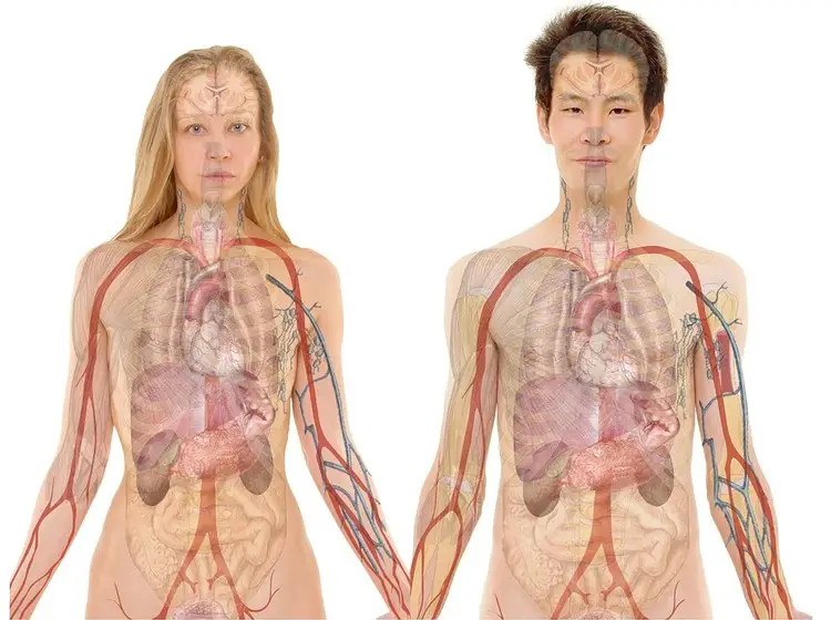
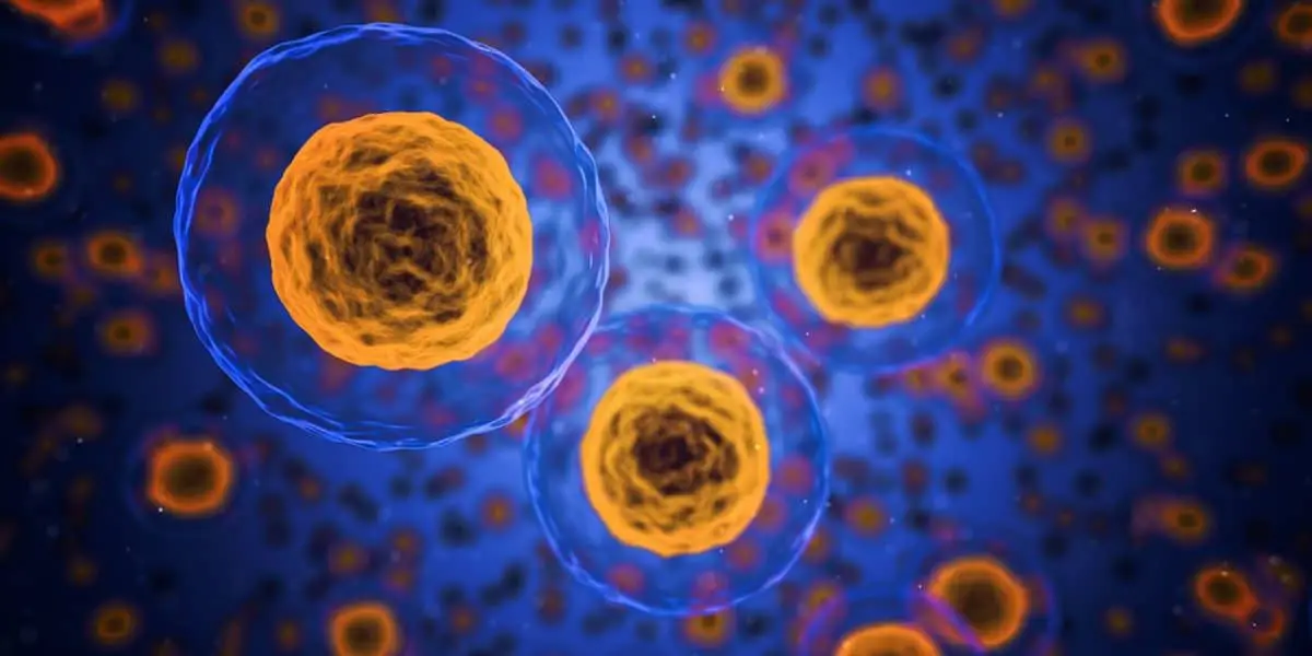
Osmosis is a type of diffusion. In biology, it is related to cells. Osmosis happens when a solvent flows through a cell membrane, to balance the concentration of a solute — such as salt. If water is a solvent, it will be affected by the amount of salt (solute) that it contains.
Understanding Diffusion
Diffusion happens when molecules move from a highly concentrated area to a less concentrated region. Solids, liquids, and gasses can all diffuse.
When a liquid such as water diffuses in cellular biology, it crosses a semipermeable membrane to balance the concentrations of substances within the cells. As water flows in or out of a cell, the concentration of solutes affects its travel.
Semipermeable Membranes
To answer the question, what is osmosis in biology, we have to understand semipermeable membranes.
Semipermeable membranes are membranes that allow specific molecules or solvents to pass through by diffusion. Every cell in the human body has a cellular membrane, and they are semipermeable.
That word breaks down: “semi” in this biology word means “partly”, and “permeable” means “able to be passed through, or permeated.” So, semipermeable membrane means a membrane partially able to be crossed.
Some things can pass through, and others cannot.
Osmosis happens as solvents pass into and out of the cell, crossing that semipermeable membrane.
Osmosis in Plants and Animals
Plant cells need more water than animal cells. Plants have thicker cell walls that can contain more solution before bursting. For that reason, plants can thrive with the diffusion of hypotonic solutions.
Hypotonic solutions have a much higher ratio of solvent to solute. Hypotonic solutions can make animal cells burst; animal cells have thinner cell walls than plant cells.
Isotonic solutions are much better for diffusion in animal cells. Isotonic solutions contain equal amounts of solvent and solute. Conversely, isotonic solutions will leave plants drooping and unhealthy.
Did you ever hear of someone pouring salt on a slug when they were a child? Hopefully not; but if you did you know the slug shriveled up and essentially disappeared. That is because the water left the slug’s cells in an attempt to balance the concentration of salt outside the cells.
That is osmosis in action.
Examples of Osmosis
Try it at home! If you are looking for an example of osmosis you can easily try at home, and you have some lettuce in your fridge (or any leafy green like kale or spinach) that has become wilted try this experiment:
Types of Solutions

image source: pixabay.com
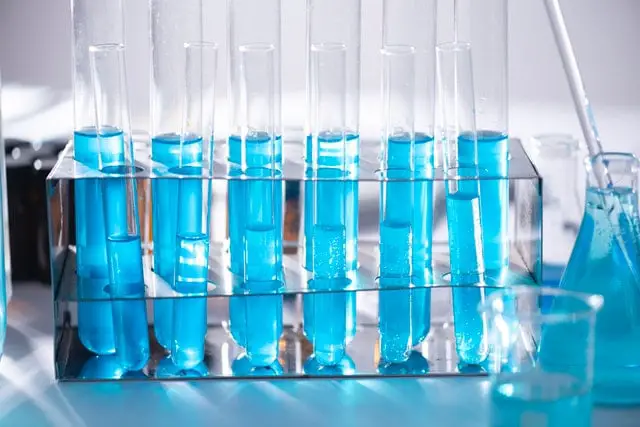
Every solution has a solvent and a solute. When you buy contact lens solution, you are essentially buying saltwater; water is the solvent and salt is the solution. The same is basically true of your tears.
Solutions
To answer the question, what is osmosis in biology, we have to understand the types of solutions in biology. Solutions include isotonic, hypotonic, and hypertonic.
Iso means “equal.”
Isotonic solutions have equal amounts of solutes inside and outside the cell. Therefore, isotonic solutions have no net movement because the concentration is already equal.
“Hypo” means “below” or “lower.”
Hypotonic solutions have lower concentrations of solutes outside of the cell than inside. This causes osmosis as solvents enter the cell to even the concentration.
Hyper means “high” or “above.”
Hypertonic solutions have higher concentrations of solutes outside the cell causing osmosis as solvents exit the cell to balance the concentration.
Osmosis Applications and Uses
We asked, what is osmosis in biology, and a logical follow-up question is, what are the applications of osmosis?
Another easy osmosis experiment to try at home:
You need two glass or ceramic cereal-sized bowls, one large carrot or two “baby” carrots, salt, and water.
- Pour water into both bowls, sufficient to cover the carrot(s).
- Stir salt into one of the bowls until it stops dissolving (hot water will dissolve the salt faster, but let it cool to room temp before adding the carrot).
- Place a baby carrot, or half of your large carrot, in both bowls.
- Wait: set a timer for one hour, and check your carrots at intervals throughout the day.
We can see something interesting when we drop a carrot into a bowl of saltwater. Within hours the carrot will have become a limp, orange piece of ribbon.
Why? Because the water left the carrot to balance the high concentration of salt surrounding the carrot.
Have you ever watched a suspense movie where the stranded travelers on a desert island are longing for something to drink and one wise traveler warns the others, “Do not drink the ocean water!” A diet of ocean water would leave your cells void of water as it traveled to counteract the salt.
Medicine
Noting the effect of osmosis on our cells, consider the role of osmosis in medicine. Our red blood cells are the giver of life to many who have undergone blood transfusions. In the meantime, red blood cells are stored in an isotonic solution. Remember the solution types?
An isotonic solution is measured to balance the concentration of solutes inside and outside the cells. If the blood cells were stored in a hypotonic or hypertonic solution, the cells would either lose their water or be overtaken by water. Either way, lives could be lost.
A similar phenomenon happens when medicine is received intravenously. If the medicine within the IV solution took on too much solution or lost too much solution, it would not achieve its intended purpose.
Fruits
Have you ever eaten a dehydrated peach chip? Or strawberry chip? Fruits are dehydrated and preserved through osmosis.
Fruits are made primarily of water, so as osmosis causes the water to leave the fruit, it becomes much less likely to spoil
Meat
The opposite is true of meats. Think of the days before refrigerators and ice boxes. How did people preserve their meat? They covered it with salt.
Why did they do that? Unlike fruits that are dehydrated, meats are preserved through drawing solvent into the meat. As the solvent enters, it brings the solute (salt) with it to prevent easy access for bacteria. Salt creates a hypertonic environment that is lethal to bacteria cells.
The Other Side of the Coin
Remember the folks on the desert island? While osmosis could lead to their death through the consumption of saltwater, osmosis could also be their best friend. Since osmosis is a two-way street, it flows into and out of cells depending on concentration levels, it can actually be used to turn saltwater into something salt-free and drinkable.
While the stranded folks wouldn’t have the proper tools to reverse osmosis on the desert island, it is not impossible for someone with an understanding of science and osmosis.
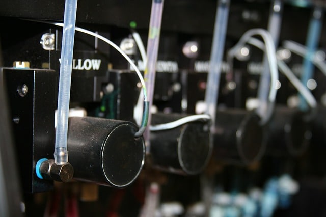
Basically, the pressure is created to push water from highly concentrated areas into an area away from the salt. Today, small units can actually be purchased to reverse osmosis and create safe drinking water.
Here’s an example of a large unit, used in Australia, to clean saltwater for drinking:
Conclusion
What is osmosis in biology? Hopefully, you can now answer that question with some thoroughness.
Osmosis is a type of diffusion that happens when a solvent moves through a semipermeable membrane. In biology, water moves through our cells based on the concentration or ratio of solvent (water) to solute (salt).
Semipermeable membranes allow some solutions to pass through, meaning cells can take on too much water or lose too much water. If a cell is in a solution more concentrated than itself (hypertonic), water will enter the cell to balance the high concentration of salt without the cell.
Osmosis also plays a key role in carrying nutrients across the cell membrane. Likewise, waste is escorted out of the cell. Osmosis allows the roots of trees and plants to get the water and nutrients they need to grow strong and healthy.
In return, the plants feed us, either directly or by sustaining the herd animals we eventually eat. Plants rely on osmosis to live, and people rely on plants to live.
Aside from plants, osmosis also is crucial to man’s survival because it expels toxins and waste from our systems.
Hopefully, you have an understanding of osmosis as you move ahead in your biology class or as you reflect on your biology class from many years ago. Osmosis in biology is more than a scientific principle in an old textbook; it is a lifeline for both plants and animals.
You can look around you each day and see it at work, from tall trees in your backyard to patients recovering in the hospital with an IV feeding their veins. Practical examples of osmosis range from accident victims receiving emergency blood transfusions to little kids pouring salt on slugs.
Take note of the osmosis that happens in front of you each day and be amazed by the science all around you.
Featured image: pixabay.com