Earthworm Worksheet

Name(s)_______________________________ Group_______ Date__________ Period_______

Earthworm Worksheet 


1. What is the name of the pumping organs of an earthworm?


2. Trace the parts of the digestive tract through which food passes.


3. Which parts of the earthworm serve as its brain?  How are these parts connected to the rest of the body?


4. Which of the parts of the worm’s body that you saw are included in the excretory system?


5. How can you find out whether an earthworm eats soil?


6. Among the earthworm’s structural adaptations are its setae. How do you think the earthworm’s setae make it well adapted to its habitat?


7. How is the earthworm’s digestive system adapted for extracting relatively small amounts of food from large amounts of ingested soil?


8. Your dissection of the earthworm did not go beyond segment 32. What will you observe if you dissect the remainder of the worm to its posterior end?


9. On a separate piece of paper, draw and label the parts of the earthworm you observed, and color code the systems. Use green for the reproductive system, yellow for the digestive system, blue for the excretory system, and red for the nervous system.


10. During mating, two earthworms exchange sperm. Fertilization is external, and cocoons are produced from which the young eventually emerge. Refer again to steps 5 and 11, where you located the earthworm’s reproductive organs. Use a reference to identify the role of each organ in the reproductive process of the earthworm. On a separate paper, summarize your findings.

BACK

Echinoderm

Echinoderms

All Materials © Cmassengale  

Phylum Echinodermata
Characteristics

  • All marine
  • Known as spiny-skinned animals
  • Endoskeleton known as the test is made of calcium plates or ossicles with protruding spines
  • Includes sea stars, brittle stars, sand dollars, sea urchins, & sea cucumbers
  • Undergo metamorphosis from bilateral, free-swimming larva to sessile or sedentary adult
  • Larval stage known as dipleurula or bipinnaria
  • Adults have pentaradial ( 5 part) symmetry
  • Lack segmentation or metamerism
  • Coelomate
  • Breathe through skin gills as adults
  • Capable of extensive regeneration


Bipinnaria Larva

  • Ventral (lower) surface called the oral surface & where mouth is located
  • Dorsal (upper) surface known as aboral surface & where anus is located
  • Have a nervous system but no head or brain in adults
  • No circulatory, respiratory, or excretory systems
  • Have a network of water-filled canals called the water vascular system to help move & feed
  • Tube feet on the underside of arms help in moving & feeding
  • One-way digestive system consists of mouth with oral spines, gut, & anus
  • Deuterostomes (blastopore becomes the anus)
  • Separate sexes
  • Reproduce sexually & asexually
  • Includes 5 classes:
    * Crinoidea – sea lilies & feather stars
    * Asteriodea – starfish
    * Ophiuroidea – basket stars & brittle stars
    * Echinoidea – sea urchins & sand dollars
    * Holothuroidea – sea cucumbers

Class Crinoidea
Characteristics

  • Sessile
  • Sea lilies & feather stars


FEATHER STAR

SEA LILY


  • Have a long stalk with branching arms that attach them to rocks & the ocean bottom
  • Can detach & move around
  • Mouth & anus on upper surface
  • May have 5 to 200 arms with sticky tube feet to help capture food (filter feeders) & take in oxygen
  • Common in areas with strong currents & usually nocturnal feeders

Class Asteroidea
Characteristics

  • Usually sedentary along shorelines
  • Starfish or sea stars
  • Come in a variety of colors
  • Prey on bivalve mollusks such as clams & oysters


Starfish Feeding on Clam

  • Have 5 arms that can be regenerated
  • Arms project from the central disk
  • Mouth on oral surface (underside)


STARFISH

Class Ophiuroidea
Characteristics

  • Largest class of echinoderms
  • Includes basket stars & brittle stars


BASKET STAR

BRITTLE STAR


  • Live on the ocean bottom beneath stones, in crevices, or in holes
  • Have long, narrow arms resembling a tangle of snakes
  • Arms readily break off & regenerate
  • Move quicker than starfish
  • Feed by raking in food with arms or trapping it with its tube feet

Class Echinoidea
Characteristics

  • Includes sea urchins & sand dollars


SEA URCHIN

SAND DOLLAR


  • Internal organs enclosed by endoskeleton or test made of fused skeletal plates
  • Body shaped like a sphere (sea urchin) or a flattened disk (sand dollar)
  • Lack arms
  • Bodies covered with movable spines
  • Have a jawlike, crushing structure called Aristotle’s lantern to grind food
  • Use tube feet to move
  • Sea Urchins:
    * Spherical shape
    * Live on ocean bottom
    * Scrape algae to feed
    * Long, barbed spines make venom for protection
  • Sand Dollars:
    * Flattened body
    * Live in sand along coastlines
    * Shallow burrowers
    * Have short spines

Class Holothuroidea
Characteristics

  • Includes sea cucumber


SEA CUCUMBER

  • Lack arms
  • Shaped like a pickle or cucumber
  • Live on ocean bottoms hiding in caves during the day 
  • Have a soft body with a tough, leathery outer skin
  • Five rows of tube feet run lengthwise on the aboral (top) surface of the body
  • Have a fringe of tentacles (modified tube feet) surrounding the mouth to sweep in food & water
  • Tentacles have sticky ends to collect plankton
  • Show bilateral symmetry
  • Can eject parts of their internal organs (evisceration) to scare predators; regenerate these structures in days

Structure & Function of Starfish
Body Plan

  • Range in size from 1 centimeter to 1 meter
  • Mouth located on oral surface (underside)
  • Have an endoskeleton made of calcium plates
  • Sharp, protective spines made of calcium plates called ossicles found under the skin on the aboral (top) surface


ABORAL SURFACE

  • Have pedicellariae or tiny, forcep-like structures surrounding their spines to help clean the body surface

Water Vascular System

  • Network of canals creating hydrostatic pressure to help the starfish move


WATER VASCULAR SYSTEM

  • Water enters through sieve plate or madreporite on aboral surface into a short, straight stone canal
  • Stone canal connects to a circular canal around the mouth called the ring canal
  • Five radial canals extend down each arm & are connected to the ring canal
  • Radial canals carry water to hundreds of paired tube feet


TUBE FEET

  • Bulb-like sacs or ampulla on the upper end of each tube foot contract & create suction to help move, attach, or open bivalves
  • Rows of tube feet on oral surface (underside) are found in ambulcaral grooves under each arm


Tube Feet in Ambulcaral Grooves

Feeding & Digestion

  • Tube feet attach to bivalve mollusk shells & create suction to pull valves apart slightly
  • Starfish everts (turns inside out) its stomach through its mouth & inserts it into prey
  • Stomach secretes enzymes to partially digest bivalve then stomach withdrawn & digestion completed inside starfish

Other Body Systems

  • No circulatory, excretory, or respiratory systems
  • Coelomic fluid bathes organs & distributes food & oxygen
  • Gas exchange occurs through skin gills & diffusion into the tube feet
  • No head or brain
  • Have a nerve ring surrounding the mouth that branch into nerve cords down each arm
  • Eyespots on the tips of each arm detect light
  • Tube feet respond to touch

Reproduction

  • Separate sexes
  • Two gonads (ovaries or testes) in each arm produce eggs or sperm
  • Have external fertilization
  • Females produce up to 200,000,000 eggs per season
  • Fertilized eggs hatch into bipinnaria larva which settles to the bottom after 2 years & changes into adult
  • Asexually reproduce by regenerating arms

Earthworm Facts

earthworm facts

How long do  worms live?
How many young are produced per year?   
Do earthworms have eyes?

How do earthworms breathe?
Can earthworms smell?
Do worms have eyes?
What do earthworms eat and how much can they eat in one day?
Can earthworms freeze?
What is the “bump” in the middle of the earthworm?
How can you determine if an earthworm is sexually mature?
Can earthworms lose their clitellum?
How do earthworms mate? 
How are cocoons produced?
How long does it take worms to hatch?
How many young worms are produced per year?
How long does it take earthworms to mature?
Can different species of worms mate creating a hybrid worm?
How long do earthworms live?
How do earthworms move?
What characteristics are used to identify earthworms?
What enemies do earthworms have?
Can earthworms regenerate themselves?
How can you distinguish the head of an earthworm from the tail?
How do earthworms obtain their food?
How big do earthworms get?

Read Our Q&A About Earthworm Facts

Q. How long do dew worms live?

A. Dew worms can live for approximately six and a half years.

Q. How many young are produced per year?

A. It is estimated that sexually mature dew worms (about one year old) produce about two cocoons per year with 1-2 young each (more research under field and laboratory conditions required).

Q. Do earthworms have eyes?

A. They do not have eyes but they do possess light- and touch-sensitive organs (receptor cells) to distinguish differences in light intensity and to feel vibrations in the ground.

Q. How do earthworms breathe?

A. Earthworms respire through their skin, and therefore require humid conditions to prevent drying out. They coat themselves in mucus to enable the passage of dissolved oxygen into their bloodstream.

Q. Can earthworms smell?

A. Worms have specialized chemoreceptors or sense organs (“taste receptors”) which react to chemical stimuli. These sense organs are located on the anterior part of the worm.

Q. What do earthworms eat and how much can they eat in one day?

A. Earthworms derive their nutrition from many forms of organic matter in soil, things like decaying roots and leaves, and living organisms such as nematodes, protozoans, rotifers, bacteria, fungi. They will also feed on the decomposing remains of other animals. They can consume, in just one day, up to one third of their own body weight.


Q. Can earthworms freeze?

A. Like all invertebrates their body processes or metabolism slow down with falling temperatures. They will hibernate at near freezing temperature. If frozen they will die. They react to advancing colder winter weather by burrowing deep (up to two meters) in the soil to avoid the extreme cold.

Q. What is the “bump” in the middle of the earthworm?

A. The bump is the clitellum, the saddle shaped swollen area 1/3 of the way back containing the gland cells which secrete a slimy material (mucus) to form the cocoon which will hold the worm embryos.

Q. How can you determine if an earthworm is sexually mature?

A. If the worm has a clitellum, it is sexually mature.

Q. Can earthworms lose their clitellum?

A. The answer is yes! During periods of drought, when soils dry up, some species of earthworms do in fact temporarily lose all secondary sexual characters such as the clitellum. When conditions become favorable, it comes back. The clitellum can also disappear at the onset of old age or senescence.

Q. How do earthworms mate?


A. Earthworms are hermaphroditic meaning each worm has organs of both sexes. The male gonopores are usually within the first 12-15 segments, and the female gonopores are further back, close to the clitellum (the swollen area in adult worms). One worm has to find another worm and they mate juxtaposing opposite gonadal openings exchanging packets of sperm, called spermatophores. Some species also appear to be either parthenogenetic (females producing all females, “virgin birth”) or may be able to self-fertilize.


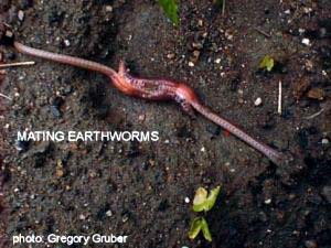
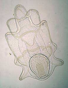
Q. How are cocoons produced?

A. The clitellum produces a mucous sheath and nutritive material, and as the sheath slides forward, it picks up ova from the earthworm’s ovaries then packets of sperm that had been transferred to the worm from another worm during mating. As the sheath slides off the worm’s head, the ends are sealed to form the cocoon. Initially, the cocoon is quite soft but soon after it is deposited in the soil it becomes slightly amber in color, leather-like and very resistant to drying and damage.Earthworm eggs
Dendrobaena rubidus cocoons (relative to a pin head).

The ova within each cocoon are fertilized, and the resulting embryos grow inside the sealed unit, much like a chick developing inside an egg. When the embryos have consumed all the nutritive material, they completely fill the lemon shaped cocoon and are ready to hatch out one end.

Q. How long does it take worms to hatch?

A. Young worms hatch from their cocoons in three weeks to five months as the gestation period varies for different species of worms. Conditions like temperature and soil moisture factor in here…if conditions are not great then hatching is delayed.

Q. How many young worms are produced per year?

A. Earthworms can produce between 3 and 80 cocoons per year depending on the species. The deeper-dwelling species don’t have to produce as many cocoons because they are protected much better from predation than surface dwelling species which tend to produce many more cocoons. The number of fertilized ova or eggs within each cocoon ranges from one to twenty. This depends on the species and also factors such as nutrition of the adults laying them and environmental conditions with soil moisture being most important. Usually, though, only few to several young worms will ever successfully emerge from each cocoon.

Q. How long does it take earthworms to mature?

A. Worms mature in 10 – 55 weeks depending on the species.

Q. Can different species of worms mate creating a hybrid worm?

A. No, this does not usually occur; hybrids can usually only occur between very closely related species and their offspring would likely be infertile.

Q. How long do earthworms live?

A. Earthworm longevity is species dependent. Various specialists report that certain species have the potential to live 4-8 years. In protected culture conditions (no predators, ideal conditions) individuals of Allolobophora longa have been kept up to 10 1/4 years, Eisenia foetida for 4½ years and Lumbricus terrestris for 6 years.

Worms continue to grow once they reach sexual maturity but once at this stage there is a much slower increase in weight until the disappearance of the clitellum indicates the onset of old age or senescence. During this period there is a slow decline in weight until the death of the worm.

Q. How do earthworms move?

A. Earthworms have bristles or setae in groups around or under their body. The bristles, paired in groups on each segment, can be moved in and out to grip the ground or the walls of a burrow. Worms travel through underground tunnels or move about on the soil surface by using their bristles as anchors pushing themselves forward or backward using strong stretching and contracting muscles.

Q. What characteristics are used to identify earthworms?

A. The external body characters used in identifying different species of earthworms are: the segmental position of the clitellum on the body, body length, body shape (cylindrical or flattened), number of body segments, type and position of body bristles or setae, the description of the tongue-like lobe, the prostomium, projecting forward above the mouth, type of peristomium or first body segment, external position and morphology of genital apertures or opening and type of glandular swellings on the clitellum. The shape and the relationship of various internal organs are also used to identify some species of worms.

Q. What enemies do earthworms have?

A. Snakes, birds, moles, toads and even foxes are known to eat earthworms. Beetles, centipedes, leeches, slugs and flatworms also feed on earthworms. Some types of mites parasitize earthworm cocoons and the cluster fly (Pollenia rudis) parasitizes worms of the species Eisenia rosea.

Q. Can earthworms regenerate themselves?

A. Yes, but only the front or head end of the earthworm will survive and the amputated tail portion will die. This remaining front portion must also be long enough to contain the clitellum and at least 10 segments behind the clitellum. This makes up about half the length of the worm. The new posterior segments grown will be slightly smaller in diameter than the original segments and sometimes a bit lighter in color.

Q. How can you distinguish the head of an earthworm from the tail?

A. The head of the worm is always located on the end of the worm closest to the clitellum and has some differentiated structures if you can view with magnification. Even though worms can move both frontward and backward they tend to travel forward more. Place a worm on a rough piece of paper and observe which direction it travels. They usually extend their “head” first when crawling.

Q. How do earthworms obtain their food?

A. Earthworms possess very strong mouth muscles – they do not have teeth. Dew worms or nightcrawlers often surface at night to pull fallen leaves down into their burrow. When the leaf decomposes or softens a little they pull small bits off at a time to munch on. They also “swallow” soil as they burrow and extract nutrients from it.

Q. How big do earthworms get?

A. Size depends on the species of worm, it’s age, diet and environmental conditions like moisture, temperature and soil conditions. Lumbricus terrestris (Nightcrawler, Dew worm) is one of North America’s largest and ranges in size from 9-30 cm with a diameter of 6-10 mm. The largest L. terrestris we’ve collected was close to 30 cm long (stretched out), weighed 11.2 g and was collected in a no-till, soybean field in Ontario up near Georgian Bay, Ontario.

The largest tropical species (Glossoscolex and Megascolides) are up to 120 cm long and the largest in the world are some Australian forms which may reach 300 cm in length. Bimastos parvus (American bark worm) is quite small at less than 2 cm long.


BACK

Clam Dissection Questions


  Clam Dissection Questions


Pre-lab:
1. Give the kingdom, phylum, and class for the clam.


2. Describe the body of bivalves.

3. How do bivalves move?

4. Why are they called bivalves?

5. Is their digestive tract complete or incomplete?  Explain your answer.


6. Do bivalves show cephalization?  Explain your answer.


7. What are siphons & what is their purpose?


8. How can you distinguish a mussel from a clam?


9. Adults are usually sessile, but the larva or _________________ is free-swimming.

10. Are bivalves protostomes or deuterostomes?

11. Their body cavity is called the _____________ cavity.

12. Body organs make up the ___________ mass and are protected by the _____________ which secretes the ___________.

13. Is circulation open or closed?

14. Give several examples of bivalves.

Lab Questions:
1. What is the oldest part of a clam’s shell called and how can it be located?


2. What do the rings on the clam’s shell indicate?

3. Name the clam’s siphons.

4. What holds the two shells together?

5. What muscles open & close the clam?

6. Describe the inside lining of the shell.

7. What is the function of the tooth-like projections at the dorsal edge of the clam’s valves?

8. Where is the mantle located in the clam?  What is its function?


9. Describe the clam’s foot.


10. What is the mantle cavity?


11. How do clams breathe?

12. What helps direct water over the gills?

13. Where are the palps found and what is their function?


14. Describe the movement of food from the current siphon through the digestive system of the clam.


15. Where is the clam’s heart located?


16. What are the parts of the clam’s nervous system?


17. Why are clam’s referred to as “filter feeders”?


18. Label the internal structures of the clam and draw arrows showing the pathway of food as it travels to the clam’s stomach:

Crayfish Dissection


Crayfish Dissection


Virtual Crayfish Dissection – CornellVirtual Crayfish Dissection – Penn State
crayfish_mini1.gif (6395 bytes) bottom.jpg (95862 bytes)


By Day:    Day 1        Day 2        Day 3

By Region: External Anatomy    Internal Anatomy

By Topic:      Skeletal       Integumentary     Cardiovascular     Muscular    Endocrine    Nervous
Reproductive     Respiratory    Excretory    Digestive


You must create a series of labeled drawings that illustrate the structures outlined below:

Day 1

I. Abdomen – Ventral View          (Day 1)         top

Place the crayfish supine (ventral surface up) on the dissecting tray and DRAW the following:


  1. Telson (What is the telson’s function?)
  2. Uropod  (Describe the location of the Uropod to the telson.   How do the add to the telson’s function?)
  3. Anus (In which of the two structures above did you find the anus? 1 or 2 way digestive system?)
  4. Swimmerets -numbered in pairs, 1-5 w/ the 5th one the most posterior (What is their function, and how is it different from the telson’s function?)
  5. Is your Crayfish a male or a female (Note the anterior-most swimmeret.   In the male, its function is to guide the sperm toward the female during copulation; as such, it will be enlarged, and pointed anteriorly in the male.  In the female there is no difference between the swimmerets)?  (Describe the appearance of the crayfish’s swimmerets in your answer.)
  6. Walking Legs (How many are there?  In terms of this feature alone, is this organism closer to an insect, or an arachnid?)
  7. Chelipeds – some people like this meat the best . . . (What is their function?)


II. Head – Ventral View             (Day 1-2)         top


  1. Mandibles – 2 – hard & white  (What are they equivalent to in humans?  How is their action – think direction of movement – different from that of humans?)
  2. Maxilla – softer w/ jagged edges  (Given the difference in texture, how is their function different from that of the mandibles?)
  3. Maxillapeds, or “mouth-feet” -3 pairs  (What is their function?  Why not use the Chelipeds?)
  4. Green Gland Ducts – (From what organ do they open out?  What is the equivalent organ in humans? What is the purpose of the duct?  Is its location at all disturbing to you?)


II. Cephalothorax – Dorsal View             (Day 1-2)         top

  1. Rostrum (What is cephalization?  Given that, what organ would you expect to be inside the rostrum?)
  2. Eyes (Does this organism have binocular vision – depth perception, why or why not?)


    Eye


  3. Carapace (What is the function of the carapace?  What two body systems in humans perform equivalent functions?  The support function is in reference to one system in particular; given the external location of the carapace, explain the name of the type of system compared to our own, internal variety.  The support function implies specifically the attachment of organs of what body system to the inside of the carapace?

Day 2

Make a Dorsal Midline Incision from the posterior end of the thorax to the posterior end of the rostrum using the rounded scissors w/ the rounded end down! Open the carapace and pin it back.

III. Thorax – Dorsal View, Part I          (Day 2)         top

  1. Heart & Ostia – the opening on the heart’s superior surface (Is this a sign of an open or closed circulatory system?  Differentiate between the two in your answer.)
  2. Gills (What are they equivalent to in humans?  To what body system do they belong?  Why are the gills so feathery – i.e., how does this aid in their function?)
  3. Cardiac Stomach -draw whole (There appear to be fibers attached to the outside of the stomach.  What is their purpose in relation to the stomach and the esophagus?)

IV. Thorax – Dorsal View, Part II         (Day 2)         top

  1. Remove one gill and draw on high power (What is the red/pink material within each “finger” of each gill?  How does this material relate to the function of the gill?)
  2. GENTLY remove one walking leg, and you will see that a gill is attached to each walking leg.  (How is this important to the function of the gills?  In your answer refer to the different requirements of the body during times of high physical activity, and how they are related to the gill-walking leg connection.)
  3. Cut open the Cardiac Stomach and draw the Gastric Mill – reddish-brown lateral “teeth” – on high power  (What is their function?  What type of digestion involves the gastric mill?  Do we accomplish that type of digestion in our own stomach?)

Day 3

V. Thorax – Dorsal View, Part III         (Day 3)         top

Gently remove the Heart.

  1. The Intestine (Given its location posterior to the stomach, what is its function?  What function of the stomach is lacking in the intestine?)
  2. The Hepatopancreas Gland (What two organs is this equivalent to in humans?  What are some of the functions of this gland?  How is its location important to its function?)
  3. The Seminifierous Tubules or Ovaries  (What is the function of each? To what body system do these belong?  Which of the two does your specimen contain?  How is this related to the swimmerets?)


VI. Thorax – Dorsal View, Part IV         (Day 3)         top

Gently remove the Cardiac Stomach.

  1. Esophagus  (Describe how it’s position relative to the stomach is different from the worm and the human.)
  2. Green Gland (What is/are the equivalent organ(s) in humans?   Do/does the analogous organ(s) appear in pairs in humans?  To what body system do the green glands belong?  What organ in our equivalent body system is missing in the crayfish?)
  3. Brain (Describe the appearance of the brain and the nerves in terms of the type of symmetry.  There are nerves that are attached to the front and the back of the brain.  Describe the function of both the anterior and the posterior nerve pairs.)

VII. Abdomen – Dorsal View, Part I       (Day 3)        top

Make a Dorsal Midline Incision from the anterior end of the abdomen to the posterior end of the abdomen using the rounded scissors w/ the rounded end down! Open the exoskeleton and pin it back.


In order for a Crayfish to determine BALANCE, it must insert a grain of sand in one of it’s appendages.
Every time it molts and makes a new exoskeleton, it must get a new grain of sand!
(In what part of the body is that function taken up by the human body?)


  1. Dorsal Blood Vessel  (Is this vessel sending the blood to, or away from, the heart?  What name would we give to that type of vessel in our body?)
  2. Large Intestine (How is the location of this organ related to the name of this section of the body [it is NOT a tail]?  What is the function of the large intestine?  Given it’s contents, is it wise, or unwise, to eat it when eating a lobster?  Explain.)
  3. Abdominal Flexor Muscles  (How do muscles function, by shortening,   lengthening, of both?  Moving the abdominal flexor muscles will cause flexion, but what is flexion?  How will the abdomen – it is NOT a tail – change shape during flexion?  What direction will the crayfish move during flexion?  Given the size and strength of the muscle, during what circumstances would the crayfish use this muscle over its walking legs?)

VIII. Abdomen – Dorsal View, Part II        (Day 3)        top

Gently remove the Abdominal Flexor Muscles.

  1. Ventral Blood Vessels  (Given that there is no main ventral blood vessel, how does the blood return to the heart?  Is this a sign of an open or closed circulatory system?)
  2. Ventral Nerve Cord  (To what phylum does the crayfish belong?   How is the location of the nerve cord different from creatures in our own phylum?   Name our own nerve cord.  How is the protection of the nerve cord different in both phyla?)

Drawings:

  1. Use a PENCIL!!

  2. Make the drawings “larger than life” size, as the specimens are so small.

  3. Draw the general shape (outline) and location of the organs, as the squiggles so many of you use to “shade” your drawings make your drawings sloppy and hard to interpret.

  4. Include Labels on all drawings.

  • Labels should start outside the drawing, and be connected to the structure by arrows with tips (===>).

  • The Tip of the arrow should be touching the structure.

  • Be sure to include the magnification for any drawings done with the dissecting microscope.

Hang on to the drawings; they will all be handed in later, together with some questions to answer!


Day 1        Day 2        Day 3         top

Modified from  Lazaroff Biology