| Chapter 18 Genetics of Viruses & Bacteria | ||||||||||||||||||||||||||||||||||||||||||||||||||||||||||||||||||||||||||||||||||
| Objectives | ||||||||||||||||||||||||||||||||||||||||||||||||||||||||||||||||||||||||||||||||||
|
||||||||||||||||||||||||||||||||||||||||||||||||||||||||||||||||||||||||||||||||||
| BACK | ||||||||||||||||||||||||||||||||||||||||||||||||||||||||||||||||||||||||||||||||||
| Chapter 19 Eukaryotic Genomes | ||
| Objectives | ||
| The Structure of Eukaryotic Chromatin 1. Compare the structure and organization of prokaryotic and eukaryotic genomes. 2. Describe the current model for progressive levels of DNA packing in eukaryotes. 3. Explain how histones influence folding in eukaryotic DNA. 4. Distinguish between heterochromatin and euchromatin. The Control of Gene Expression 5. Explain the relationship between differentiation and differential gene expression. 6. Describe at what level gene expression is generally controlled. 7. Explain how DNA methylation and histone acetylation affect chromatin structure and the regulation of transcription. 8. Define epigenetic inheritance. 9. Describe the processing of pre-mRNA in eukaryotes. 10. Define control elements and explain how they influence transcription. 11. Distinguish between general and specific transcription factors. 12. Explain the role that promoters, enhancers, activators, and repressors may play in transcriptional control. 13. Explain how eukaryotic genes can be coordinately expressed and give some examples of coordinate gene expression in eukaryotes. 14. Describe the process and significance of alternative RNA splicing. 15. Describe factors that influence the life span of mRNA in the cytoplasm. Compare the longevity of mRNA in prokaryotes and in eukaryotes. 16. Explain how gene expression may be controlled at the translational and post-translational level. The Molecular Biology of Cancer 17. Distinguish between proto-oncogenes and oncogenes. Describe three genetic changes that can convert proto-oncogenes into oncogenes. 18. Explain how mutations in tumor-suppressor genes can contribute to cancer. 19. Explain how excessive cell division can result from mutations in the ras proto-oncogenes. 20. Explain why a mutation knocking out the p53 gene can lead to excessive cell growth and cancer. Describe three ways that p53 prevents a cell from passing on mutations caused by DNA damage. 21. Describe the set of genetic factors typically associated with the development of cancer. 22. Explain how viruses can cause cancer. Describe several examples. 23. Explain how inherited cancer alleles can lead to a predisposition to certain cancers. Genome Organization at the DNA Level 24. Describe the structure and functions of the portions of eukaryotic DNA that do not encode protein or RNA. 25. Distinguish between transposons and retrotransposons. 26. Describe the structure and location of Alu elements in primate genomes. 27. Describe the structure and possible function of simple sequence DNA. 28. Using the genes for rRNA as an example, explain how multigene families of identical genes can be advantageous for a cell. 29. Using a-globin and b-globin genes as examples, describe how multigene families of nonidentical genes may have evolved. 30. Define pseudogenes. Explain how such genes may have evolved. 31. Describe the hypothesis for the evolution of a-lactalbumin from an ancestral lysozyme gene. 32. Explain how exon shuffling could lead to the formation of new proteins with novel functions. 33. Describe how transposition of an Alu element may allow the formation of new genetic combinations while retaining gene function.
|
||
| BACK | ||
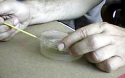
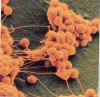
| Caught Red-Handed |  |
Introduction:
Bacteria are everywhere. They have evolved the ability to inhabit almost every surface on the planet; however, they are invisible to the naked eye due to their small size. Bacteria have been found living in the deepest part of the ocean, in volcanic vents, in boiling hot springs, and even deep in polar ice caps. Many species of bacteria live inside of other organisms in a harmless commensalistic way such as the intestinal bacteria, Escherichia coli. Bacteria can reproduce at very rapid rates whenever conditions are favorable, as often as every 20 minutes doubling in number. The bacterial population is kept in check by the natural defenses of the host, such as the immune system and proper washing habits. When these natural defenses fail, bacteria can quickly become a problem. Some bacteria produce poisons or toxins that can be life-threatening if the bacterial population isn’t controlled by our natural defenses.
The United States Centers for Disease Control (CDC) states that the best way to prevent bacterial spread and infection is through the use of proper sanitary techniques. Perhaps the most critical step in this prevention is the use of proper hand washing. When improperly washed, your hands are one of the most easily colonized areas of your body and many of our behaviors involve the use of our hands. Proper hand washing requires the use of water as hot as you can stand, soap, and lots of rubbing. The soap and water serve to destroy bacteria, and the rubbing helps slough off dead skin cells along with lots of bacteria.
Objective:
Students will examine:
Materials (Part A):
Black light, Glo-Germ powder, lotion or Glo-Germ oil, hand soap, water, paper towels, pencil, lab sheet
Procedure (Part A):
Data Table 1
| Time of Wash in Seconds | Percent of Hand Covered with “germs” |
| 0 (initial observation) | |
| 10 | |
| 30 | |
| 60 | |
| 120 |
Materials (Part B):
Tennis ball, “play” money, stuffed toy, pencil, lab sheet
Procedure (Part B):
Data Table 2
| Name of Item | Percent Coverage |
| Initial Hand Coverage | |
| Tennis Ball | |
| “Play” money | |
| Toy |
Questions:
Optional:
Create a graph based on the data from table 1.
Title _____________________________


| Where are Bacteria Found? |
Introduction:
They’re everywhere. Bacteria are the huddled masses of the microbial world, performing tasks that include everything from causing disease to fixing nitrogen in the soil. The estimated number of bacteria on Earth is five million trillion trillion — that’s a five with 30 zeroes after it. When people think of bacteria, they likely first consider the nasty ones that cause disease, but the bacteria inside all animals combined — including humans — makes up less than one percent of the total amount. By far the greatest numbers are in the subsurface, soil and oceans.
Objectives:
Materials:
Petri dish, pencil, incubator, hot water bath, nutrient agar, thermometer
Procedure (Part A): Petri Dish Preparation
Materials:
Petri dish with nutrient agar, sterile cotton swabs, permanent marker, index card with sample location, pencil, incubator
Procedure (Part B): Collecting Bacteria
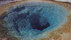
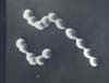
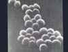
Example of Bacterial Colonies on Plate

Data:
Reminder — Fuzzy Colonies = Fungus not Bacteria
Figure 1
Day 1 Day 2 Day 5

Table 1: Number of Colonies on petri dish
| Location: | |
| Day | Number of Colonies |
Analysis:
Dispose of the petri dishes carefully! Place them in a biohazard bag to be autoclaved.
KINGDOMS ARCHAEBACTERIA & EUBACTERIA

All Materials © Cmassengale
Bacterial Evolution & Classification
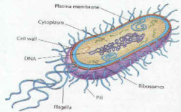
| STRUCTURE | FUNCTION |
| Cell Wall | protects the cell and gives shape |
| Outer Membrane | protects the cell against some antibiotics (only present in Gram negative cells) |
| Cell Membrane | regulates movement of materials into and out of the cell; contains enzymes important to cellular respiration |
| Cytoplasm | contains DNA, ribosomes, and organic compounds required to carry out life processes |
| Chromosome | carries genetic information inherited from past generations |
| Plasmid | contains some genes obtain through genetic recombination |
| Capsule, and slime layer | protects the cell and assist in attaching the cell to other surfaces |
| Endospore | protects the cell against harsh environmental conditions, such as heat or drought |
| Pilus (Pili) | assist the cell in attaching to other surfaces, which is important for genetic recombination |
| Flagellum | moves the cell |
Kingdom Archaebacteria
Methanogens
Extreme Halophiles
Thermoacidophiles (Thermophiles)
Kingdom Eubacteria (true bacteria)

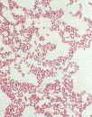
Gram Staining
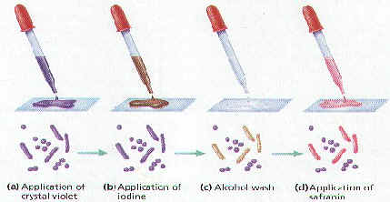
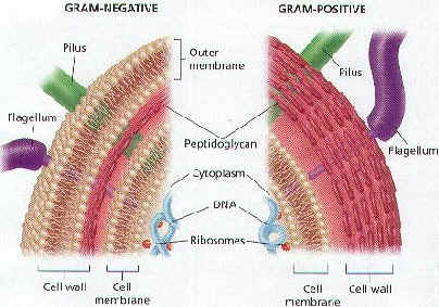
Gram-positive bacteria (Gram +)

Gram-negative bacteria (Gram -)
Phylum Cyanobacteria

OSCILLATORIA
Phylum Spirochetes
Phylum Gram Positive bacteria
Phylum Proteobacteria
Enteric bacteria
Chemoautotrophs
Nitrogen-Fixing bacteria
Methods of Nutrition
Methods of Respiration
Bacterial Reproduction & Genetic Recombination
Conjugation
Transformation
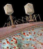
Transduction
Pathogenic bacteria