Natural Selection Peanut Activity


Natural Selection Within a Species


Introduction:

Natural selection is the evolutionary process by which the most adaptable individuals survive. An adaptation is an inherited variation that helps an organism to survive. When the organism survives, its chances of reproduction are increased as well as its ability to pass on its inherited traits.  All members of a species are different from one another.  In this activity, you will investigate two variations among peanut plants — length of shell and number of seeds per shell.   Most shells contain a certain number of seeds which is an adaptation to its survival.  

Objective:

Students will investigate natural selection in peanuts.

Materials:

50 peanuts in shells, metric ruler, pencil, graph paper

Procedure:

  1. Count out 50 peanuts in their shells.
  2. Use a metric ruler to measure the length of each shell in millimeters.  Put a check mark in Table 1 in the space indicating the length of the shell.
  3. Open the shell and count the number of seeds inside.  Put a check mark in Table 2 in the space next to the number of seeds that you counted. 
  4. Repeat steps 1 – 3 for the other 49 peanuts.
  5. Set up two bar graphs using the headings from each table as the axes for each graph.
  6. Plot a bar graph from the data in each table.

Data:

Table 1


Length of Shell
(mm)
Number of Shells
5-10
10-15
15-20
20-25
25-30
30-35
35-40
40-45
45-50
50-55
55-60
60-65
65-70
70-75


Table 2


Number of Seeds Per ShellNumber of Shells
1
2
3


Title: ________________________________________________________


Title: ________________________________________________________


Questions:

  1. What is the most common length of the shells you measured?
  2. What was the most common number of seeds in the peanut shells that you opened?
  3. What might happen if each shell contained fewer than normal seeds and why?
  4. What might happen if each shell contained more than the normal number of seeds and why?
  5. Was there a relationship between the most common length of shells and the number of seeds they contained? Explain your answer.
  6. Write 1-2 paragraphs explaining how shell length and seed number in peanuts illustrates natural selection within this species.

 

Origin of life PPT Qs

Origin Of Life
ppt Questions

Early Thoughts on Life

1. What was Aristotle’s idea about how life arose called?

2. What is another name for spontaneous generation?

3. Explain spontaneous generation of life.


4. How long did the idea of abiogenesis or spontaneous generation last?

5. The idea of abiogenesis lasted so long because, instead of testing their ideas, people based their beliefs on what?


6. Were their observations tested?

7. Did they use the scientific method for their observations?

Examples of Spontaneous Generation

8. What observation about new life did Egyptians make when the Nile River flooded each year?


9. What observation about new life did Medieval farmers make when they stored their grain each year?


10. The English people centuries ago, threw their garbage and sewage out on the streets. What observation about new life did these people make?


11. This practice led to a plague that killed many Europeans. What was this plague called and what carried the disease organism?


12.Before refrigerators, large slabs of meat were hung after being purchased. What observation about new life was made from this practice?


13. People believed so strongly in abiogenesis that they had recipes for making living things. Name two organisms that had accepted recipes.


Disproving Spontaneous Generation

14. Francesco ____________ was an early scientists who conducted experiments to try and disprove spontaneous generation.

15. What was Redi’s hypothesis?


16. Explain how Redi tried to prove this.


17. What were the results Redi found in the closed jars & why?


18. What were the results in the open jars?


19. How did maggots appear in the open jars?


20. Complete this table summarizing Redi’s experiment:


Evidence Against Spontaneous Generation
Unsealed Jar
Sealed Jar
Gauze Covered jar


21. Redi’s experiment disproved spontaneous generation for _____________ organisms.

Use of the Scientific Method

22. Did Francesco Redi use the scientific method in his experiment?

23. What served as the control in Redi’s experiment?


24. What jars served as the experimental groups?

25. What was Redi’s conclusion?


Disproving Spontaneous Generation of Microbes

26. Anton Van _______________ made one of the first simple microscopes.

27. Leeuwenhoek called the living things he saw in pond water ______________.

28. By the end of the 19th century, these organisms were known as ______________.

29. John _____________ did experiments with microorganisms growing in broths.

30. Needham believed there was a __________ __________ present in nonliving substances like air.

31. Why were bacteria able to grow in Needham’s soups?


32. What could have been done to the broths to kill the bacteria already present?

33. What scientists repeated this experiment but with boiled broth?

34. After boiling, what did Spallanzani do to the tops of the bottles? how did this help?


35. Critics of Spallanzani’s experiment said there was not enough _______ for the bacteria to survive and that boiling had destroyed the _________ __________.

The Theory Changes

36. What did the Paris Academy of Science do in 1860 to solve the problem?


37.Who won the prize? 

38. What was Pasteur’s experimental hypothesis?


39. What was the shape of Pasteur’s flasks? Include a sketch.


40. What was the special S-shaped neck intended to do?


41. Did Pasteur boil the broth in his flasks? Why?


42. The flasks were left at ___________ locations.

43. Did the broth change cloudy because microbes were growing in it?


44. What was visible in the neck of the flask after collecting there?

45. Once the S-shaped stem was broken off the top of the flasks, what happened to the broth and why?


46. Pasteur’s S-shaped flasks kept ___________ out but let ______ inside.

47. Pasteur’s experiment proved that living things only come from other _________ ___________.

48. What is the name of Pasteur’s theory?

Review

49. Where did the maggots come from in Redi’s experiment?

50. What was the purpose of the sealed jars?

51. Redi was trying to disprove – spontaneous generation or biogenesis?

52. Where did the microbes come from in Needham’s broth?

53. Needham & Spallanzani were trying to disprove – spontaneous generation or biogenesis?

54.Who proved biogenesis?


Pasteur Experiment

Recreation of Pasteur’s Experiment

Introduction:

Today, we take many things in science for granted. Many experiments have been performed and much knowledge has been accumulated that people didn’t always know. For centuries, people based their beliefs on their interpretations of what they saw going on in the world around them without testing their ideas to determine the validity of these theories — in other words, they didn’t use the scientific method to arrive at answers to their questions. Rather, their conclusions were based on untested observations.

Among these ideas, for centuries, since at least the time of Aristotle (4th Century BC), people (including scientists) believed that simple living organisms could come into being by spontaneous generation. This was the idea that non-living objects can give rise to living organisms. It was common “knowledge” that simple organisms like worms, beetles, frogs, and salamanders could come from dust, mud, etc., and food left out, quickly “swarmed” with life. For example:

Observation: Every year in the spring, the Nile River flooded areas of Egypt along the river, leaving behind nutrient-rich mud that enabled the people to grow that year’s crop of food. However, along with the muddy soil, large numbers of frogs appeared that weren’t around in drier times. Conclusion: It was perfectly obvious to people back then that muddy soil gave rise to the frogs.

Objective:

In this experiment, you will conduct an experiment similar to the one done by Pasteur whenever he disproved spontaneous generation.


Materials Needed:Experiment Set-Up

  • Low-salt broth (chicken or beef, home-made or purchased)
  • 2  250-mL Erlenmeyer flasks
  • 2  1-hole rubber stoppers with bent glass tubing inserted (see diagram)
  • Glycerine
  • Hot plate & pot holders
  • 50-ml Graduated Cylinder
  • Marker

Procedure:

  1. Students should work in teams of 2 to 3 people. Each team should perform the following steps.
  2. Use glycerine and a twisting motion to insert glass tubing into the stoppers. be sure to rinse off excess glycerine with water.
  3. Mark Erlenmeyer flasks accordingly:
    1. Flask 1 with stopper and glass tube going straight up
    2. Flask 2 with stopper and glass tube bent in S-curve
  4. Using a graduated cylinder, place about 50-mL of broth in each Erlenmeyer flask.
  5. Place appropriate lids on flasks.
  6. Use a hot plate to boil broth in flasks with appropriate lids on them for 30 min., then let cool.
  7. For the next ten days, observe the flasks and record any changes in color, turbidity, smell, etc. (Be careful to NOT remove the stoppers from the flasks.)

Data:

Microbial Growth Record
Record the appearance of the flask contents.

DayFlask 1 with Straight TubingDayFlask 2 with S-shaped Tubing
11
22
33
44
55
66
77
88
99
1010

Conclusion:

  1. What was the appearance on the broth in each flask on Day 1?
  2. Was their an observed appearance change in flask 1 over the 10 days? Describe the change, if any.
  3. Was their an observed appearance change in flask 2 over the 10 days? Describe the change, if any.
  4. Explain why there was or was not a change in the appearance of the broth in each flask.
  5. Why do you think the idea of spontaneous generation was believed to be true for so long (1000+ years)?
  6. Did your experiment support spontaneous generation of organisms? Explain why or why not?

Nucleic Acids & Protein Synthesis

Nucleic Acids and Protein Synthesis
All Materials © Cmassengale

Cell   à   Nucleus    à    Chromosomes   à   Genes    à     DNA 

Proteins

  • Organic molecules (macromolecules) made by cells
  • Make up a large part of your body
  • Used for growth, repair, enzymes, etc.
  • Composed of long chains of small units called amino acids bonded together by peptide bonds
  • Twenty amino acids exist

DNA

  • Deoxyribonucleic acid is a coiled double helix carrying hereditary information of the cell

  • Contains the instructions for making proteins from 20 different amino acids
  • Appears as chromatin when cell not dividing

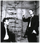
  • Structure discovered by Watson & Crick in 1953
  • Sides made of pentose (5-sided) sugars attached to phosphate groups by phosphodiester bonds
  • Pentose sugar called Deoxyribose

  • Steps or rungs of DNA made of 4 nitrogen-containing bases held together by weak hydrogen bonds
  • Purines (double carbon-nitrogen rings) include adenine (A) and guanine (G)
  • Pyrimidines (single carbon-nitrogen rings) include thymine (T) and cytosine (C)

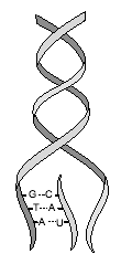
  • Base pairing means a purine bonds to a pyrimidine   (Example:  A — T   and   C — G)
  • Coiled, double stranded molecule known as double helix
  • Make up chromosomes in the nucleus
  • Subunits of DNA called nucleotides
  • Nucleotides contain a phosphate, a Deoxyribose sugar, and one nitrogen base (A,T,C, or G)

  • Free nucleotides also exist in nucleus
  • Most DNA is coiled or twisted to the right
  • Left twisted DNA is called southpaw or Z-DNA
  • Hot spots which can result in mutations occur where right & left twisted DNA meet


History of DNA discovery

  • Freidrich Miescher (1868) found nuclear material to be ½ protein & ½ unknown substance
  • 1890’s, unknown nuclear substance named DNA
  • Walter Sutton (1902) discovered DNA in chromosomes
  • Fredrick Griffith (1928) working with Streptococcus pneumoniae conducted transformation experiments of virulent & nonvirulent bacterial strains
  • Levene (1920’s) determined 3 parts of a nucleotide
  • Hershey & Chase (1952) used bacteriophages (viruses) to show that DNA, not protein, was the cell’s hereditary material
  • Rosalind Franklin (early 1950’s) used x-rays to photograph DNA crystals


Click for larger picture!


  • Erwin Chargraff (1950’s) determined that the amount of A=T and amount of C=G in DNA; called Chargaff’s Rule
  • Watson & Crick discovered double helix shape of DNA & built the 1st model

Click for larger picture!

 DNA Replication

  •  Process by which DNA makes a copy of itself
  • Occurs during S phase of interphase before cell division
  • Extremely rapid and accurate (only 1 in a billion are incorrectly paired)
  • Requires many enzymes & ATP (energy)
  • Begins at special sites along DNA called origins of replication where 2 strands open & separate making  a replication fork


  • Nucleotides added & new strand forms at replication forks
  • DNA helicase (enzyme) uncoils & breaks the weak hydrogen bonds between complementary bases (strands separate)
  •  DNA polymerase adds new nucleotides to the exposed bases in the 5’ to 3’ direction

  •  Leading strand (built toward replication fork) completed in one piece
  • Lagging strand (built moving away from the replication fork) is made in sections called Okazaki fragments


OKAZAKI FRAGMENTS

  •  DNA ligase helps join Okazaki segments together

  • DNA polymerase proofreads the new DNA checking for errors & repairing them; called excision repair
  • Helicase recoils the two, new identical DNA molecules

RNA

  • Ribonucleic acid
  • Single stranded molecule  

  • Found in nucleus & cytoplasm
  • Contains ribose sugar
  • Contains the nitrogen base uracil (U) instead of thymine so A pairs with U
  • Base pairings are A-U and C-G
  • Three types of RNA exist (mRNA, TRNA, & rRNA)

mRNA

  • Messenger RNA
  • Single, uncoiled, straight strand of nucleic acid
  • Found in the nucleus & cytoplasm
  • Copies DNA’s instructions & carries them to the ribosomes where proteins can be made
  • mRNA’s base sequence is translated into the amino acid sequence of a protein
  • Three consecutive bases on mRNA called a codon (e.g. UAA, CGC, AGU)
  • Reusable

tRNA

  • Transfer RNA
  • Single stranded molecule containing 80 nucleotides in the shape of a cloverleaf
  • Carries amino acids in the cytoplasm to ribosomes for protein assembly
  • Three bases on tRNA that are complementary to a codon on mRNA are called anticodons (e.g. codon- UUA; anticodon- AAU)
  • Amino Acid attachment site across from anticodon site on tRNA
  • Enters a ribosome & reads mRNA codons and links together correct sequence of amino acids to make a protein
  • Reusable  

rRNA

  • Ribosomal RNA
  • Globular shape
  • Helps make up the structure of the ribosomes  
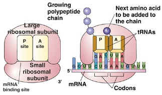
  • rRNA & protein make up the large & small subunits of ribosomes
  • Ribosomes are the site of translation (making polypeptides)

  • Aids in moving ribosomes along the mRNA strand as amino acids are linked together to make a protein

Amino Acids

  • 20 exist
  • Linked together in a process called protein synthesis in the cytoplasm to make polypeptides (subunits of proteins)
  • DNA contains the instructions for making proteins but is too large to leave the nucleus
  • Three consecutive bases on DNA called a triplet (e.g. TCG, ATG, ATT)
  • mRNA codon table tells what 3 bases on mRNA code for each amino acid (64 combinations of 3 bases)
  • Methionine (AUG) on mRNA is called the start codon because it triggers the linking of amino acids
  • UAA, UGA,  & UAG on mRNA signal ribosomes to stop linking amino acids together

Genetic Code (RNA)


 Amino Acid 3 Letter
Abbreviation
 Codons
 Alanine Ala GCA GCC GCG GCU
 Arginine Arg AGA AGG CGA CGC CGG CGU
 Aspartic Acid Asp GAC GAU
 Asparagine Asn AAC AAU
 Cysteine Cys UGC UGU
 Glutamic Acid Glu GAA GAG
 Glutamine Gln CAA CAG
 Glycine Gly GGA GGC GGG GGU
 Histidine His CAC CAU
 Isoleucine Ile AUA AUC AUU
 Leucine Leu UUA UUG CUA CUC CUG CUU
 Lysine Lys AAA AAG
 Methionine Met AUG
 Phenylalanine Phe UUC UUU
 Proline Pro CCA CCC CCG CCU
 Serine Ser AGC AGU UCA UCC UCG UCU
 Threonine Thr ACA ACC ACG ACU
 Tryptophan Trp UGG
 Tyrosine Tyr UAC UAU
 Valine Val GUA GUC GUG GUU
 Start AUG
 Stop UAA UAG UGA


  Practice Table:

DNA
Codon
mRNA
Codon
tRNA
Anticodon
Amino
Acid

GCU

TAC    
    AUU
  UUU  
TCA    
    UCU
CTT    
  ACU
ACU  

Protein Synthesis

  • Consists of 2 parts — Transcription & Translation
  • Begins in the nucleus with mRNA copying DNA’s instructions for proteins (transcription)
  • Completed in the cytoplasm when tRNA enters ribosomes to read mRNA codons and link together amino acids (translation)

 Steps in Transcription

  1. DNA helicase (enzyme) uncoils the DNA molecule
  2. RNA polymerase  (enzyme) binds to a region of DNA called the promoter which has the start codon TAC to code for the amino acid methionine
  3. Promoters mark the beginning of a DNA chain in prokaryotes, but mark the beginning of 1 to several related genes in eukaryotes
  4. The 2 DNA strands separate, but only one will serve as the template & be copied
  5. Free nucleotides are joined to the template by RNA polymerase in the 5’ to 3’ direction to form the mRNA strand
  6. mRNA sequence is built until the enzyme reaches an area on DNA called the termination signal
  7. RNA polymerase breaks loose from DNA and the newly made mRNA
  8. Eukaryotic mRNA is modified (unneeded sections snipped out by enzymes & rejoined) before leaving the nucleus through nuclear pores, but prokaryotic RNA isn’t
  9. All 3 types of RNA called transcripts are produced by this method

Steps in Translation

  1. mRNA brings the copied DNA code from the nucleus to the cytoplasm
  2. mRNA attaches to one end of a ribosome; called initiation
  3. tRNA’s attach the correct amino acid floating in the cytoplasm to themselves
  4. tRNA with its attached amino acid have 2 binding sites where they join the ribosome
  5. The tRNA anticodon “reads” & temporarily attaches to the mRNA codon in the ribosome
  6. Two amino acids at a time are linked together by peptide bonds to make polypeptide -chains (protein subunits); called elongation
  7. Ribosomes) move along the mRNA strand until they reach a stop codon (UAA, UGA, or UAG); called termination

  1. tRNA’s break loose from amino acid, leave the ribosome, & return to cytoplasm to pick up another amino acid

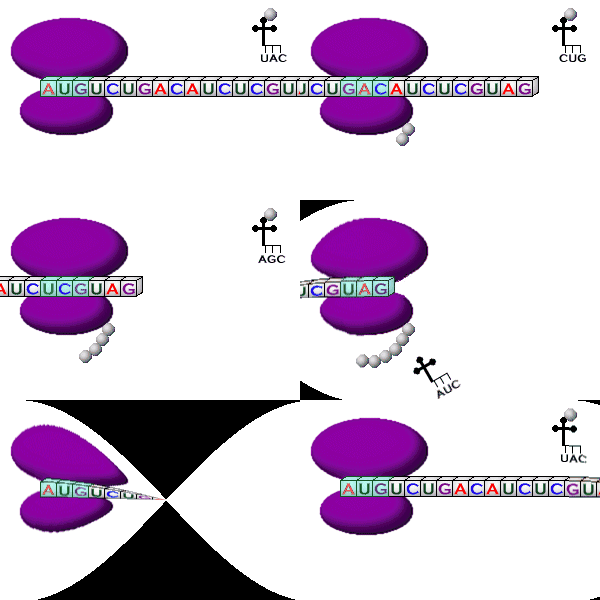
Click here for an animation of Translation 

Osmosis & Diffusion in Egg Lab


Osmosis & Diffusion in an Egg


Objective:
In this investigation, you will use a fresh hen’s egg to determine what happens during osmosis & diffusion across membranes.

Materials: (per lab group)
1-2 fresh hen eggs in their shells, masking tape & marker, distilled water, clear sugar syrup (Karo, for example), vinegar, clear jar with lid, tongs, electronic balance, paper towels, paper, pencil

Procedure:

Day 1   

  1. Label the jar with your lab group & the word “vinegar”.
  2. Mass the egg with the electronic balance & record in the data table.
  3. Carefully place the raw egg into the jar & cover the egg with vinegar.
  4. Loosely re-cap the jar & allow the jar to sit for 24 to 48 hours until the outer calcium shell is removed.

Day 2   

  1. Open the jar & pour off the vinegar.
  2. Use tongs to carefully remove the egg to a paper towel & pat it dry.
  3. Record the size & appearance of your egg in your data table.
  4. Mass the egg on an electronic balance & record.
  5. Clean and re-label the jar with your lab group & the word “distilled water”.
  6. Carefully place the egg into the jar & cover the egg with distilled water.
  7. Loosely re-cap the jar & allow it to sit for 24 hours.

Day 3   

  1. Open the jar & discard the distilled water.
  2. Use tongs to carefully remove the egg to a paper towel & pat it dry.
  3. Record the size & appearance of your egg in your data table.
  4. Mass the egg on an electronic balance & record.
  5. Clean and re-label the jar with your lab group & the word “syrup”.
  6. Carefully place the egg into the jar & cover the egg with clear syrup.
  7. Loosely re-cap the jar & allow it to sit for 24 hours.

Day 4   

  1. Open the jar & pour off the syrup.
  2. Use tongs to very carefully remove the egg & rinse off the excess syrup under slow running water.
  3. Pat the egg dry on a paper towel.
  4. Record the size & appearance of your egg in your data table.
  5. Mass the egg on an electronic balance & record.
  6. Clean up your work area & put away all lab equipment.

Data:


RESULTS OF DIFFUSION

Original MassFinal MassAppearance of Egg
VINEGAR
WATER
SYRUP


Questions & Conclusion:

1. Vinegar is made of acetic acid & water. Explain how it was able to remove the calcium shell.


2. (a) What happened to the size of the egg after remaining in vinegar?

(b) Was there more or less liquid left in the jar?

   (c) Did water move into or out of the egg? Why?


3. (a) What happened to the size of the egg after remaining in distilled water?

(b) Was there more or less liquid left in the jar?

   (c) Did water move into or out of the egg? Why?


4. (a) What happened to the size of the egg after remaining in syrup?

(b) Was there more or less liquid left in the jar?

   (c) Did water move into or out of the egg? Why?


5. Was the egg larger after remaining in water or vinegar? Why?


6. Why are fresh vegetables sprinkled with water at markets?


7. Roads are sometimes salted to melt ice. What does this salting do to the plants along roadsides & why?