Insects & Flowers


The Flower and the Fly: Long Insect Mouthparts and Deep Floral Tubes
Natural History,  March, 2005  by Laura A. Session,  Steven D. Johnson

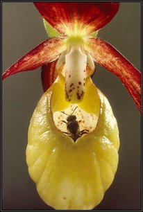
The mega-nosed fly (Moegistorhynchus longirostris) of southern Africa, like its literary counterpart, Pinocchio, has a bizarre appearance that reveals an underlying truth. Its proboscis, which looks like a nose but is actually the longest mouthpart of any known fly, protrudes as much as four inches from its head–five times the length of its bee-size body. In flight the ungainly appendage dangles between the insect’s legs and trails far behind its body.

To an airborne fly, an elongated proboscis might seem a severe handicap (imagine walking down the street with a twenty-seven-foot straw dangling from your mouth). Apparently, though, the handicap can be well worth its aerodynamic cost. The outlandish proboscis gives the mega nosed fly access to nectar pools in long, deep flowers that are simply out of reach to insects with shorter mouthparts.

But that poses a conundrum: why would natural selection favor such a deep tube in a flower? After all, nectar itself has evolved because it attracts animals that carry pollen, the sperm of the floral world, from one plant to another. And since pollinators perform such an essential service for the flower, shouldn’t evolution have favored floral geometries that make nectar readily accessible to the pollinators?

Yet the story of the long proboscis of the mega-nosed fly and the long, deep tubes of the flowers on which it feeds is not quite so straightforward. There are subtle advantages, it turns out, to making nectar accessible to only a few pollinators, and nature factors those advantages into the evolutionary equation as well. In fact, the evolution of those two kinds of organisms, pollinator and pollinated, presents an outstanding example of an important evolutionary phenomenon known as coevolution. Coevolution can explain the emergence of bizarre or unusual anatomies when no simple evolutionary response to natural selection is really adequate. It can help conservationists identify species that could be vital in maintaining a given habitat. And it can help naturalists investigating novel plants predict what kinds of animals might pollinate their flowers.

The coevolution of the mega nosed fly and the plants it pollinates is a tale of extreme specialization. Each species has adapted to changes in the other in ways that have left each of them, to some degree, reliant on the other. The idea that a plant species might become dependent for pollination on a single species of animal goes back to the writings of Charles Darwin. For example, Darwin noted, the flower spur of the Malagasy orchid (Angraecum sesquipedale) contains a pool of nectar that is almost a foot inside the opening of the flower. (A flower spur is a hollow, hornlike extension of a flower that holds nectar in its base.) In pondering the evolutionary significance of those unusual flowers, Darwin predicted that the orchid must be adapted to a moth pollinator with a long proboscis.

Critical to Darwin’s prediction was his suspicion that pollination could take place only if the depth of a plant’s flowers matched or exceeded the length of a pollinator’s tongue. Only then would the body of the pollinator be pressed firmly enough against the reproductive parts of the flower to transfer pollen effectively as the pollinator fed. Thus, as ever deeper flowers evolved through enhanced reproductive success, moths with ever longer proboscises would also, preferentially, live long enough to reproduce, because they would most readily reach the available supplies of nourishing nectar. Longer proboscises would lead yet again to selection for deeper flower tubes.

The result would be the reciprocal evolution of flowers and pollinator mouthparts. That coevolutionary process would cease only when the disadvantages of an exaggerated trait balanced or outweighed its benefits. Given enough time, the process might even produce new species: an insect the specializes in feeding on nectar from deep flowers, and a deep-flowered plant specialized for being pollinated by insects with long mouthparts.

In the early twentieth century it seemed that Darwin’s prediction had been borne out. A giant hawk moth from Madagascar, Xanthopan morganii praedicta, was captured, with a proboscis that measured more than nine inches long. Although no one has actually seen the insect feeding on the flower, the discovery is still remarkable, and strongly suggestive of the coevolution of the orchid and moth. Other insects that have relationships with highly specific plants, such as the mega nosed fly and other, related long-nosed fly species of southern Africa, provide even better evidence of the reciprocal links between planes and their pollinators.

Darwin would have been amazed that some flies in southern Africa have longer tongues than most hawk moths do. After all, the flies’ bodies are several times smaller than the hawk moths’ are. Flies are described as long-nosed if their mouthparts are longer than three quarters of an inch. By that criterion, more than a dozen long-nosed fly species are native to southern Africa. They belong to two families. The nemestrinids, or tangle-veined flies (which include the mega-nosed fly), feed solely on nectar, whereas the tabanids, or horseflies, feed mostly on nectar, though female tabanids have separate mouthparts to suck blood for their developing eggs.

Like all other long-nosed flies, the mega nosed fly is the sole pollinator to a group of unrelated plant species; such a group is known as a guild. The plant guild of the mega nosed fly includes species from a wide variety of plant families, including geraniums, irises, orchids, and violets.

Even though guild members may be only distantly related, all of them have roughly the same characteristics. For example, plants in the long-nosed fly guild all have long, straight floral tubes or spurs; brightly colored flowers that are open during the day; and no scent. The defining traits of a guild together form what botanists call a pollination syndrome. For example, bird-pollinated flowers are typically large, red, and unscented, whereas moth-pollinated flowers are more likely to be long, narrow, white, and scented in the evening.

The most important trait in the pollination syndrome of the long-nosed fly (and indeed, in all pollination syndromes of long-nosed insects) is a deep, tubular flower or floral spur. One of us (Johnson) and Kim E. Steiner of the Compton Herbarium in Claremont, South Africa, studied the orchid Disadraconis, a southern African plant with a deep, tubular floral spur. The two investigators artificially shortened the spurs of some orchids in a habitat where the only pollinators present were long-nosed flies. The plants whose spurs remained long got more pollen, and were more likely to produce fruits, than the ones whose spurs were shortened.

Yet short floral spurs are not necessarily a reproductive disadvantage. Shorter spurs would make it possible for a wider range of pollinators to access the nectar, if various potential pollinators are present. Instead, longer spurs only seem to be an advantage when long-tongued insects are the sole pollinators. Johnson and Steiner found that differences in spur length among populations cannot be blamed on differences in moisture or temperature, thus reinforcing their conclusion that spur length was an adaptation to the local distributions of long-tongued flies.

Not only does spur length correlate statistically with pollinator traits, but a direct causal connection can be demonstrated. Johnson and Ronny Alexandersson, a botanist at Uppsala University in Sweden, studied South African Gladiolus flowers pollinated by long-tongued hawk moths. When the hawk moth proboscises were long compared to the length of the flower tube, the hawk moths did not efficiently pick up pollen, and the flowers did not reproduce well. When the hawk moth proboscises were relatively short, pollen was more readily transferred, and the plants were more likely to be fertilized and bear fruit. Thus the length of the pollinator’s proboscis exerts a strong pressure on the reproductive success of the flowers.

Those studies and others suggest that what Darwin predicted of the Malagasy orchid is a rather general phenomenon: hawk moths and long-nosed flies coevolved with their plant partners. As floral tubes became longer, so did the pollinators’ proboscises, and those led, in turn, to even longer flowers. As the lengths of the flower tube and the insect proboscis converge, a remarkable degree of specialization develops. The plants come to rely for pollination on the few insect species that can reach their flowers’ nectar supplies.

There are advantages for the specialists on both sides of this relationship. The long-nosed flies obviously get privileged access to pools of nectar. And the plants pollinated by long-nosed flies benefit from a near-exclusive pollen courier service–or at least one that minimizes the risk of delivery to the wrong address. But specializing can also be a risky strategy for the plants if the pollinators are less interested in fidelity than the plants are. Long-nosed flies could not survive on the nectar they could get by visiting just one plant species; the flies must visit several plant species to gather the energy they need. Johnson and Steiner observed mega nosed flies visiting at least four species with deep flowers.

Such promiscuous behavior could be detrimental to the plants. A fly might end up carrying pollen from one species to a different species in the guild, thereby wasting the pollen. Worse, the foreign pollen could end up clogging the stigmata, the female reproductive structures, of the receiving flowers, preventing them from getting the “right” pollen. But the stigmata of plants in the guild of the mega nosed fly do not clog, because among those plants yet another clever adaptation to specialized pollination has evolved. Each plant species arranges its anthers, the male reproductive structures, in a characteristic position. That way, the pollen from each species sticks to the pollinator’s body in a distinct but consistent, plant-specific location. The fly becomes an even more efficient courier, carrying pollen from various plant species simultaneously, say, on its head, legs, and thorax.

The risks of specialization are not confined to the flowers. Just as the flies are unfaithful partners, some flowers are dishonest about signaling a nectar reward. The orchid D. draconis, for instance, is not the mutualistic partner it seems. The flower attracts the mega-nosed fly because it looks like other members of the fly’s guild. But, whereas the fly carries the orchid’s pollen, the orchid offers no nectar in return.

The risk of falling for such a trick seems a small price for the flies to pay for the benefits of specialization. But specialization also carries a much graver risk–in fact the ultimate risk–for both members of the partnership because the disappearance of either partner is likely to doom the other one, as well. Some plant species have mechanisms, such as vegetative reproduction or self-pollination, that may help sustain their populations in the short run. But in the long run, without their pollinators, the species will slowly and irrevocably decline. Pollinating insects may be more flexible in some cases, but are still vulnerable if a key food source disappears.

Unfortunately, in southern Africa that is just what is happening to many plants and their long-nosed fly partners. Often not even closely related insect species can help in pollination. For affected plants, the loss of a single fly species means extinction. And examples of that gloomy cascade have already been observed. Peter Goldblatt of the Missouri Botanical Garden in St. Louis and John C. Manning of the Compton Herbarium have ‘reported that many populations of long-nosed flies are threatened by the loss of their wetland breeding habitat, and also, possibly, by the loss of other insects they parasitize during their larval stages. In some habitats, flowers in the long-nosed fly guild already produce no seeds, because their pollinator is locally extinct.

Naturalists have accepted the concepts of guilds and pollinator syndromes for many years, and predicting which pollinators regularly visit which plants has become something of a cottage industry. But just how common is pollinator specialization in southern Africa? Promiscuity could turn out to be a more successful–and more widespread–strategy than specialization, even among plants that seem to fit into identifiable guilds.

In recent years ecologists have discovered that just because plants and insects appear to form a pollination guild does not guarantee they never venture outside it. For example, ecologists have noted that in years when hummingbird populations are low, flowers ordinarily pollinated by hummingbirds can fill up with nectar and become pollinated effectively by bees. Likewise, bees once thought to specialize in only one or two plant species turn out to forage on a variety of plants.

The take-home lesson has been that the syndrome concept is no substitute for careful field observation. Some investigators even think that the concept has caused botanists to overlook generalists. In the Northern Hemisphere, for instance, studies suggest that generalization is the norm, not the exception. Johnson and Steiner recently completed a study showing that members of the orchid and asclepiad families in the Northern Hemisphere tend to rely on between three and five pollinators each. In contrast, plants from the same families in the Southern Hemisphere rely on just one pollinator each.

So why might generalization be more common in the Northern Hemisphere than it is in the Southern Hemisphere? Perhaps the reason is that social bees, which are largely opportunistic, dominate pollinator faunas in northern regions. In the Southern Hemisphere, by contrast, social bees are mostly absent, replaced instead by more specialized pollinators such as the long-nosed flies and hawk moths.

But that is just a broad generalization itself. More data on the geographic distribution of pollinator specialization needs to be gathered, particularly in tropical countries. The data is vital, not only to advance the specialization debate, but also to protect as many of these unique species and relations as possible, lest they disappear forever.


Homeostasis & Transport


HOMEOSTASIS AND TRANSPORT
All Materials © Cmassengale


I. Cell Membranes


A. Cell membranes help organisms maintain homeostasis by controlling what substances may enter or leave cells

B. Some substances can cross the cell membrane without any input of energy by the cell

C. The movement of such substances across the membrane is known as passive transport


D. To stay alive, a cell must exchange materials such as food, water, & wastes with its environment

E. These materials must cross the cell or plasma membrane

F. Small molecules like water, oxygen, & carbon dioxide can move in and out freely

G. Large molecules like proteins & carbohydrates cannot move easily across the plasma membrane

H. The Cell Membrane is semipermeable or selectively permeable only allowing certain molecules to pass through


II. Diffusion


A. Diffusion is the movement of molecules from an area of higher concentration to an area of lower concentration

B. Small molecules can pass through the cell membrane by a process called diffusion


C. Diffusion across a membrane is a type of passive transport because it does not require energy

D. This difference in the concentration of molecules across a membrane is called a concentration gradient


E. Diffusion is driven by the kinetic energy of the molecules

F. Kinetic energy keeps molecules in constant motion causing the molecules to move randomly away from each other in a liquid or a gas

G. The rate of diffusion depends on temperature, size of the molecules, & type of molecules diffusing


H. Molecules diffuse faster at higher temperatures than at lower temperatures

I. Smaller molecules diffuse faster than larger molecules

J. Most short-distance transport of materials into & out of cells occurs by diffusion

K. Solutions have two parts — the solute which is being dissolved in the solvent


L. Water serves as the main solvent in living things

M. Diffusion always occurs down a concentration gradient (water moves from an area where it is more concentrated to an area where it is less concentrated)

N. Diffusion continues until the concentration of the molecules is the same on both sides of a membrane


O. When a concentration gradient no longer exists, equilibrium has been reached but molecules will continue to move equally back & forth across a membrane


III. Osmosis


A. The diffusion of water across a semipermeable membrane is called osmosis

B. Diffusion occurs from an area of high water concentration (less solute) to an area of lower water concentration (more solute)


C. Movement of water is down its concentration gradient & doesn’t require extra energy

D. Cytoplasm is mostly water containing dissolved solutes

E. Concentrated solutions have many solute molecules & fewer water molecules

F. Water moves from areas of low solute concentration to areas of high solute concentration

G. Water molecules will cross membranes until the concentrations of water & solutes is equal on both sides of the membrane; called equilibrium


H. At equilibrium, molecules continue to move across membranes evenly so there is no net movement

I. Hypertonic Solution
1. Solute concentration outside the cell is higher (less water)
2. Water diffuses out of the cell until equilibrium is reached
3. Cells will shrink & die if too much water is lost
4. Plant cells become flaccid (wilt); called plasmolysis

J. Hypotonic Solution
1. Solute concentration greater
inside the cell (less water)
2. Water moves into the cell until equilibrium is reached
3. Animal cells swell & burst (lyse) if they take in too much water
4. Cytolysis is the bursting of cells
5. Plant cells become turgid due to water pressing outward against cell wall
6. Turgor pressure in plant cells helps them keep their shape
7. Plant cells do best in hypotonic solutions

K. Isotonic Solutions
1. Concentration of solutes same inside & outside the cell
2. Water moves into & out of cell at an equal rate so there is no net movement of water
3. Animal cells do best in isotonic solutions


IV. How Cells Deal With Osmosis


A. The cells of animals on land are usually in isotonic environment (equilibrium)

B. Freshwater organisms live in hypotonic environments so water constantly moves into their cells

C. Unicellular freshwater organisms use energy to pump out excess water by contractile vacuoles

D. Plant cell walls prevent plant cells from bursting in hypotonic environments

E. Some marine organisms can pump out excess salt


V. Facilitated Diffusion


A. Faster than simple diffusion

B. Considered passive transport because extra energy not used

C. Occurs down a concentration gradient

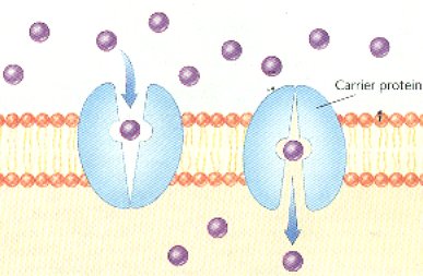
D. Involves carrier proteins embedded in a cell’s membrane to help move across certain solutes such as glucose


E. Carrier molecules change shape when solute attaches to them

F. Change in carrier protein shape helps move solute across the membrane

G. Channel proteins in the cell membrane form tunnels across the membrane to move materials

H. Channel proteins may always be open or have gates that open & close to control the movement of materials; called gated channels


I. Gates open & close in response to concentration inside & outside the cell


VI. Active Transport


A. Requires the use of ATP or energy

B. Moves materials against their concentration gradient from an area of lower to higher concentration

C. May also involve membrane proteins

D. Used to move ions such as Na+, Ca+, and K+ across the cell membrane

E. Sodium-Potassium pump moves 3 Na+ out for every 2 K+ into the cell
1. Causes a difference in charge inside and outside the cell
2. Difference in charge is called membrane potential


F. Ion pumps help muscle & nerve cells work


G. Plants use active transport to help roots absorb nutrients from the soil (plant nutrients are more concentrated inside the root than outside)


VII. Bulk Transport


A. Moves large, complex molecules such as proteins across the cell membrane

B. Large molecules, food, or fluid droplets are packaged in membrane-bound sacs called vesicles


C. Endocytosis moves large particles into a cell

D. Phagocytosis is one type of endocytosis
1. Cell membrane extends out forming pseudopods (fingerlike projections) that surround the particle
2. Membrane pouch encloses the material & pinches off inside the cell making a vesicle
3. Vesicle can fuse with lysosomes (digestive organelles) or release their contents in the cytoplasm
4. Used by ameba to feed & white blood cells to kill bacteria
5. Known as “cell eating”


E. Pinocytosis is another type of endocytosis
1. Cell membrane surrounds fluid droplets
2. Fluids taken into membrane-bound vesicle
3. Known as “cell drinking”


F. Exocytosis is used to remove large products from the cell such as wastes, mucus, & cell products

G. Proteins made by ribosomes in a cell are packaged into transport vesicles by the Golgi Apparatus

H. Transport vesicles fuse with the cell membrane and then the proteins are secreted out of the cell (e.g. insulin)

BACK

Human Genetics Notes BI


Home      General Biology 1      General Biology 2      Human Biology      Anatomy and Physiology
Chapter 16 – Genetics, Part 3: Human Genetics Introduction

This chapter is a review of patterns of inheritance in humans including a review of genetic diseases.

The genetic diseases are divided into two categories: chromosomal abnormalities and gene abnormalities.  Chromosomal abnormalities are caused by cells that have extra or missing chromosomes or parts of chromosomes.  Gene abnormalities (gene mutations) occur when the genetic instructions stored in the DNA are altered so that the protein product coded for by the gene is less functional or nonfunctional.

Prenatal Diagnosis

The techniques listed below enable physicians to diagnose many kinds of genetic abnormalities by examining some of the cells from the developing fetus.


Amniocentesis


The fetus is surrounded by a layer of liquid called amniotic fluid. Amniocentesis is a technique in which a sample of amniotic fluid is removed and cells that it contains are grown on a culture dish. Because these cells are of fetal origin, any chromosomal abnormalities present in the fetus will also be present in the cells.

In addition to chromosomal analysis, a number of biochemical tests can be done on the fluid to determine if any problems exist.

Amniocentesis cannot be done until the 14th to 16th week of pregnancy. Cells must then be cultured on a laboratory culture dish for 2 weeks to obtain sufficient numbers of cells.

The risk of inducing a spontaneous abortion by this procedure is 0.5 to 1% above the background rate of spontaneous abortion.


Chorionic Villi Sampling


Chorionic villi sampling is a procedure in which a small amount of the placenta is removed.

It is normally done during the 10th to 12th week but it can be done as early as the 5th week of pregnancy. Karyotype analysis can be performed on these cells immediately after sampling.

Although Chorionic villi sampling can be performed earlier in the pregnancy than amniocentesis, the risk of inducing a spontaneous abortion is 1 to 2% higher than the background rate.

Karyotypes

Karyotypes are prepared using cells from amniocentesis, chorionic villi sampling, or white blood cells.

Cells are photographed while dividing. cells are normally stained so that banding patterns appear on the chromosomes. The bands make it easier to identify the chromosomes. Banding patterns are not visible in the photograph below due to the staining technique.

Pictures of the chromosomes are cut out and arranged in pairs according to size and banding patterns.

Karyotypes can be used to determine if there is an abnormality in chromosome number or structure.

Nondisjunction

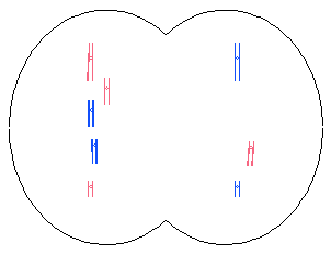
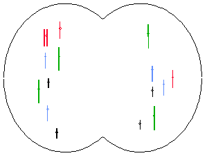
Nondisjunction occurs when chromosomes fail to “disjoin” during meiosis or mitosis.

Meiosis

Metaphase I

img017.gif (3781 bytes)

Anaphase I

img018.gif (3680 bytes)

Telophase I

img019.gif (3168 bytes)

Prophase II

img020.gif (4544 bytes)

Meiosis II and Mitosis

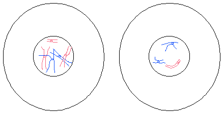
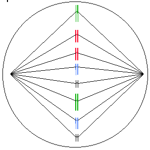
The diagrams below show nondisjunction during mitosis in a hypothetical species with 2N=8 chromosomes.

Metaphase

img013.gif (5064 bytes)

Anaphase

img014.gif (5337 bytes)

Telophase

img015.gif (3798 bytes)

G1 Interphase

img016.gif (4787 bytes)

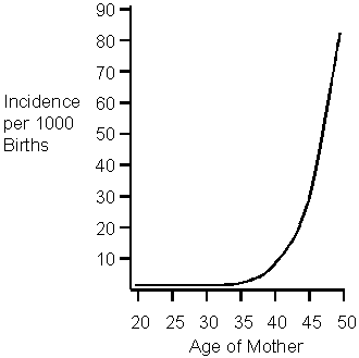
The probability of nondisjunction increases with age. It increases rapidly after age 35 years in women and after 55 years in men.

Aneuploidy

Cells that have extra chromosomes or chromosomes missing are aneuploid. Two types of aneuploidy are discussed below.

Monosomy refers to a condition in which there is one chromosome is missing. It is abbreviated 2N – 1. For example, monosomy X is a condition in which cells have only one X chromosome.

A trisomy has one extra chromosome and is abbreviated 2N + 1. Trisomy 21 is an example of a trisomy in which cells have an extra chromosome 21.

Monosomies and trisomies usually result from nondisjunction during meiosis but can also occur in mitosis. They are more common in meiosis 1 than meiosis 2.

They are generally lethal except monosomy X (female with one X chromosome) and trisomy 21 (Down’s Syndrome).

Affected indivisuals have a distinctive set of physical and mental characteristics called a syndrome. For example, trisomy 21 is Down syndrome.

Incidence of Genetic Abnormalities

Maternal Age

At 25 years, 17% of secondary oocytes may have chromosomal abnormalities. At 40 years, up to 74% may contain abnormalities.

Spontaneous Abortion (Miscarriage)

Two-thirds of all pregnancies are lost. These miscarriages are called spontaneous abortions.

Genetic mutation causes an estimated 60% of these spontaneous abortions.

Autosomal Abnormalities

Nine percent of spontaneous abortions are trisomy 13, 18, or 21; but 0.1% of newborns have these trisomies.


Down Syndrome


Down syndrome is trisomy 21. It is characterized by mental retardation, an abnormal pattern of palm creases, a flat face, sparse, straight hair, and short stature. People with Down syndrome have a high risk of having cardiac anomalies, leukemia, cataracts, and digestive blockages.

Life expectancy of Down syndrome individuals is in the middle teens but some live much longer.

The gene responsible for Alzheimer’s is on chromosome 21. Down’s are at increased risk for developing Alzheimer’s.

Down Syndrome is associated with maternal age. Older women, particularly those older than 40, are more likely to have a Down Syndrome child.


Translocation Down Syndrome


A translocation is the movement of a chromosomal segment from one chromosome to another nonhomologous chromosome.

Five percent of Down Syndrome cases involve a translocation.

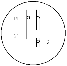
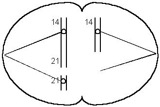
The translocation often involves chromosome 14.

In the translocation diagrammed below, chromosome #21 has become fused with chromosome #14.

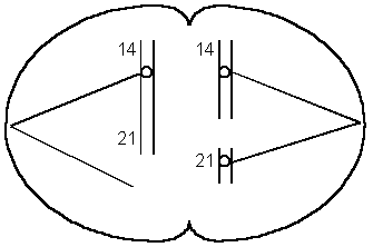
During meiosis, the two chromosomes might align so that each daughter cell receives one chromosome 21 as shown below. This will produce a normal egg.

If the chromosomes align as illustrated below, one daughter cell will receive two chromosome 21s and the other will not receive any.  When a gamete with two 21s fuses with a normal gamete, the result is a zygote with three chromosome 21s.

This form runs in families and is not age-related.


Mosaic Down Syndrome


Some of the cells of mosaic Down’s sydrome are trisomy 21 but others are normal.

This is due to nondisjunction that occurs during mitosis (after fertilization).

Mosaic Down Syndrome is likely to be less severe because some of the cells are normal.


Trisomy 18 (Edward Syndrome)


Trisomy 18 is associated with mental and physical retardation, skull and facial abnormalities, defects in all organ systems, and poor muscle tone.

Mean survival is 2 to 4 months.


Trisomy 13 (Patau Syndrome)


Trisomy 13 produces mental and physical retardation, skull and facial abnormalities, and defects in all organ systems. It is also associated with a left lip, a large, triangular nose, and extra digits.

One half die in first month; the mean survival time is 6 months.

Polyploidy

Polyploidy is a condition in which there is more than 2 sets of chromosomes.

Triploids (3N), tetraploids (4N), pentaploids (5N) etc. are polyploids.


Polyploidy in Plants


Polyploidy is a major evolutionary mechanism in plants. Approximately 47% of all flowering plants are polyploid.

Some examples of polyploid plant species are corn, wheat, cotton, sugarcane, apples, bananas, watermelons, and many flowers.

Polyploid plants are often more vigorous than the diploid parent species.

Polyploid plants are fertile.


Polyploidy in Humans


Polyploids have defects in nearly all organs.

Most die as embryos or fetuses. Occasionally an infant survives for a few days.

Abnormalities of the Sex Chromosomes

Turner Syndrome – XO

Characteristics of Turner syndrome include the following:

Sexually underdeveloped

Short stature

Folds of skin on the back of the neck

Wide-spaced nipples

Narrow aorta

Pigmented moles

97% die before birth

Malformed elbows

Infertile

Normal Intelligence

The incidence of Turner syndrome is 1 in 2000 female births.

Turner syndrome individuals that are treated with hormones lead fairly normal lives.

XXX – Triple-X Syndrome (also XXXX and XXXXX)

Triple-X individuals are tall and thin and have menstrual irregularities. Their IQ is in the normal range but it is slightly reduced.

The incidence of Triple-X Syndrome is 1 in 1,500 female births.

Additional X chromosomes are associated with an increased mental handicap.


XXY – Klinefelter Syndrome (also XXXY)


Males with two or more X chromosomes have Klinefelter Syndrome.

The incidence of Klinefelter Syndrome is 1 in 1000 male births.

Symptoms include reduced sexual maturity and secondary sexual characteristics, breast swelling, and no sperm. Klinefelter males are slow to learn and individuals with additional X’s (XXXY) may be mentally retarded.


XYY – Jacob Syndrome


XYY males are tall, have acne, speech, and reading problems.

Although there are a disproportionate number in penal institutions, 96% of Jacob’s Syndrome men are normal.

In the early 1970’s screening began in hospitals in England, Canada, Denmark and US. Families with XYY boys were offered “anticipatory guidance”. These types of programs were stopped because they were self-fulfilling prophesies.

Other Chromosomal Abnormalities

Deletions


Deletions are fragments of chromosomes that are missing. They are usually lethal when homozygous and cause abnormalities when heterozygous.

Radiation, viruses, chemicals, and unequal crossing-over may cause them.


Cri du Chat Syndrome


Cri du chat syndrome is due to a deletion of a portion of chromosome 5.

Cri du chat individuals are mentally retarded.

“Cri du chat” is French for “cry of the cat”. The infants cry sounds like a cat.


Duplication


A chromosome segment that is repeated is called a duplication.

It can be due to unequal crossing over which produces a deletion on one chromosome and a duplication on the other.

Often, multiple copies of genes from duplication can mutate without harming the individual because they still have one good copy of the gene. This type of mutation may be a source of variation for species. For example, the gene for human globin has given rise to several different genes that produce similar types of proteins. The different globins produced by these genes have very similar amino acid sequences.

An example of a family of genes that have been produced by duplication is the beta globin family. This family contains five functioning genes and a pseudogene.

Epsilon globin

G-gamma globin

A-gamma globin

delta globin

beta globin

a pseudogene

All of these genes have similar amino acid sequences due to their evolution from the same ancestral gene.

Some families of genes contain hundreds of genes.


Repeated Sequences


Repeated sequences are short segments of DNA that are repeated hundreds or thousands of times. For example: In the segment of DNA illustrated below, CCG is repeated several times.


The cause is unknown.

Fragile X Syndrome

This is the second most common cause of mental retardation (Down Syndrome is first).

The characteristic long, narrow face becomes more pronounced with age.

The symptoms of fragile-X syndrome appear to be caused by an abnormal number of repeats (CCG) on the X chromosome. Normal DNA has 6 – 50 copies of “CCG” at the locus in question. Carrier males have 50 – 230 copies. This is referred to as a premutation (pre-fragile-X). The full mutation involves more than 230 repeats of CCG.

The chance of being affected increases in successive generations because extra copies of CCG are added during the gamete-formation process.

Females are more likely to add repeats than males. At most, males pass on 230 repeats to their children but females pass on more than 230 repeats.

Mental problems are more common if the fragile X is inherited from the mother. This is an example of genomic imprinting discussed in the previous chapter. Fragile-X is more common in males because males inherit their X chromosome from their mother.

The repeats cause the X to have a thread-like portion. It is called a fragile site because it breaks if cultured under certain conditions in the laboratory.


Translocation


Chromosomes that break usually rejoin at the same place but sometimes the broken ends rejoin in different places.

Translocation is the movement of a chromosome or part of a chromosome to another (nonhomologous) chromosome.


Inversion


A segment of a chromosome may become turned around forming an inversion.

This can cause altered gene activity, a loss of crossing-over, or a duplication/deletion if crossing-over does occur.

Pedigrees

It is often easy to visualize relationships within an extended family by using symbols to represent people and relationships. A family tree which uses these symbols is called a pedigree. A sample pedigree is below.

In a pedigree, squares represent males and circles represent females. Horizontal lines connecting a male and female represent mating. Vertical lines extending downward from a couple represent their children. Subsequent generations are therefore written underneath the parental generations and the oldest individuals are found at the top of the pedigree.

If the purpose of a pedigree is to analyze the pattern of inheritance of a particular trait, it is customary to shade in the symbol of all individuals that possess this trait.

In the pedigree above, the grandparents had two children, a son and a daughter. The son had the trait in question. One of his four children also had the trait.

Autosomal Recessive

Characteristics of autosomal recessive inheritance


It often skips generations; children that have the trait can have parents that do not.

Heterozygotes (carriers) do not have the trait. People with the trait have two copies of the genes.

If both parents are have the trait, all offspring will.

Males and females are affected equally.

Inbreeding results in a greater-than-expected number of rare autosomal recessive phenotypes.


Cystic Fibrosis


Thick mucous forms in the digestive tract and lungs of people with CF. As a result, they have difficult breathing and are susceptible to lung infections.

People with cystic fibrosis have a life expectancy of approximately 30 years.

The gene that causes the disease is on chromosome 7. One particular mutation of this allele causes 70-75% of the cases.

It is somewhat difficult to detect prenatally.

Gene therapy may be a possibility in the future. The normal gene was inserted into cells in laboratory cultures.

Viruses have been engineered to deliver the gene. An aerosol spray is used to deliver the virus to the lungs.

There has been some success reported in treating human patients in 1994.

Cystic fibrosis is the most common lethal genetic disease among Caucasians in the US.

One in 25 is a carrier; one in 2500 is affected.


Tay Sachs


A fatty substance builds up in the neurons (nerve cells) of people with Tay Sachs. This causes a gradual paralysis and loss of nervous function that leads to death by age 4 or 5.

It is due to a single defective enzyme which normally digests the fatty material.

Heterozygotes (Aa) are not affected and are resistant to tuberculosis.

Prenatal diagnosis is available.

It is a common genetic disease among the Jewish population in the US (central and eastern European descent). Up to 11% are carriers. It is also common in people of French-Canadian or Cajun descent.


PKU – Phenylketonuria


PKU is a recessive genetic disease in which the person does not have the ability to break down the amino acid phenylalanine. The level of phenylalanine in the persons blood builds up and interferes with the development of the nervous system.

Children that are raised on a phenylalanine-restricted diet may develop normally but children that are not raised on a special diet will become severely mentally retarded. The diet should be followed for life because high phenylalanine levels affect cognitive functioning.

Genetic screening is the routine testing of individuals for specific genotypes. Newborns in U.S. hospitals are screened for PKU.

PKU women must resume the diet several months before conception

The incidence of PKU in the United States is 1 in 13,500 to 1 in 19,000.


Sickle-Cell Anemia


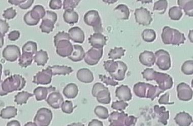
Sickle-cell anemia is an abnormality of hemoglobin, the molecule that carries oxygen in our blood. Hemoglobin is contained within red blood cells. When the oxygen concentration in the hemoglobin molecules becomes low, the molecules stick together forming long rods that distort the cell (picture below). The cells break down or clog blood vessels causing pain, poor circulation, jaundice, anemia, internal hemorrhaging, low resistance, and damage to internal organs. Death usually occurs before age 50.

Heterozygotes (carriers) are not affected with anemia and are resistant to malaria.

Eight to ten percent of African Americans carry the allele (have sickle-cell trait).

Hemochromatosis

Hemochromatosis is a disease that causes the body to absorb more iron from food than normal. High iron levels can lead to organ damage if it is left untreated for many years.

Symptoms include joint pain, fatigue, and abdominal pain.

There are two different mutations of the gene that causes hemochromatosis (the HFE gene) and the severity of symptoms depends on the mutations that are inherited.

One in 200 people in the United States carry the gene and it is the most common genetic disease in people of northern European descent.

There is also a form of this disease that is not due to genetic factors, it is acquired.

Autosomal Dominant

Severe dominant diseases are rare because carriers die before they get a chance to reproduce and pass on the disease to their offspring.

Heterozygotes (Aa) have the trait.

Children with the trait have at least one parent that has the trait.

Two parents with the trait can produce a child that does not have the trait.

Both males and females are affected equally.


Neurofibromatosis (NF)


Neurofibromatosis is sometimes called elephant man disease.

People with this gene have 6 or more large tan spots on the skin which may increase in size, number and darkness. The nerve cells form benign tumors which may vary in size. There may be learning disabilities and hyperactivity.

The disease is usually mild but may be severe causing deformities and even death.

The incidence is 1 in 3000 newborns.

The gene is on chromosome 17.


Huntington’s Disease


The brain cells of Huntington’s victims slowly degenerate, producing jerking muscles, slurred speech, swallowing difficulty, loss of balance, mood swings, reasoning and memory loss, incapacitation, and eventually death (usually from pneumonia or heart failure).

The onset of Huntington’s disease is typically 35 to 45 years.

It is caused by a repeated DNA sequence (AGC).  The normal allele has 11-34 copies; affected people have 42 – 120 copies.

The severity and time of onset depends on the number of repeats.

People who are most at risk inherit the gene from their father.  This is an example of genomic imprinting.

The gene is on chromosome 4.  A diagnostic test is available.

X-Linked Recessive

More males than females have x-linked recessive traits.

A son with the trait can have parents that do not have the trait.

There is no father to son transmission of the gene.

The trait can skip generations; grandfather to grandson transmission can occur.

If a female has the trait, her father has it, her mother is a carrier (or has it), and all her sons will have it.


Color Blindness


3 different kinds

2 X-linked forms: 1 for green insensitivity (6% of all males), one for red insensitivity (2% of all males); 1 in 12 males have some form of colorblindness.


Hemophilia


People with hemophilia lack a clotting factor in their blood and as a result, their blood does not form clots normally. This results in excessive bleeding from even minor cuts. Internal hemorrhaging from bruises is common and leads to painful complications.

The incidence is in 1,500 newborn males. Most (75%) have hemophilia A, a lack of clotting factor VIII. Hemophilia B- “Christmas Disease” is a defect in clotting factor IX.

Transfusions of fresh whole blood or plasma or factor concentrates control bleeding but have previously caused AIDS infections.

The human gene has been isolated and cloned using recombinant DNA techniques. This is leading to improved treatment.


Royal Families of Europe


Victoria (granddaughter of George III) was a carrier and spread the gene to the royal families of Europe. Her granddaughter Alix- married Czar Nicholas II of Russia. The Czar’s son Alexis, heir to the throne, had hemophilia.

The Czar’s preoccupation with Alexis’ health contributed to the revolution that overthrew the throne and eventually led to the communist government.


Duchenne Muscular Dystrophy


There are four different kinds of X-linked muscular dystrophy. They are multiple alleles at a single locus.

Duchenne’s is the most common and most severe form of muscular dystrophy.

1 in 5,000 live male births (Duchenne’s)

One in 4000 newborn males have some form of muscular dystrophy. One third of these are new mutations.

Muscular deterioration begins between ages 3 to 5. Affected individuals are confined to a wheelchair by age12 and rarely survive past age 20. Death is usually due to breathing or heart problems.

It is transmitted primarily by female carriers (males rarely reproduce)

Sex-Influenced Inheritance

Sex-influenced traits are those that are dominant in one sex but recessive in the other

This difference is due to the different hormonal environments between the sexes.

Sex-influenced genes are not necessarily located on the X chromosomes. Don’t confuse this with X-linked inheritance.


Examples

Pattern baldness is male dominant.

A gene that causes the index finger to be longer than the third finger is female dominant.


Home      General Biology 1      General Biology 2      Human Biology      Anatomy and Physiology

Homeostasis Worksheet Ch5 BI


Homeostasis & Transport


Section 5-1 Passive Transport

1. What is the purpose of the cell membrane?

2. Explain passive transport.

3. What is the simplest type of passive transport?

4. In which direction does diffusion occur?

5. What is a concentration gradient?

6. Sugar dissolving in water is an example of _______________________.

7. What supplies the energy for diffusion?

8. Molecules are constantly _____________________.

9. What is meant by equilibrium?

10. Do molecules stop moving when equilibrium is reached? Explain.

11. List three things that determine if a molecule will be able to diffuse across a membrane.

12. Name the 2 parts of a solution.

13. Define osmosis. Is it passive or active transport?

14. The direction water moves across a cell membrane depends on the concentration of what on either side of the cell membrane?

15. Explain what is true about solutes if the outside of the cell is hypotonic to the cytosol? Which way does water move?

16. Explain the solute conditions if the outside is hypertonic to the cytosol. Which way does water move?

17. What occurs if the solute concentration on each side of the cell membrane is isotonic?

18. If the inside & outside of a cell are both isotonic, does water still move across the cell membrane? Explain.

19. If the inside of the cell is hypotonic, the outside will be _________________________.

20. Water tends to diffuse from ____________________ to ___________________ solutions.

21. How does a unicellular paramecium get rid of its excess water? Is energy used?

22. Many cells in multicellular organisms have _________________ pumps to prevent them from taking in too much water in hypotonic solutions.

23. What structure around the outside of plant cells keeps hem from rupturing from too much water?

24. What is turgor pressure & how does it help plant cells?

25. What happens to plant cells placed in a hypertonic solution? Name this process.

26. What is cytolysis & what causes it?

27. Another type of passive transport is __________________________ diffusion.

28. Explain how carrier proteins help in facilitated.

29. Sketch the changes that take place in a carrier protein as it helps molecules move across the cell membrane.

30. What sugar moves across the cell membrane by facilitated diffusion?

31. What are ion channels & are they used in passive or active transport?

32. Name 4 ions that cross the cell membrane through ion channels.

33. Why can’t these ions diffuse across the lipid bilayer of the cell membrane?

34. Ion channels may be always ________________ or have ___________________.

35. Name 3 stimuli that open & close gated channels.

Section 5-2 Active Transport

36. Define active transport.

37. Why are carrier proteins in the cell membrane that are used for active transport called “pumps”?

38. What is the best-known carrier protein pump in animal cells?

39. What 2 ions move up their concentration gradient in this pump?

40. ___________________ ions are pumped out, while ______________ ions are pumped into the cell.

41. Is energy required for active transport? Explain.

42. Sodium ions are exchanged for potassium ions at a ____________ to ____________ ratio.

43. Name 2 processes used to move macromolecules & food particles across the cell membrane. Is energy required?

44. Explain how cells move large particles into the cell by endocytosis.

45. Name & describe the 2 types of endocytosis.

46. How do phagocytes protect cells?

47. What process moves large materials such as wastes & proteins out of the cell?

Human Hand Adaptations


Human Hand Adaptation

Introduction:        Living things have bodies that are adapted for the places they live and the things they do. Fish have gills so that they can remove oxygen that is dissolved in water. Most plants have green leaves which contain chlorophyll so that they can make food. Jellyfish have stinging cells to capture prey. Birds have hollow spongy bones so that they will be light enough to fly. Arctic animals have layers of fat and thick coats of fur to keep warm in the frigid Arctic climate. There are hundreds of examples of ways that organisms are adapted for a successful lifestyle.       Humans, too, are adapted for the things they do. One of our adaptations is our hand. Humans, as well as monkeys, gorillas, and other primates, have a hand that can grasp objects. We are able to grasp objects because of our opposable thumb. When students first hear or read about the opposable thumb during discussions of human evolution, they may perceive it as an anatomical fact with little seeming importance. In this activity, students will discover which of their simplest daily activities are possible only because of their opposable thumbs, which activities take longer without the use of an opposable thumb, and what sort of human activities would not be likely in the absence of an opposable thumb.   In this lab exercise, you will perform several common actions. Then you will change your hand so that it resembles that of a non-primate animal. You will determine whether or not you can successfully perform the same actions. This will demonstrate how the human hand is adapted for the actions it performs. You will work with a partner to do this exercise.   Materials: (per group)

  • masking tape
  • scissors
  • paper clips
  • zip-lock storage bag
  • plastic fork and knife
  • small amounts of food items to be cut
  • pencil
  • jar with screw-on lid
  • paper
  • roll of tape
  • balloons
  • comb
  • book
  • lace-up shoe
  • clock with a second hand
  • Piece of yarn or string
  • balloon
  • clothes with zippers & buttons

Procedure: Using masking tape, have your partner tightly tape each of your thumbs to the palm of the hand. Then, try to complete the tasks that are listed below. Be careful not to use your thumbs. Have your partner record on your data table how long it takes to do each task with your thumb taped and then with your thumb free. If an activity takes longer than 2 minutes, record the event as unsuccessful . After completing each item, write out the answers to the following questions:

  • Is the task more difficult with or without an opposable thumb?
  • How did you have to change your usual technique in order to complete this task?
  • Do you think organisms without opposable thumbs would carry out this task on a regular basis? Why or why not?

Tasks:

  1. Pick up a single piece of paper. Put it down on your desk.
  2. Pick up a pen or pencil from the table top. Use it to write your name on the piece of paper.
  3. Open a book. Turn single pages in the book.
  4. Unscrew a bottle cap or jar cover.
  5. Use a fork and knife to cut a food item into small pieces.
  6. Tear off a small piece of tape.
  7. Turn on the water faucet. (Complete activity #8!) Turn it off.
  8. Moisten a paper towel and wash and dry the desktop.
  9. Sharpen a pencil.
  10. Cut a circle out of a piece of paper using scissors.
  11. Pick up all the scraps from activity #10 and throw them into the recycling box.
  12. Comb your hair.
  13. Open a door.
  14. Pick up one paper clip. Clip a pile of papers together.
  15. Tie your shoelaces.
  16. Button several buttons.
  17. Zip up your jacket.
  18. Blow up a balloon and tie it.
  19. Tie a knot in a piece of string.
  20. Close a zip-lock bag.

Data:

Table 1 – Time It Took To Perform Various Tasks


TaskTime Taken for Event:Task Difficulty With Taped Thumb
(More/Less)
Modification Made to complete Task
Thumb FreeThumb Taped
Pick up paper
Write name
Turn book pages
Open jar
Use knife & fork
Tear off tape
Turn faucet on & off
Clean desk top
Sharpen a pencil
Cut out a circle
Pick up the scraps of paper
Comb hair
Open door
Clip papers together
Tie shoelaces
Button & unbutton garment
Use zipper
Blow up & tie balloon
Knot string
Close zip-lock bag

Conclusion:   1. Explain why dog and cat paws are not adapted for doing the six actions you tested.     2. What are cat and dog paws adapted for?     3. Describe how your hand is adapted for doing the actions you tested.       4. You have an opposable thumb. Explain what this means.     5. Why do you feel that human hand adaptations have helped to make humans such a successful species on earth?