| Chapter 16 – Genetics, Part 3: Human Genetics Introduction
This chapter is a review of patterns of inheritance in humans including a review of genetic diseases.
The genetic diseases are divided into two categories: chromosomal abnormalities and gene abnormalities. Chromosomal abnormalities are caused by cells that have extra or missing chromosomes or parts of chromosomes. Gene abnormalities (gene mutations) occur when the genetic instructions stored in the DNA are altered so that the protein product coded for by the gene is less functional or nonfunctional.
Prenatal Diagnosis
The techniques listed below enable physicians to diagnose many kinds of genetic abnormalities by examining some of the cells from the developing fetus.
Amniocentesis
The fetus is surrounded by a layer of liquid called amniotic fluid. Amniocentesis is a technique in which a sample of amniotic fluid is removed and cells that it contains are grown on a culture dish. Because these cells are of fetal origin, any chromosomal abnormalities present in the fetus will also be present in the cells.
In addition to chromosomal analysis, a number of biochemical tests can be done on the fluid to determine if any problems exist.
Amniocentesis cannot be done until the 14th to 16th week of pregnancy. Cells must then be cultured on a laboratory culture dish for 2 weeks to obtain sufficient numbers of cells.
The risk of inducing a spontaneous abortion by this procedure is 0.5 to 1% above the background rate of spontaneous abortion.
Chorionic Villi Sampling
Chorionic villi sampling is a procedure in which a small amount of the placenta is removed.
It is normally done during the 10th to 12th week but it can be done as early as the 5th week of pregnancy. Karyotype analysis can be performed on these cells immediately after sampling.
Although Chorionic villi sampling can be performed earlier in the pregnancy than amniocentesis, the risk of inducing a spontaneous abortion is 1 to 2% higher than the background rate.
Karyotypes
Karyotypes are prepared using cells from amniocentesis, chorionic villi sampling, or white blood cells.
Cells are photographed while dividing. cells are normally stained so that banding patterns appear on the chromosomes. The bands make it easier to identify the chromosomes. Banding patterns are not visible in the photograph below due to the staining technique.

Pictures of the chromosomes are cut out and arranged in pairs according to size and banding patterns.
Karyotypes can be used to determine if there is an abnormality in chromosome number or structure.
Nondisjunction
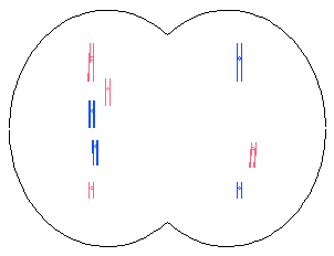
Nondisjunction occurs when chromosomes fail to “disjoin” during meiosis or mitosis.
Meiosis
Metaphase I
Anaphase I
Telophase I
Prophase II
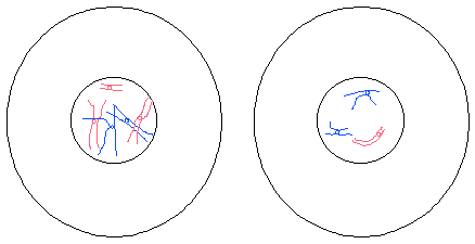
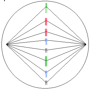
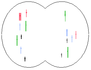
The diagrams below show nondisjunction during mitosis in a hypothetical species with 2N=8 chromosomes.
Metaphase
Anaphase

Telophase
G1 Interphase
The probability of nondisjunction increases with age. It increases rapidly after age 35 years in women and after 55 years in men.
Aneuploidy
Cells that have extra chromosomes or chromosomes missing are aneuploid. Two types of aneuploidy are discussed below.
Monosomy refers to a condition in which there is one chromosome is missing. It is abbreviated 2N – 1. For example, monosomy X is a condition in which cells have only one X chromosome.
A trisomy has one extra chromosome and is abbreviated 2N + 1. Trisomy 21 is an example of a trisomy in which cells have an extra chromosome 21.
Monosomies and trisomies usually result from nondisjunction during meiosis but can also occur in mitosis. They are more common in meiosis 1 than meiosis 2.
They are generally lethal except monosomy X (female with one X chromosome) and trisomy 21 (Down’s Syndrome).
Affected indivisuals have a distinctive set of physical and mental characteristics called a syndrome. For example, trisomy 21 is Down syndrome.
Incidence of Genetic Abnormalities
Maternal Age
At 25 years, 17% of secondary oocytes may have chromosomal abnormalities. At 40 years, up to 74% may contain abnormalities.
Spontaneous Abortion (Miscarriage)
Two-thirds of all pregnancies are lost. These miscarriages are called spontaneous abortions.
Genetic mutation causes an estimated 60% of these spontaneous abortions.
Autosomal Abnormalities
Nine percent of spontaneous abortions are trisomy 13, 18, or 21; but 0.1% of newborns have these trisomies.
Down syndrome is trisomy 21. It is characterized by mental retardation, an abnormal pattern of palm creases, a flat face, sparse, straight hair, and short stature. People with Down syndrome have a high risk of having cardiac anomalies, leukemia, cataracts, and digestive blockages.
Life expectancy of Down syndrome individuals is in the middle teens but some live much longer.
The gene responsible for Alzheimer’s is on chromosome 21. Down’s are at increased risk for developing Alzheimer’s.
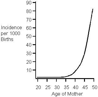
Down Syndrome is associated with maternal age. Older women, particularly those older than 40, are more likely to have a Down Syndrome child.
Translocation Down Syndrome
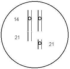
A translocation is the movement of a chromosomal segment from one chromosome to another nonhomologous chromosome.
Five percent of Down Syndrome cases involve a translocation.
The translocation often involves chromosome 14.
In the translocation diagrammed below, chromosome #21 has become fused with chromosome #14.
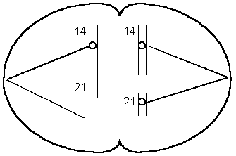
During meiosis, the two chromosomes might align so that each daughter cell receives one chromosome 21 as shown below. This will produce a normal egg.
If the chromosomes align as illustrated below, one daughter cell will receive two chromosome 21s and the other will not receive any. When a gamete with two 21s fuses with a normal gamete, the result is a zygote with three chromosome 21s.
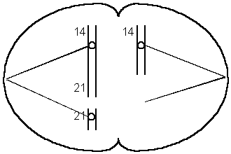
This form runs in families and is not age-related.
Mosaic Down Syndrome
Some of the cells of mosaic Down’s sydrome are trisomy 21 but others are normal.
This is due to nondisjunction that occurs during mitosis (after fertilization).
Mosaic Down Syndrome is likely to be less severe because some of the cells are normal.
Trisomy 18 is associated with mental and physical retardation, skull and facial abnormalities, defects in all organ systems, and poor muscle tone.
Mean survival is 2 to 4 months.
Trisomy 13 produces mental and physical retardation, skull and facial abnormalities, and defects in all organ systems. It is also associated with a left lip, a large, triangular nose, and extra digits.
One half die in first month; the mean survival time is 6 months.
Polyploidy
Polyploidy is a condition in which there is more than 2 sets of chromosomes.
Triploids (3N), tetraploids (4N), pentaploids (5N) etc. are polyploids.
Polyploidy in Plants
Polyploidy is a major evolutionary mechanism in plants. Approximately 47% of all flowering plants are polyploid.
Some examples of polyploid plant species are corn, wheat, cotton, sugarcane, apples, bananas, watermelons, and many flowers.
Polyploid plants are often more vigorous than the diploid parent species.
Polyploid plants are fertile.
Polyploidy in Humans
Polyploids have defects in nearly all organs.
Most die as embryos or fetuses. Occasionally an infant survives for a few days.
Abnormalities of the Sex Chromosomes
Characteristics of Turner syndrome include the following:
Sexually underdeveloped
Short stature
Folds of skin on the back of the neck
Wide-spaced nipples
Narrow aorta
Pigmented moles
97% die before birth
Malformed elbows
Infertile
Normal Intelligence
The incidence of Turner syndrome is 1 in 2000 female births.
Turner syndrome individuals that are treated with hormones lead fairly normal lives.
Triple-X individuals are tall and thin and have menstrual irregularities. Their IQ is in the normal range but it is slightly reduced.
The incidence of Triple-X Syndrome is 1 in 1,500 female births.
Additional X chromosomes are associated with an increased mental handicap.
Males with two or more X chromosomes have Klinefelter Syndrome.
The incidence of Klinefelter Syndrome is 1 in 1000 male births.
Symptoms include reduced sexual maturity and secondary sexual characteristics, breast swelling, and no sperm. Klinefelter males are slow to learn and individuals with additional X’s (XXXY) may be mentally retarded.
XYY males are tall, have acne, speech, and reading problems.
Although there are a disproportionate number in penal institutions, 96% of Jacob’s Syndrome men are normal.
In the early 1970’s screening began in hospitals in England, Canada, Denmark and US. Families with XYY boys were offered “anticipatory guidance”. These types of programs were stopped because they were self-fulfilling prophesies.
Other Chromosomal Abnormalities
Deletions are fragments of chromosomes that are missing. They are usually lethal when homozygous and cause abnormalities when heterozygous.
Radiation, viruses, chemicals, and unequal crossing-over may cause them.
Cri du chat syndrome is due to a deletion of a portion of chromosome 5.
Cri du chat individuals are mentally retarded.
“Cri du chat” is French for “cry of the cat”. The infants cry sounds like a cat.
A chromosome segment that is repeated is called a duplication.
It can be due to unequal crossing over which produces a deletion on one chromosome and a duplication on the other.
Often, multiple copies of genes from duplication can mutate without harming the individual because they still have one good copy of the gene. This type of mutation may be a source of variation for species. For example, the gene for human globin has given rise to several different genes that produce similar types of proteins. The different globins produced by these genes have very similar amino acid sequences.
An example of a family of genes that have been produced by duplication is the beta globin family. This family contains five functioning genes and a pseudogene.
Epsilon globin
G-gamma globin
A-gamma globin
delta globin
beta globin
a pseudogene
All of these genes have similar amino acid sequences due to their evolution from the same ancestral gene.
Some families of genes contain hundreds of genes.
Repeated sequences are short segments of DNA that are repeated hundreds or thousands of times. For example: In the segment of DNA illustrated below, CCG is repeated several times.

The cause is unknown.
This is the second most common cause of mental retardation (Down Syndrome is first).
The characteristic long, narrow face becomes more pronounced with age.
The symptoms of fragile-X syndrome appear to be caused by an abnormal number of repeats (CCG) on the X chromosome. Normal DNA has 6 – 50 copies of “CCG” at the locus in question. Carrier males have 50 – 230 copies. This is referred to as a premutation (pre-fragile-X). The full mutation involves more than 230 repeats of CCG.
The chance of being affected increases in successive generations because extra copies of CCG are added during the gamete-formation process.
Females are more likely to add repeats than males. At most, males pass on 230 repeats to their children but females pass on more than 230 repeats.
Mental problems are more common if the fragile X is inherited from the mother. This is an example of genomic imprinting discussed in the previous chapter. Fragile-X is more common in males because males inherit their X chromosome from their mother.
The repeats cause the X to have a thread-like portion. It is called a fragile site because it breaks if cultured under certain conditions in the laboratory.
Chromosomes that break usually rejoin at the same place but sometimes the broken ends rejoin in different places.
Translocation is the movement of a chromosome or part of a chromosome to another (nonhomologous) chromosome.
A segment of a chromosome may become turned around forming an inversion.
This can cause altered gene activity, a loss of crossing-over, or a duplication/deletion if crossing-over does occur.
Pedigrees
It is often easy to visualize relationships within an extended family by using symbols to represent people and relationships. A family tree which uses these symbols is called a pedigree. A sample pedigree is below.

In a pedigree, squares represent males and circles represent females. Horizontal lines connecting a male and female represent mating. Vertical lines extending downward from a couple represent their children. Subsequent generations are therefore written underneath the parental generations and the oldest individuals are found at the top of the pedigree.
If the purpose of a pedigree is to analyze the pattern of inheritance of a particular trait, it is customary to shade in the symbol of all individuals that possess this trait.
In the pedigree above, the grandparents had two children, a son and a daughter. The son had the trait in question. One of his four children also had the trait.
Autosomal Recessive
Characteristics of autosomal recessive inheritance
It often skips generations; children that have the trait can have parents that do not.
Heterozygotes (carriers) do not have the trait. People with the trait have two copies of the genes.
If both parents are have the trait, all offspring will.
Males and females are affected equally.
Inbreeding results in a greater-than-expected number of rare autosomal recessive phenotypes.
Thick mucous forms in the digestive tract and lungs of people with CF. As a result, they have difficult breathing and are susceptible to lung infections.
People with cystic fibrosis have a life expectancy of approximately 30 years.
The gene that causes the disease is on chromosome 7. One particular mutation of this allele causes 70-75% of the cases.
It is somewhat difficult to detect prenatally.
Gene therapy may be a possibility in the future. The normal gene was inserted into cells in laboratory cultures.
Viruses have been engineered to deliver the gene. An aerosol spray is used to deliver the virus to the lungs.
There has been some success reported in treating human patients in 1994.
Cystic fibrosis is the most common lethal genetic disease among Caucasians in the US.
One in 25 is a carrier; one in 2500 is affected.
A fatty substance builds up in the neurons (nerve cells) of people with Tay Sachs. This causes a gradual paralysis and loss of nervous function that leads to death by age 4 or 5.
It is due to a single defective enzyme which normally digests the fatty material.
Heterozygotes (Aa) are not affected and are resistant to tuberculosis.
Prenatal diagnosis is available.
It is a common genetic disease among the Jewish population in the US (central and eastern European descent). Up to 11% are carriers. It is also common in people of French-Canadian or Cajun descent.
PKU – Phenylketonuria
PKU is a recessive genetic disease in which the person does not have the ability to break down the amino acid phenylalanine. The level of phenylalanine in the persons blood builds up and interferes with the development of the nervous system.
Children that are raised on a phenylalanine-restricted diet may develop normally but children that are not raised on a special diet will become severely mentally retarded. The diet should be followed for life because high phenylalanine levels affect cognitive functioning.
Genetic screening is the routine testing of individuals for specific genotypes. Newborns in U.S. hospitals are screened for PKU.
PKU women must resume the diet several months before conception
The incidence of PKU in the United States is 1 in 13,500 to 1 in 19,000.
Sickle-cell anemia is an abnormality of hemoglobin, the molecule that carries oxygen in our blood. Hemoglobin is contained within red blood cells. When the oxygen concentration in the hemoglobin molecules becomes low, the molecules stick together forming long rods that distort the cell (picture below). The cells break down or clog blood vessels causing pain, poor circulation, jaundice, anemia, internal hemorrhaging, low resistance, and damage to internal organs. Death usually occurs before age 50.
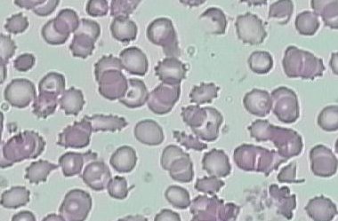
Heterozygotes (carriers) are not affected with anemia and are resistant to malaria.
Eight to ten percent of African Americans carry the allele (have sickle-cell trait).
Hemochromatosis
Hemochromatosis is a disease that causes the body to absorb more iron from food than normal. High iron levels can lead to organ damage if it is left untreated for many years.
Symptoms include joint pain, fatigue, and abdominal pain.
There are two different mutations of the gene that causes hemochromatosis (the HFE gene) and the severity of symptoms depends on the mutations that are inherited.
One in 200 people in the United States carry the gene and it is the most common genetic disease in people of northern European descent.
There is also a form of this disease that is not due to genetic factors, it is acquired.
Autosomal Dominant
Severe dominant diseases are rare because carriers die before they get a chance to reproduce and pass on the disease to their offspring.
Heterozygotes (Aa) have the trait.
Children with the trait have at least one parent that has the trait.
Two parents with the trait can produce a child that does not have the trait.
Both males and females are affected equally.
Neurofibromatosis is sometimes called elephant man disease.
People with this gene have 6 or more large tan spots on the skin which may increase in size, number and darkness. The nerve cells form benign tumors which may vary in size. There may be learning disabilities and hyperactivity.
The disease is usually mild but may be severe causing deformities and even death.
The incidence is 1 in 3000 newborns.
The gene is on chromosome 17.
The brain cells of Huntington’s victims slowly degenerate, producing jerking muscles, slurred speech, swallowing difficulty, loss of balance, mood swings, reasoning and memory loss, incapacitation, and eventually death (usually from pneumonia or heart failure).
The onset of Huntington’s disease is typically 35 to 45 years.
It is caused by a repeated DNA sequence (AGC). The normal allele has 11-34 copies; affected people have 42 – 120 copies.
The severity and time of onset depends on the number of repeats.
People who are most at risk inherit the gene from their father. This is an example of genomic imprinting.
The gene is on chromosome 4. A diagnostic test is available.
X-Linked Recessive
More males than females have x-linked recessive traits.
A son with the trait can have parents that do not have the trait.
There is no father to son transmission of the gene.
The trait can skip generations; grandfather to grandson transmission can occur.

If a female has the trait, her father has it, her mother is a carrier (or has it), and all her sons will have it.

3 different kinds
2 X-linked forms: 1 for green insensitivity (6% of all males), one for red insensitivity (2% of all males); 1 in 12 males have some form of colorblindness.
People with hemophilia lack a clotting factor in their blood and as a result, their blood does not form clots normally. This results in excessive bleeding from even minor cuts. Internal hemorrhaging from bruises is common and leads to painful complications.
The incidence is in 1,500 newborn males. Most (75%) have hemophilia A, a lack of clotting factor VIII. Hemophilia B- “Christmas Disease” is a defect in clotting factor IX.
Transfusions of fresh whole blood or plasma or factor concentrates control bleeding but have previously caused AIDS infections.
The human gene has been isolated and cloned using recombinant DNA techniques. This is leading to improved treatment.
Royal Families of Europe
Victoria (granddaughter of George III) was a carrier and spread the gene to the royal families of Europe. Her granddaughter Alix- married Czar Nicholas II of Russia. The Czar’s son Alexis, heir to the throne, had hemophilia.
The Czar’s preoccupation with Alexis’ health contributed to the revolution that overthrew the throne and eventually led to the communist government.
There are four different kinds of X-linked muscular dystrophy. They are multiple alleles at a single locus.
Duchenne’s is the most common and most severe form of muscular dystrophy.
1 in 5,000 live male births (Duchenne’s)
One in 4000 newborn males have some form of muscular dystrophy. One third of these are new mutations.
Muscular deterioration begins between ages 3 to 5. Affected individuals are confined to a wheelchair by age12 and rarely survive past age 20. Death is usually due to breathing or heart problems.
It is transmitted primarily by female carriers (males rarely reproduce)
Sex-Influenced Inheritance
Sex-influenced traits are those that are dominant in one sex but recessive in the other
This difference is due to the different hormonal environments between the sexes.
Sex-influenced genes are not necessarily located on the X chromosomes. Don’t confuse this with X-linked inheritance.
Examples
Pattern baldness is male dominant.
A gene that causes the index finger to be longer than the third finger is female dominant.
|