Cell Membrane
STRUCTURE AND FUNCTION OF THE CELL

All Materials © Cmassengale
I. All Organisms are Made of Cells
A. The cell is the basic unit of structure & function
B. The cell is the smallest unit that can still carry on all life processes
C. Both unicellular (one celled) and multicellular (many celled) organisms are composed of cells
D. Before the 17th century, no one knew cells existed
E. Most cells are too small to be seen with the unaided eye
F. In the early 17th century microscopes were invented & cells were seen for the 1st time
G. Anton Von Leeuwenhoek, a Dutchman, made the 1st hand-held microscope & viewed microscopic organisms in water & bacteria from his teeth
| Leeuwenhoek’s microscope consisted simply of:
|
H. In 1665, an English scientist named Robert Hooke made an improved microscope and viewed thin slices of cork viewing plant cell walls
I. Hooke named what he saw “cells”

J. In the 1830’s, Matthias Schleiden (botanist studying plants) & Theodore Schwann (zoologist studying animals) stated that all living things were made of cells
K. In 1855, Rudolf Virchow stated that cells only arise from pre-existing cells
L. Virchow’s idea contradicted the idea of spontaneous generation (idea that nonliving things could give rise to organisms)
M. The combined work of Schleiden, Schwann, & Virchow is known as the Cell Theory
 |
 |
 |
| Schwann | Schleiden | Virchow |
II. Principles of the Cell Theory
A. All living things are made of one or more cells
B. Cells are the basic unit of structure & function in organisms
C. Cells come only from the reproduction of existing cells
III. Cell Diversity
A. Not all cells are alike
B. Cells differ in size, shape, and function
C. The female egg cell is the largest cell in the body & can be seen without a microscope

D. Bacterial cells are some of the smallest cells & are only visible with a microscope

E.coli Bacterial Cells
E. Cells need surface area of their cell membrane large enough to adequately exchange materials with the environment (wastes, gases such as O2 & CO2, and nutrients)
F. Cells are limited in size by the ratio between their outer surface area & their volume
G. Small cells have more surface area for their volume of cytoplasm than large cells
H. As cells grow, the amount of surface area becomes too small to allow materials to enter & leave the cell quickly enough
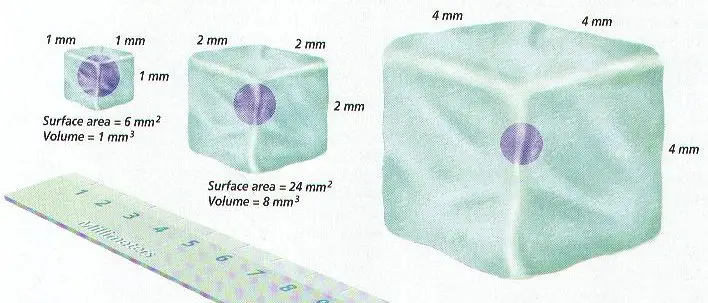
I. Cell size is also limited by the amount of cytoplasmic activity that the cell’s nucleus can control
J. Cells come in a variety of shapes, & the shape helps determine the function of the cell (e.g. Nerve cells are long to transmit messages in the body, while red blood cells are disk shaped to move through blood vessels)

IV. Prokaryotes
A. Prokaryotic cells are less complex
B. Unicellular
C. Do not have a nucleus & no membrane-bound organelles
D. Most have a cell wall surrounding the cell membrane & a single, looped chromosome (genetic material) in the cytoplasm
E. Include bacteria & blue-green bacteria
F. Found in the kingdom Monera

V. Eukaryotes
A. More complex cells
B. Includes both unicellular & multicellular organisms
C. Do have a true nucleus & membrane-bound organelles
D. Organelles are internal structures in cell’s that perform specific functions
|
|
|||
| a. Nucleus | b. Chloroplast | c. Golgi | d. Mitochondria |
E. Organelles are surrounded by a single or double membrane
F. Entire eukaryotic cell surrounded by a thin cell membrane that controls what enters & leaves the cell
G. Nucleus is located in the center of the cell
H. The nucleus contains the genetic material (DNA) & controls the cell’s activities
I. Eukaryotes include plant cells, animal cells, fungi, algae, & protists
J. Prokaryotes or bacteria lack a nucleus

K. Found in the kingdoms Protista, Fungi, Plantae, & Animalia

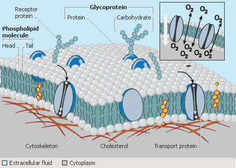
VI. Cell Membrane
A. Separates the cytoplasm of the cell from its environment
B. Protects the cell & controls what enters and leaves
C. Cell membranes are selectively permeable only allowing certain materials to enter or leave
D. Composed of a lipid bilayer made of phospholipid molecules

E. The hydrophilic head of a phospholipid is polar & composed of a glycerol & phosphate group and points to the aqueous cytoplasm and external environment.
F. The two hydrophobic tails are nonpolar point toward each other in the center of the membrane & are composed of two fatty acids

G. When phospholipids are placed in water, they line up on the water’s surface with their heads sticking into the water & their tails pointing upward from the surface.
H. The inside of the cell or cytoplasm is an aqueous or watery environment & so is the outside of the cell. Phospholipid “heads” point toward the water.

I. Phospholipid “tails” are sandwiched inside the lipid bilayer.

J. The cell membrane is constantly breaking down & being reformed inside living cells.
K. Certain small molecules such as CO2, H2O, & O2 can easily pass through the phospholipids
VII. Membrane Proteins
A. A variety of protein molecules are embedded in the cell’s lipid bilayer.

B. Some proteins called peripheral proteins are attached to the external & internal surface of the cell membrane
C. Integral proteins or transmembrane proteins are embedded & extend across the entire cell membrane. These are exposed to both the inside of the cell & the exterior environment.
D. Other integral proteins extend only to the inside or only to the exterior surface.
E. Cell membrane proteins help move materials into & out of the cell.
F. Some integral proteins called channel proteins have holes or pores through them so certain substances can cross the cell membrane.
G. Channel proteins help move ions (charged particles) such as Na+, Ca+, & K+ across the cell membrane
H. Transmembrane proteins bind to a substance on one side of the membrane & carry it to the other side. e.g. glucose
I. Some embedded, integral proteins have carbohydrate chains attached to them to serve as chemical signals to help cells recognize each other or for hormones or viruses to attach

VIII. Fluid Mosaic Model
A. The phospholipids & proteins in a cell membrane can drift or move side to side making the membrane appear “fluid”.
B. The proteins embedded in the cell membrane form patterns or mosaics.

C. Because the membrane is fluid with a pattern or mosaic of proteins, the modern view of the cell membrane is called the fluid mosaic model.
IX. Internal Cell Structure & Organelles of Eukaryotes
A. Cytoplasm includes everything between the nucleus and cell membrane.
B. Cytoplasm is composed of organelles & cytosol (jellylike material consisting of mainly water along with proteins.
C. Eukaryotes have membrane-bound organelles; prokaryotes do not

D. Mitochondria are large organelles with double membranes where cellular respiration (breaking down glucose to get energy) occurs
1. Energy from glucose is used to make ATP or adenosine triphosphate
2. Cells use the ATP molecule for energy
3. More active cells like muscle cells have more mitochondria
4. Outer membrane is smooth, while inner membrane has long folds called cristae
5. Have their own DNA to make more mitochondria when needed

E. Ribosomes are not surrounded by a membrane & are where proteins are made in the cytoplasm (protein synthesis)
1. Most numerous organelle
2. May be free in the cytoplasm or attached to the rough ER (endoplasmic reticulum)
F. Endoplasmic reticulum are membranous tubules & sacs that transport molecules from one part of the cell to another

1. Rough ER has embedded ribosomes on its surfaces for making proteins

2. Smooth ER lacks ribosomes & helps break down poisons, wastes, & other toxic chemicals
3. Smooth ER also helps process carbohydrates & lipids (fats)
4. The ER network connects the nucleus with the cell membrane
G. Golgi Apparatus modifies, packages, & helps secrete cell products such as proteins and hormones
1. Consists of a stack of flattened sacs called cisternae
2. Receives products made by the ER

H. Lysosomes are small organelles containing hydrolytic enzymes to digest materials for the cell
1. Single membrane
2. Formed from the ends of Golgi that pinch off
3. Found in most cells except plant cells
I. Cytoskeleton consists of a network of long protein tubes & strands in the cytoplasm to give cells shape and helps move organelles
1. Composed of 2 protein structures — microtubules, intermediate filaments, & microfilaments
2. Microfilaments are ropelike structures made of 2 twisted strands of the protein actin capable of contracting to cause cellular movement (muscle cells have many microfilaments)
3. Microtubules are larger, hollow tubules of the protein called tubulin that maintain cell shape, serve as tracks for organelle movement, & help cells divide by forming spindle fibers that separate chromosome pairs
| Cytoskeleton Element | General Function |
| Microtubules | Move materials within the cell Move the cilia and flagella |
| Actin Filaments | Move the cell |
| Intermediate Filaments | Provides mechanical support |
J. Cilia are short, more numerous hair like structures made of bundles of microtubules to help cells move
1. Line respiratory tract to remove dust & move paramecia

Cross section of Cilia & Flagella
K. Flagella are long whip like tails of microtubules bundles used for movement (usually 1-3 in number)
1. Help sperm cells swim to egg
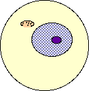
L. Nucleus (nuclei) in the middle of the cell contains DNA (hereditary material of the cell) & acts as the control center
1. Most cells have 1 nucleolus, but some have several
2. Has a protein skeleton to keep its shape
3. Surrounded by a double layer called the nuclear envelope containing pores
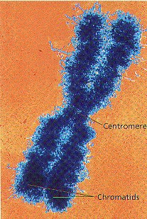
4. Chromatin is the long strand of DNA in the nucleus, which coils during cell division to make chromosomes
5. Nucleolus (nucleoli) inside the nucleus makes ribosomes & disappears during cell division

M. Cell walls are nonliving, protective layers around the cell membrane in plants, bacteria, & fungi

1. Fungal cell walls are made of chitin, while plant cell walls are made of cellulose
2. Consist of a primary cell wall made first and a woody secondary cell wall in some plants
N. Vacuoles are the largest organelle in plants taking up most of the space
1. Serves as a storage area for proteins, ions, wastes, and cell products such as glucose
2. May contain poisons to keep animals from eating them
3. Animal vacuoles are smaller & used for digestion
O. Plastids in plants make or store food & contain pigments to trap sunlight
1. Chloroplast is a plastid that captures sunlight to make O2 and glucose during photosynthesis; contains chlorophyll
a. Double membrane organelle with an inner system of membranous sacs called thylakoids
b. Thylakoids made of stacks of grana containing chlorophyll
2. Other plastids contain red, orange, and yellow pigments

3. Found in plants, algae, & seaweed
X. Multicellular Organization
A. Cells are specialized to perform one or a few functions in multicellular organisms
B. Cells in multicellular organisms depend on each other
C. The levels of organization include:
Cells –> Tissues –> Organs –> Systems –> Organism

D. Tissues are groups of cells that performs a particular function (e.g. Muscle)
E. Organs are groups of tissues working together to do a job (e.g. heart, lungs, kidneys, brain)
F. Systems are made of several organs working together to carry out a life process (e.g. Respiratory system for breathing)
G. Plants have specialized tissues & organs different from animals
1. Dermal tissue forms the outer covering of plants
2. Ground tissue makes up roots & stems
3. Vascular tissue transports food & water
4. The four plant organs are the root, stem, leaf, & flower
H. Colonial organisms are made of cells living closely together in a connected group but without tissues & organs (e.g. Volvox)

| Cell Cycle & Division All Materials © Cmassengale |
 |
Cell Division:
Reasons for Cell Division:
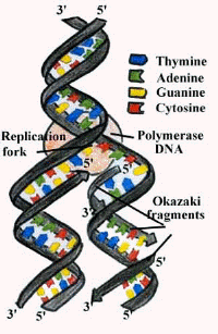
Copying DNA:
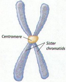
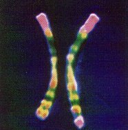
Chromosomes & Their Structure:
Chromosome Numbers:
Homologs
| Organism | Chromosome Number (2n) |
| Human | 46 |
| Fruit fly | 8 |
| Lettuce | 14 |
| Goldfish | 94 |

Human Male Karyotype
Genes:
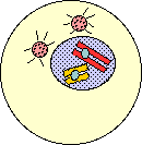
Cell Cycle:

Interphase:
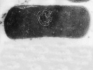
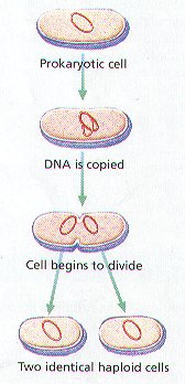
Cell division in Prokaryotes:
Cell Division in Eukaryotes:
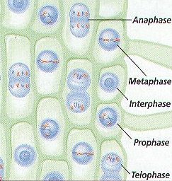
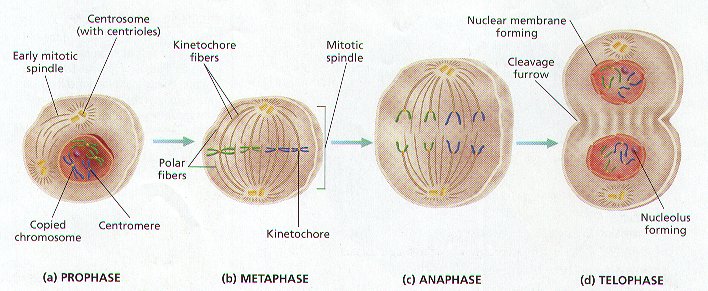
Stages of Mitosis:
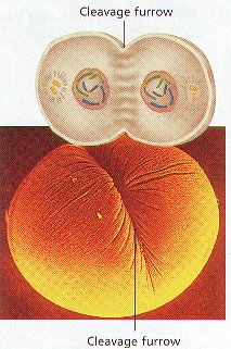
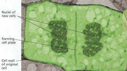
Cytokinesis:
Summary of Mitosis:
 |
 |
| Interphase
|
Early Prophase
|
 |
 |
Late Prophase
|
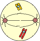
Metaphase
|
 |
 |
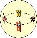
Anaphase
|
Telophase/Cytokinesis
|
Cancer is Uncontrolled Mitosis:
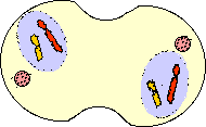
Meiosis & Sexual Reproduction
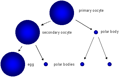
Oogenesis
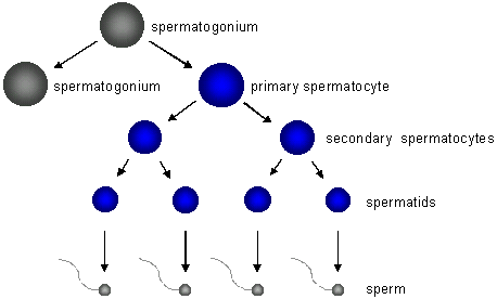
Spermatogenesis
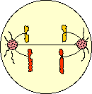
Meiosis I:


Asexual & Sexual reproduction:

Calorimetry – Measuring the energy in Foods

Introduction:
There are two processes that organisms use to make usable energy. The process by which autotrophs convert sunlight to a usable form of energy is called photosynthesis. Photosynthesis supports all life on earth. Products from photosynthesis include food, textiles, fuel, wood, oils, and rubber. During photosynthesis, light energy is used to make organic compounds from inorganic water and carbon dioxide. Photosynthesis goes through light dependent reactions and the light independent reactions which include the Calvin cycle.
The process where heterotrophs break down food molecules to release energy for work is called cellular respiration. Cellular respiration is the reverse of photosynthesis; the reactants of one are the products of the other. The reactants of cellular respiration are glucose and oxygen, and the products are carbon dioxide, water, and energy. Cellular respiration breaks down glucose to form carbon dioxide and water, while releasing energy usable by the cells. The first step, glycolysis is the process that converts glucose to pyruvate and releases a small amount of cellular energy. The second step may be aerobic or anaerobic depending on the amount of oxygen available. Aerobic respiration is the breakdown of pyruvate in the presence of oxygen. A larger amount of cellular energy or ATP is produced during the Kreb’s cycle and electron transport chain. Anaerobic respiration is the breakdown of food molecules in the absence of oxygen. Less ATP is produced by anaerobic respiration or fermentation.
Hypothesis:
If the heat given off by a burning pecan is measured by how much the temperature increases in a given amount of water, then the number of calories of energy stored in the nut during photosynthesis can be determined.
Materials:
Items needed for the lab included a large paper clip, a 100 ml graduated cylinder, thermometer, 2 soft drink cans, electronic balance, butane lighter, plastic tray, scissors, paper, and pencil.
Procedure:
Use a graduated cylinder to measure 100 ml of water and add this to an empty soft drink can. Cut holes on two sides of a second soft drink can so there is room to place a large bent paper clip. Measure and record the mass of one pecan using the electronic balance. Bend a large paper clip to make a “nut stand” and measure and record the mass of this clip. Place the pecan on the nut stand and put the stand inside the cut-out drink can. Use a thermometer to measure and record the temperature of the water in the second can. Place this can on top of the can with the nut. Use a butane lighter to ignite the nut. Record the temperature of the water when the nut is completely burned. Complete the data table by calculating the the total calories in the pecan.
Data:
Data Table 1 |
|||
| Before Burning | After Burning | Difference | |
| Mass of Nut | 1.7 g | 0.1g | 1.6g |
| Temperature of Water | 20 | 40.1 | 20.1 |
| Mass of Paper Clip | 1.4g | 1.4g | 0g |
| Data Table 2 | |
| Mass of pecan | 0.1 g |
| Temperature change of 100 ml of water | 20.1 |
| Calories required to produce temperature change in 100 ml water | 2010 |
| Calories per gram contained in the pecan | 1182.4 |
Error Analysis:
Errors may have occurred in several ways during this experiment. One error that may have occurred is that some of the energy may have been lost during the burning. Some of the pecan’s energy was lost as light instead of heat energy. Also some of the heat measured in the water could have been due to the butane lighter used to ignite the pecan.
Conclusion:
The temperature of the 100 ml of water in the can above the burning pecan was changed by the energy given off by the pecan when it was burned. The energy given off by the burning pecan was great enough to increase the water temperature by 20.1 degrees Celsius. The mass of the unburned pecan was 1.7g. It takes 100 calories to raise the temperature of 1 ml of water by 1 degree Celsius. The temperature of 100 ml of water was recorded to have increased by 20.1 degrees Celsius; therefore, the total number of calories in the pecan equals 20.1 x 100 or 2010 calories. Since the nut had a mass of 1.7g, the number of calories per gram equals 2010 divided by 1.7 or 1182.4 calories per gram.
The increase of temperature in the water showed that energy had been stored in the pecan. In this experiment, the amount of calories of heat energy stored in a pecan during photosynthesis was measured by a process known as calorimetry.
Experiment 3. (Frequency/Distance between T and S)
Determine the recombination frequency for the genes controlling Tallness and Snout:
| 40 tall-upturned snout | = 40% | expected |
| 42 dwarf-downturned snout | = 42% | expected |
| 9 dwarf-upturned snout | = 9% | recombinant |
| 9 tall-downturned snout | = 9% | recombinant |
Total = 100%
Therefore this recombination frequency between genes T and S is 18%
One can determine the relative frequency between genes using the percent frequencies as distances.
The Recombinant relationships from experiments 1-3 are:
Exp. 1 T-A = 12 map units Exp. 2 A-S = 5 map units Exp. 3 T-S = 18 map units
An arrangement that fits the data would be:

