|
Ecology |
Chapter 19 Ecology
1. What is ecology?
2.. What is the most significant environmental change that is taking place today?
3. What is the sixth mass extinction?
4. What is the ozone layer, what does it do for earth, & what is happening to this layer & why?
5. Explain the green house effect.
6. List in order the ecological levels of organization.
7. What is the biosphere, tell where it extends, & tell why it is so important?
8. Define ecosystems & give an example.
9. What is a community?
10. What is a population?
11. What is the simplest ecological level of organization?
12. Use figure 19-6 on page 364 & explain how Lyme disease affects organisms in an ecosystem.
13. What are biotic factors & list them?
14. What are abiotic factors & list them?
15. Are abiotic factors constant? Explain by giving an example.
16.Organisms are able to survive within a _____________ range of environmental conditions.
17. Graphing the range of conditions an organism can survive is called a __________________ Curve.
18.When organisms adjust their tolerance to abiotic factors, the process is called ___________.
19. Explain how dormancy & migration help organisms escape unsuitable environmental conditions.
20. Define niche
Chapter 20 Populations
21. What is meant by population size?
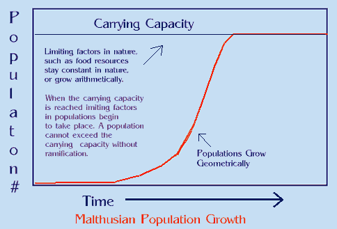
22. What is meant by population density?
23. Name the 4 processes that determine whether a population will grow, shrink, or remain the same size.
24. What are immigration & emigration & how do they affect population size?
25. What are limiting factors & give some examples?
26. What affect does inbreeding have on small populations?
Chapter 21 Community Ecology
27. Interactions among species are called ____________.
28. List the 5 types of symbioses.
29. Define predator & prey & give an example.
30. What is mimicry & give an example?
31. Define these terms — parasitism, parasite, host, ectoparasites, & endoparasites.
32. When niches overlap, _________________________ results so more than one species are using the limited resources.
33. What are mutualism & commensalism?
34. Define succession.
35. Name & describe the 2 types of succession.
36. What are pioneer species & why are they important?
37. What is a climax community?
Chapter 22 Ecosystems
38. What are producers & what is another name they may be called?
39. What is biomass, why is it important, how does it accumulate, & what is its rate of accumulation called?
40. What is gross primary productivity?
41. All heterotrophs would be ______________________.
42. Define & give an example of each of these consumers — herbivore, carnivore, omnivore, detritivores, & decomposer.
43. Whenever one organism eats another, ________________ is transferred.
44. What are trophic levels?
45. All _______________ belong to the first trophic level, _______________ belong to the
Second trophic level, and the _______________ of herbivores belong to the third trophic level.
46. How many trophic levels do most ecosystems contain?
47. What is a food chain & what always begins the chain?
48. Write an example of a food chain.
49. What is a food web?
50. Draw a diagram of a food web that has at least 4 food chains.
51. Approximately __________ percent of the total energy consumed at one trophic level is incorporated into the organisms in the next level.
52. In terms of energy passage, why will there be many more producers than herbivores and fewer large carnivores than small carnivores?
53. What are biogeochemical cycles, why are they important, & name three?
54. Draw & explain the water cycle. Be sure to color your diagram!
55. List & define the 3 important processes in the water cycle.
56. What is groundwater?
57. What 2 processes form the basis for the carbon cycle?
58. Draw & explain the carbon cycle. Be sure to color your diagram!
59. What purpose do decomposers have in the carbon cycle?
60. Why do organisms need nitrogen?
61. Draw & explain the nitrogen cycle. Be sure to color your diagram!
62. Organisms such as ________________ convert _________________ gas into compounds
Called __________________ during the process known as________________________.
63. Bodies of dead organisms contain mainly in _________________ & _________________.
64. Wastes such as __________________ & _______________ also contain nitrogen that must be recycled.
65. ________________ recycle nitrogen from dead organisms & wastes by changing it into
______________________. The process is called ________________________.
66. Explain nitrification & denitrification.
67. Plants can absorb ____________________ from the soil, but animals obtain nitrogen from
their ___________________.
68. Define biome.
69. List the 7 major biomes.
70. Why don’t mountains belong to any one biome?
71. What is a tundra, where are they found, & tell organisms that would be found tree?
72. What is permafrost & how does it control plant life in the tundra?
73. What are taigas, where would they be found, & what type of vegetation dominates this area?
74. Plants & animals in the taiga must be adapted for long __________________, short
_________________, & ________________________ soil.
75. List some typical animals of the taiga.
76. What characterizes a temperate deciduous forest?
77. Deciduous forests have 4 pronounced ____________________ with _________________
summers, _______________________ winters, and__________________________ than the
taiga.
78. Grasses dominate what biome?
79. Why aren’t there more trees on grassland?
80. What are grasslands called in each of these areas —– North America, Asia, South America, & southern Africa?
81. Describe the soil of grasslands. Because of the soil condition, how is much of the grassland used?
82.What type of animals would be found on grassland?
83. What periodically occurs across grasslands & why doesn’t it kill the grasses?
84. Approximately how much rainfall do deserts receive each year?
85. Are deserts always hot? Explain.
86. What adaptation must desert vegetation make to survive?
87. What types of adaptations must desert animals make to conserve water?
88. What are savannas & where are the best known savannas found?
89. Describe temperature & rainfall on savannas?
90. Name some herbivores & carnivores found on a savanna.
91. Describe the rainy season on a savanna & tell what special problem this poses for the animals & plants there?
92. What are tropical rain forests & where are they located?
93. Rain forests have stable, year-round ______________________ & abundant ____________.
94. Plants in the rainforest must constantly compete for what?
95. Explain the canopy & epiphytes in a rainforest.
96. Describe the plant & animal life in a rainforest.
97. Tropical rainforests are more commonly called _____________________.
98.Oceans cover what percent of the earth’s surface?
99. Draw, label, & color the zones found in the ocean (see figure 22-16). Define each term labeled on your drawing.
100. What are intertidal organisms exposed to & name some intertidal organisms.
101. Which zone in the ocean is the most productive & why?
102. What small organisms are found in the neritic zone & why are they important?
103. In tropical areas, what forms in the neritic zone & why are they important?
104. Which ocean zone has fewer species & why?
105. Where does most of the earth’s photosynthesis take place?
106. Animals in the aphotic zone feed on what?
107. Organisms living deep in the ocean must cope with what 2 problems? Give some examples of deep ocean animals & explain how they adapt to their environmental problems.
108. What are volcanic vents, when were they discovered, & describe the organisms found there?
109. What are estuaries & what special problem do estuary organisms face?
110. What characterizes freshwater zones & give several examples?
111. Name & describe the 2 categories into which ecologists divide lakes 7 ponds?
112. Define a river & describe organisms found there?
Chapter 23 Environmental Science
113. Where do upwellings occur & how are they helpful?
114. Describe the event known as El Nino & tell its effect.
115. Describe chlorofluorocarbons effect on the ozone layer & tell why we should be concerned?
116. Define biodiversity.
117. Define conservation biology & use migratory birds to explain an example of this new discipline?
118. Sometimes species are reintroduced into areas. Use the Gray wolf & describe its reintroduction in the United States.
119. Where are the Everglades located & what is being done to restore them?