| Chromosomes Linkage
Genes on the same chromosome are linked.
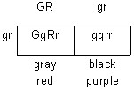
G = gray body
g = black (ebony) body
R = red eyes
r = purple eyes
The diagrams below show that the locus for body color (G or g) is on a different chromosome than the locus for eye color (R or r). These two loci will assort independently to produce either GR and gr gametes or Gr and gR gametes.
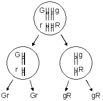
cross: GgRr X ggrr
gametes: GR, Gr, gR, gr X gr
Ratio expected: 1:1:1:1
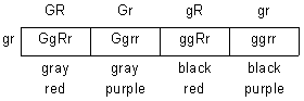
Suppose G and R are linked as shown below. If the body color and eye color loci are on the same chromosome, they will not assort independently unless crossing-over occurs frequently.
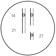
In this case, GgRr can produce only two kinds of gametes: GR and gr.
GgRr X ggrr
gametes: GR, gr X gr
If G and R are linked, then whenever you have a G, you have an R. Any gray, purple offspring (G-rr) would result from crossing over because a Gr gamete is needed.
Suppose out of 100 offspring, you got 46 gray, red, 46 black purple, 4 gray purple and 4 black red. Eight percent of the offspring resulted from crossing over. These offspring are recombinant.
Crossing Over
Crossing over is more likely to occur between genes that are far apart. The farther apart genes are, the greater the probability that crossing over will occur between them.
In the example above, we had 8% crossing over.
The percent of recombination (crossing over) can beused as a measure of how far apart genes are. 1% crossing over = 1 map unit.
Example
G = gray body
g = black (ebony) body
R = red eyes
r = purple eyes
Suppose that G and R are linked (on the same chromosome) in a particular individual and g and r are also linked
P1 GgRr X ggrr
If there is no crossing-over, possible gametes for the first parent are GR and gr.
If there is crossing-over, possible gametes are gR and Gr.
the following results were obtained:

How far apart are the G and R loci?
Sex Chromosomes
Humans have 23 pairs of chromosomes (46 total) chromosomes. Two of these are called sex chromosomes, the other 44 are called autosomes.
There are two kinds of sex chromosomes, called the X chromosome and the Y chromosome. The X chromosome is larger and contains many genes. The Y chromosome is much smaller and contains very few genes.
Normally, human females have two X chromosomes (XX) and males have one X and one Y chromosome (XY).
Occasionally, an accident happens in which a person is born with too many or too few sex chromosomes. In these cases, the person will be male if they inherit a Y chromosome and female if they do not.
Examples of four different possibilities that produce males are shown below. The last three are abnormal.
XY
XXY
XXXY
XYY
Examples of four different possibilities that produce females are shown below. Normal females are XX.
X
XX
XXX
XXXX
The cross below shows that normal females produce eggs that have one X chromosome. Half of the sperm produced by normal males have an X chromosome and the other half have a Y chromosome.
XX x XY
¯
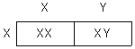
This analysis shows that half of the offspring are expected to be male, half are expected to be female.
Chromosomal Determination of Sex
Males
The Y chromosome contains a gene called SRY (for sex-determining region of Y).
Females
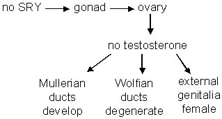
Testicular Feminization
The body cells of people with testicular feminization are insensitive to testosterone and therefore develop the female phenotype even though they have a Y chromosome.
It has an X-linked recessive mode of inheritance.
Guevodoces
Guevodoces refers to a condition in which the male phenotype develops after puberty.
It is due to delayed testosterone production.
X-Linkage
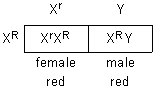
Morgan (Columbia U):
P1 red-eyed X white-eyed
¯
F1 all red-eyed
F2 3:1 (red:white) but all white were male
explanation:
These genes are found on the X chromosome but not on the Y chromosome. An XrY male will therefore have red eyes. Details of this cross are below.
| P1 |
XRXR |
X |
XrY |
| female |
male |
gametes: XR (female) and Xr, Y (male)
The offspring produced from the above cross are crossed with each other (below):
F1 XRXr X XRY
¯
gametes: XR and Xr (from female); XR and Y (from male)
F2:
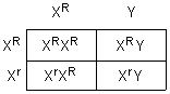
Notice that there are three possible genotypes for females and two possible genotypes for males.
| Females |
Males |
| Genotypes |
Phenotypes |
Genotypes |
Phenotypes |
| XRXR |
red |
XRY |
red |
| XRXr |
red |
XrY |
white |
| XrXr |
white |
|
|
Males inherit their X chromosome from their mother. Their Y chromosome comes from their father. A male, therefore, cannot pass an X-linked trait to his sons. Males inherit all of their X-linked traits from their mother.
If a male inherits an X-linked recessive trait, it will be expressed because males do not have a homologous X chromosome.
Females can be carriers of X-linked traits without expressing them because they might carry the dominant allele on the other X chromosome. For example, the following genotype will have a dominant phenotype: XAXa.
Dosage Compensation
Although females have twice as many X-linked genes, the amount of protein produced by these genes is the same in females as it is in males.
Reduced protein production (called dosage compensation) occurs as a result of inactivating one X chromosome by coiling and condensing it. When condensed, it cannot be transcribed, that is, it cannot be used to produce mRNA.
Condensed X chromosomes, called Barr bodies, are visible using ordinary light microscope techniques.
The table below shows the number of Barr bodies in normal cells and in the cells of people with an abnormal number of X chromosomes. Normal males do not have Barr bodies because they only have one X chromosome.
| Genetic Condition |
# Barr Bodies per Cell |
| normal male |
0 |
| normal female |
1 |
| XXX female |
2 |
| XXXX female |
3 |
| XXY (Klinefelter male) |
1 |
In summary, one X chromosome remains active, the others are inactivated by forming Barr bodies.
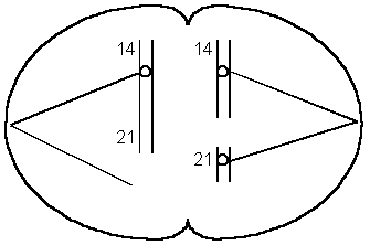
Inactivation
Inactivation occurs early in embryonic development (12-16 days).
In females, each cell normally contains two X chromosomes. The X chromosome that is inactivated is determined randomly.
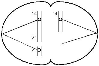
Once inactivation occurs, all daughter cells of a particular cell have the same X chromosome inactivated.
All of the “pink” chromosomes in the drawing below (left side of diagram) have been inactivated. All future cells produced by this cell will have the pink chromosome inactivated. In the diagram on the right, all of the blue chromosomes have been inactivated. All future generations of this cell will have the blue chromosome inactivated.
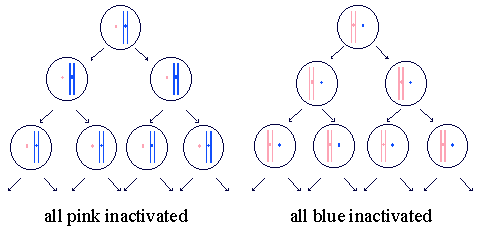
Females are therefore mosaics with respect to the X chromosome. Patches of body cells will have the maternally inherited X chromosome inactivated and other patches will have the paternally inherited one inactivated.
Example of Mosaicism: Calico Cats
A calico cat has patches of orange and patches of black
X = orange
X1 = black
MALES:
XY = orange
X1Y = black
FEMALES:
XX = orange
X1 X1 = black
X X1 = orange or black patches
All cells descended from an X1 cell (X is inactive) are orange-yellow.
All cells descended from an X cell (X1 is inactive) are black.
Human Example – Anhydrotic Dysplasia
Anhydrotic dysplasia is a disease that results in the absence of sweat glands.
It is inherited as an X-linked recessive disease.
Let X = normal sweat glands and X’ = absence of sweat glands. Normal males are XY. Affected males are X’Y and do not have sweat glands.
Normal females are XX, heterozygous females are XX’ and have patches of skin with sweat glands and patches of skin without sweat glands. Females that are X’X’ do not have sweat glands.
Other Information
Should heterozygous females for colorblindness be able to see color?
Suppose: X = color vision
x = colorblind
The Retina of a heterozygous (Xx) female will have some cells with the “X” inactivated and other cells with the “x” inactivated.
A heterozygous carrier of red-green colorblindness has some colorblind cells in her retina. The non-colorblind cells enable her to see color.
Turner’s syndrome is an abnormality in females where there is only one X chromosome; the other is missing. These people have abnormalities that will be discussed in the next chapter. Why aren’t Turners syndrome females normal? Evidence indicates that some genes in the Barr body remain active. Their DNA is uncoiled and extends from the Barr body. If the Barr bodies of a normal female were missing, she would exhibit Turners Syndrome.
|
 Crayfish Dissection Worksheet