AP Materials
Pre AP Biology
|
| BACK |
AP Materials
Pre AP Biology
|
| BACK |
Recreation of Pasteur’s Experiment
Introduction:
Today, we take many things in science for granted. Many experiments have been performed and much knowledge has been accumulated that people didn’t always know. For centuries, people based their beliefs on their interpretations of what they saw going on in the world around them without testing their ideas to determine the validity of these theories — in other words, they didn’t use the scientific method to arrive at answers to their questions. Rather, their conclusions were based on untested observations.
Among these ideas, for centuries, since at least the time of Aristotle (4th Century BC), people (including scientists) believed that simple living organisms could come into being by spontaneous generation. This was the idea that non-living objects can give rise to living organisms. It was common “knowledge” that simple organisms like worms, beetles, frogs, and salamanders could come from dust, mud, etc., and food left out, quickly “swarmed” with life. For example:
Observation: Every year in the spring, the Nile River flooded areas of Egypt along the river, leaving behind nutrient-rich mud that enabled the people to grow that year’s crop of food. However, along with the muddy soil, large numbers of frogs appeared that weren’t around in drier times. Conclusion: It was perfectly obvious to people back then that muddy soil gave rise to the frogs.
Objective:
In this experiment, you will conduct an experiment similar to the one done by Pasteur whenever he disproved spontaneous generation.
Materials Needed:
Procedure:
Data:
Microbial Growth Record
|
|||
| Day | Flask 1 with Straight Tubing | Day | Flask 2 with S-shaped Tubing |
| 1 | 1 | ||
| 2 | 2 | ||
| 3 | 3 | ||
| 4 | 4 | ||
| 5 | 5 | ||
| 6 | 6 | ||
| 7 | 7 | ||
| 8 | 8 | ||
| 9 | 9 | ||
| 10 | 10 | ||
Conclusion:
AP Materials
Pre AP Biology
Biology I
|
| BACK |
Nucleic Acids, Protein Synthesis, & DNA Technology Study Guide
1. What are purines & pyrimidines and give examples of each?
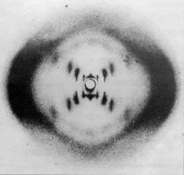
2. Which scientists determined the structure of DNA?
3. DNA and RNA are named by their __________.
4. What three things make up a nucleotide?
5. Describe the structure of DNA.
6. An organism’s characteristics are coded for by molecules of __________.
7. What are the subunits called that make up DNA?
8. Sketch the basic structure of a nucleotide.
9. What 2 things are found on RNA, but are not found on DNA molecules?
10. What is the primary function of DNA?
11.What did Rosalind Franklin’s x-ray photographs of DNA crystals tell us about this molecule?
12. State Chargaff’s rule.
13. What happens to tRNA anticodons during translation?
14. What is a codon & where are they found?
15. What is the function of rRNA?
16. What bases pair with each other on: a) DNA? b) RNA?
17. Name the 3 types of RNA & tell the function of each.
18. What is the function of DNA polymerase?
19. If the code on DNA is TTAGCCTGA, what will be the code on the complementary section of DNA when it’s copied during replication?
20. List all the ways that RNA differs from DNA?
21. Where does mRNA go for proteins to be made in a cell?
22. What is transcription?
23. What is translation?
24. Which RNA carries instructions for making proteins?
25. What is the function of DNA helicases?
26. What is the job of restriction enzymes?
27. What are “sticky ends” and how are they helpful?
28. What is the difference between introns & exons?
29. What is an operon and in what type of cell would they be found?
30. What does RFLP stand for? How is this process used?
31. What is meant by cloning?
32.What is DNA fingerprinting and how can it be used?
33. How is recombinant DNA formed?
Nucleic Acids and Protein Synthesis
All Materials © Cmassengale

Cell à Nucleus à Chromosomes à Genes à DNA
Proteins
DNA


 |
 |
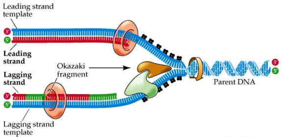
DNA Replication


OKAZAKI FRAGMENTS


mRNA
tRNA

rRNA
Amino Acids
Genetic Code (RNA)
| Amino Acid | 3 Letter Abbreviation |
Codons |
| Alanine | Ala | GCA GCC GCG GCU |
| Arginine | Arg | AGA AGG CGA CGC CGG CGU |
| Aspartic Acid | Asp | GAC GAU |
| Asparagine | Asn | AAC AAU |
| Cysteine | Cys | UGC UGU |
| Glutamic Acid | Glu | GAA GAG |
| Glutamine | Gln | CAA CAG |
| Glycine | Gly | GGA GGC GGG GGU |
| Histidine | His | CAC CAU |
| Isoleucine | Ile | AUA AUC AUU |
| Leucine | Leu | UUA UUG CUA CUC CUG CUU |
| Lysine | Lys | AAA AAG |
| Methionine | Met | AUG |
| Phenylalanine | Phe | UUC UUU |
| Proline | Pro | CCA CCC CCG CCU |
| Serine | Ser | AGC AGU UCA UCC UCG UCU |
| Threonine | Thr | ACA ACC ACG ACU |
| Tryptophan | Trp | UGG |
| Tyrosine | Tyr | UAC UAU |
| Valine | Val | GUA GUC GUG GUU |
| Start | AUG | |
| Stop | UAA UAG UGA |
Practice Table:
| DNA Codon |
mRNA Codon |
tRNA Anticodon |
Amino Acid |
GCU |
|||
| TAC | |||
| AUU | |||
| UUU | |||
| TCA | |||
| UCU | |||
| CTT | |||
| ACU | |||
| ACU |
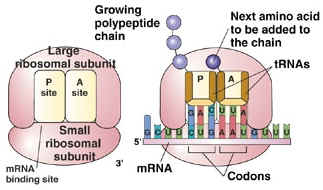
Protein Synthesis
Steps in Transcription
Steps in Translation