Bacteria
ppt Q’s
Prokaryote & Eukaryote Evolution
1. What does our current evidence tell us about the evolution of prokaryotes and eukaryotes?
2. About how long ago did eukaryotes evolve from prokaryotes?
3. Name the 2 theories of cellular evolution.
4. Explain the infolding theory.
5. What does endosymbiosis mean?
6. Explain the endosymbiotic theory of cell organelle formation.
7. Name 2 organelles thought to have arisen in this way.
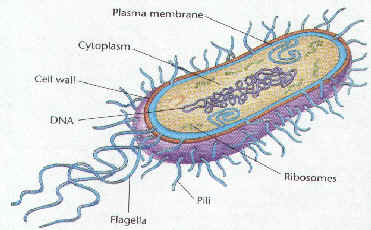
Prokaryotic & Eukaryotic Cells
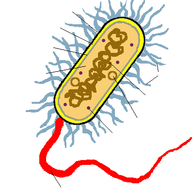
8. Label the parts of this prokaryotic cell.
9. Name several structures that are found in eukaryotic, but NOT prokaryotic cells.
10. What type of cells are the most numerous on Earth?
11. What are the most common type of prokaryotic cells?
12. How old are the earliest prokaryotic fossils?
Classification of Life
13. Name the 3 domains and the organisms found in each.
a.
b.
c.
14. ______________ are found in harsh environments.
15. Give 3 examples of harsh environments in which Archaebacteria can be found.
16. What group is referred to as the true bacteria?
17. What photosynthetic member is in this group?
Characteristics of Bacteria
18. What must be used to view prokaryotic cells?
19.What cell structures are lacking in prokaryotes?
20. Do bacteria have ribosomes like other types of cells?
21. Describe the genetic material of the bacteria. be sure to tell where it is found.
22. What surrounds the cytoplasm of bacterial cells?
23.What surrounds the outside of all bacterial cells?
24. Cell walls of true bacteria contain ____________________.
25. Some bacteria have a sticky ____________ around the cell wall to attach to __________ or other bacteria.
26. Besides the circular chromosome, where else can DNA be found inside a bacterial cell?
27. What is the size of most bacterial cells?
28. Compare the size of bacteria to the tip of a pin.
29. ____________ of the bacterial cell membrane are called _______________.
30. What two cellular processes can take place in mesosomes?
31. At what pH do bacteria do best?
32. Most bacteria act as ________________. Why is this so important?
33. How can some bacterial be harmful? Give an example.
34. name two other important uses for bacteria.
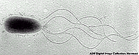
35. What does motile mean?
36. Motile bacteria may have one or more ______________ for movement.
37. Flagella attach to the bacteria by the ___________ ___________.
38. The basal body attaches to the cell through both the cell _________and the cell ___________.
39. What protein makes up bacterial flagella?
40. Tell how these types of bacteria differ from each other:
a. Monotrichous
b. Lophotrichous
c. Amphitrichous
d. Peritrichous
41. What type of bacteria is this?
42. What are bacterial pili?
43. How do pili compare to flagella in size?
44. Give three functions of pili.
a.
b.
c.
Bacterial Shapes
45. Name and describe 5 shapes used to classify bacteria.
a.
b.
c.
d.
e.
46. What does each of these prefixes tell you about the bacteria’s shape:
a. Diplo-
b. Strepto-
c. Staphylo-
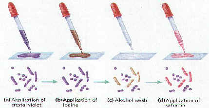
47. Sketch the shape of these bacteria:
a. Coccus
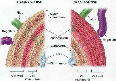
b. Bacillus
c. Spirillium
d. Diplococcus
e. Streptococcus
f. Staphylococcus
g. Diplobacillus
48. E. coli is classified as what shape bacteria?
Bacterial Kingdoms
49. How do the cell walls of Archaebacteria differ from the true bacteria?
50. How do the cell membranes differ?
51. Are the ribosomes the same?
52. Are the gene sequences the same?
53. Do Archaebacteria require oxygen?
54. How is there environment different from true bacteria?
55. What are they commonly called?
56. How many groups make up the ancient bacteria and name them?
57. Methanogens live in _____________ environments. What is lacking in this environment?
58. How do methanogens get their energy?
59. Name 3 environments in which methanogens are found.
60. How do methanogens help cows?
61. How did the methanogens get their name?
62. The __________ ___________ live in very salty environments.
63. How do they get their energy?
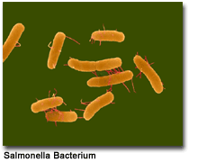
64. Name two bodies of water in which halophiles are found.
65. ______________ live in extremely hot environments.
66. Thermophiles that also live in acidic conditions are called _____________________.
67. Name 3 habitats in which thermophiles are found.
Kingdom Eubacteria
68. Most true bacteria are ____________ and come in ________ basic shapes. Name the shapes.
69. Do eubacteria require oxygen?
70. How are they identified?
71. When was gram staining developed?
72. Describe Gram staining.
73. What colors do bacterial cell walls stain?
74. Describe the cell wall of Gram positive bacteria.
75. What color do they stain?
76. Can Gram positive bacteria be treated with antibiotics?
77.Name 5 Gram positive bacteria and tell how they’re used or what they may cause.
a.
b.
c.
d.
e.
78. Describe the cell walls of Gram negative bacteria.
79. Are antibiotics effective against Gram negative bacteria?
80. Some photosynthetic Gram negative bacteria make ___________ instead of oxygen.
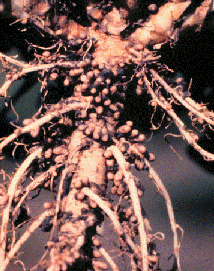
81. How do some Gram negative bacteria help plants?
82. Where can Rhizobacteria be found and what is their job?
83. _____________ are parasitic bacteria carried by ticks that may cause ___________ disease or _____________ _______________ _____________ fever.
84. Cyanobacteria are Gram ____________ and carry on ______________ to make food.
85. What is the common name for cyanobacteria?
86. What two main pigments do cyanobacteria contain?
87. What colors are cyanobacteria?
88. _______________ is a cyanobacterium that grows in chains.
89. Name the specialized structures on cyanobacteria that help fix nitrogen.
90. How do cyanobacteria cause eutrophication?
91. Spirochetes are Gram __________ bacteria that move by ___________.
92. Describe the motion of spirochetes.
93. Do all spirochetes need oxygen?
94. Spirochetes may be _______________, _______________, or symbiotic.
95. What are enteric bacteria? Give an example.
96. _______________ is an enteric bacterium that causes food poisoning.
97. How do chemoautotrophic bacteria get their energy?
Nutrition, Respiration, and Reproduction
98. Name and describe 4 modes of nutrition in bacteria.
a.
b.
c.
d.
99. Explain each of the following methods of respiration in bacteria.
a. Obligate Aerobes-
b. Obligate Anaerobes-
c. Facultative Anaerobes-
100. Anaerobes carry on ______________ to release energy from food, while aerobes carry on ____________ _______________.
101. Bacteria reproduce asexually by what method?
102. Before the cell can divide, what must happen?
103. Is binary fission a slow or fast process?
104. How do the new cells compare with each other after binary fission? What are they called?
105. Bacteria can reproduce sexually by ________________.
106. Describe how conjugation occurs.
107. What is the function of pili in conjugation?
108. How do the new cells compare to each other after conjugation?
109. When can bacteria produce spores and why?
110. What are the spores called?
111. How long can an endospore survive?
112. Why are endospores such a problem in health care facilities and in the canning industry?
113. Bacteria can genetically change by _________________ and ____________________.
114. Disease-causing bacteria may become ______________ _____________ when they genetically change.
115. How do bacteria transform?
116. Describe transduction in bacteria and give an example of a product made by bacteria using this method.
Pathenogenic Bacteria
117. What are pathogens?
118. Pathogens may cause ____________.
119. What are toxins?
120. What is the difference between endotoxins and exotoxins?
121. Name a bacterium that produces each type of toxin.
a. Endotoxin?
b. Exotoxin?