

Cells and Transport Teaching Resources · High school biology; AP extensions where noted.
Use these plant and animal cell diagrams to connect each structure with its function. Start with the labeled references, then use the student sheet to practice. These are simplified models of selected structures; they do not show every organelle.
Student handout: Download the printable student sheet (PDF). Use the reference below to check labels. Teacher explanations are separate at the end of this page.
Plant and animal cell diagrams
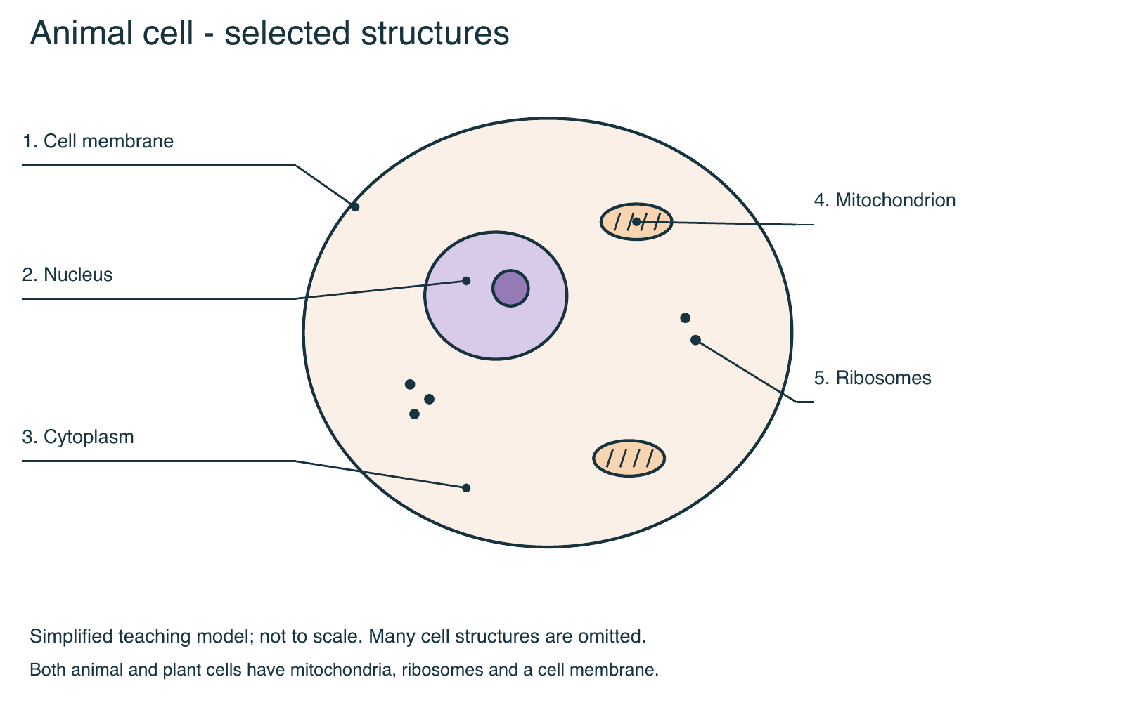
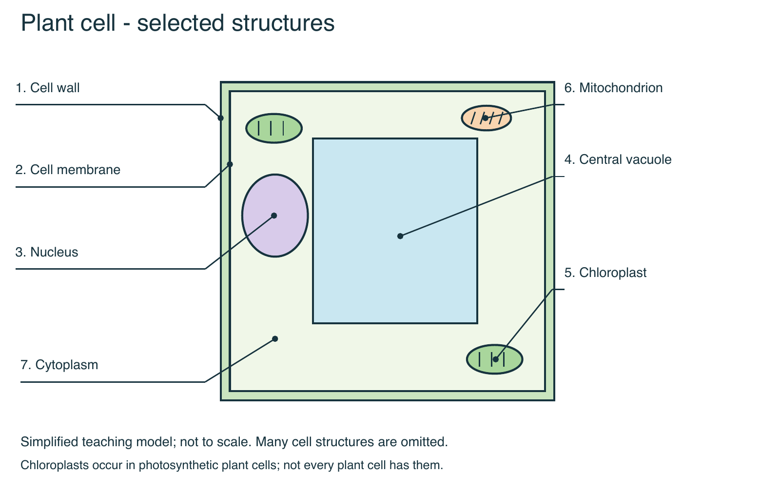
Plant and animal cells: compare the structures
| Structure | Typical plant cell | Typical animal cell | Main role |
|---|---|---|---|
| Cell membrane | Present, inside the wall | Present | Regulates exchange and signaling |
| Cell wall | Present; contains cellulose | Absent | Support and protection |
| Nucleus | Present in a typical living cell | Present in a typical cell | Contains most cellular DNA |
| Mitochondria | Present | Present | Aerobic respiration and ATP production |
| Chloroplasts | Present in photosynthetic cells | Absent | Photosynthesis |
| Vacuoles | Often one large central vacuole | Smaller compartments can occur | Storage; plant vacuoles also support water balance and breakdown |
| Ribosomes | Present | Present | Protein synthesis |
There are specialized exceptions, such as mature mammalian red blood cells lacking a nucleus. Plant cells have ribosomes even though the simplified plant diagram omits them.
Learning objectives
- Connect a cell structure to a function rather than memorize a label alone.
- Compare a typical plant cell, animal cell, and bacterial cell.
- Explain how several structures cooperate to produce and export a protein.
Structures to know
- Plasma membrane: the cell boundary that regulates exchange and carries proteins involved in transport and signaling. Plant cells have a plasma membrane inside the cell wall.
- Cytosol and cytoplasm: cytosol is the fluid portion; in a eukaryotic cell, cytoplasm includes cytosol and structures outside the nucleus.
- Nucleus: contains most of a eukaryotic cell’s DNA. The nucleolus helps assemble ribosomal subunits.
- Ribosomes: assemble proteins using instructions carried by messenger RNA. They are not membrane-bound; bacteria have them too.
- Rough endoplasmic reticulum: ribosomes associated with this membrane system make many proteins destined for secretion or membranes. The ER helps process those proteins.
- Smooth endoplasmic reticulum: participates in lipid production; its other functions depend on cell type.
- Golgi apparatus: modifies and sorts proteins and lipids for delivery.
- Mitochondria: support aerobic cellular respiration and ATP production. Plant cells have mitochondria as well as animal cells.
- Chloroplasts: carry out photosynthesis in photosynthetic plant cells. A root cell need not contain chloroplasts.
- Vacuoles and lysosomes: plant central vacuoles support storage, breakdown, and water balance. Animal lysosomes carry out digestion and recycling. Vacuoles do more than store water.
- Cell wall: a supporting layer outside the membrane. Plant walls contain cellulose; bacterial walls differ in composition.
- Cytoskeleton: protein filaments contribute to cell shape, internal organization, movement, and division.
To decode cell vocabulary using word parts, start with cyto- for cell and endo- for within. Then check the full definition: a word’s parts do not explain the whole structure or process.
Compare cells with evidence
Typical plant and animal cells have a nucleus and membrane-bound organelles; bacteria lack a membrane-bound nucleus. All three have DNA, ribosomes, cytoplasm, and a plasma membrane. Size and outline alone cannot reliably identify a cell. Both plants and animals grow through cell division and cell enlargement.
Use this in a lesson
- Ask students to label only structures supported by the selected diagram; a simplified picture may omit real structures.
- For each label, require one function and one connection to another structure.
- Trace a secreted protein from an ER-associated ribosome through the ER and Golgi to a vesicle that fuses with the plasma membrane.
- Finish with an unfamiliar example: why would a cell that secretes many proteins need extensive rough ER?
Check understanding
Question: Is a cell without chloroplasts necessarily an animal cell? Explanation: No. Many plant cells, including many root cells, lack chloroplasts. Look for other evidence such as a cellulose wall. Question: Do plants respire only at night? Explanation: No. Plant cells carry out respiration during the day and night; photosynthesis depends on suitable light.
Teaching note: Distinguish observations from inferences. A classroom light microscope does not normally resolve ribosomes or the detailed membrane bilayer. For AP biology, ask students to connect internal membranes to compartmentalization.
Teacher explanations for the new student sheet
Show the checked diagram labels and explanations
Animal labels: 1 cell membrane; 2 nucleus; 3 cytoplasm; 4 mitochondrion; 5 ribosomes. The membrane regulates exchange; the nucleus contains most DNA; many reactions take place in the cytoplasm; mitochondria support aerobic ATP production; ribosomes synthesize proteins.
Plant labels: 1 cell wall; 2 cell membrane; 3 nucleus; 4 central vacuole; 5 chloroplast; 6 mitochondrion; 7 cytoplasm.
Question 2: Yes. Plant cells use mitochondria for aerobic respiration, including during daylight. Question 4: A cell wall and chloroplasts are acceptable examples; a large central vacuole is another characteristic distinction. Question 5: Yes. Many root cells lack chloroplasts; use the wall and other evidence, not a single missing organelle, to identify the cell.
These explanations apply to the new sheet only, not every legacy worksheet.
Continue the unit
- Practice with the cell structure study guide
- Build and evaluate a cell analogy
- Create a labeled cell model
Sources and further reading
New diagrams and student sheets: original Biology Junction teaching resources, 2026. Teachers may print these new sheets for classroom use. Diagrams are simplified models, not photomicrographs.
Further reference: OpenStax Biology 2e: eukaryotic cells.