Toothpick-ase: Introduction to Enzymes

Enzymes are used in all metabolic reactions to control the rate of reactions and decrease the amount of energy necessary for the reaction to take place. Enzymes are specific for each reaction and are reusable. Enzymes have an area called the active site to which a specific substrate will bond temporarily while the reaction is taking place. Enzymes are proteins that are used as catalysts in biochemical reactions. A catalyst is a factor that controls the rate of a reaction without itself being used up. In biological systems, enzymes are used to speed up the rate of a reaction. However, there are a number of factors that can affect the rate of an enzyme-facilitated reaction, in addition to the presence of the enzyme, amongst them are:
- Substrate concentration
- Temperature

Here is a set of quick activities designed to simulate how substrate concentration and temperature affect enzyme function. In the activities that follow:
- One person’s fingers are the enzyme TOOTHPICKASE
- The toothpicks are the SUBSTRATE
- Toothpickase is a DIGESTIVE ENZYME. It breaks down toothpicks into two units. To hydrolyse the toothpick, place a toothpick between the thumb and the first finger of each hand. Break the toothpick in two pieces.
Materials:
100 toothpicks per team
bowl
clock/watch with a second hand
Pencil
Procedure:
Part A – rate of Product Formation in an Enzyme-Facilitated reaction
In this activity, the toothpicks represent a substrate and your thumbs and index fingers represent the enzyme, toothpick-ase. When you break a toothpick, the place where the toothpick fits between your fingers represents the active site of the enzyme.
1. Count out 100 unbroken toothpicks into a bowl on your desk.
2. Have one person in the group serve as the timer, have one person serve as the recorder, and have another person in your group act as the enzyme or toothpick-ase.
3. The person acting as the enzyme is to break toothpicks without looking at the bowl and all of its products (broken toothpicks). All broken toothpicks must remain in the bowl along with the unbroken toothpicks, & you cannot re-break a broken toothpick!.
4. The experiment is conducted in 10 second intervals.
5. WITHOUT LOOKING AT THE BOWL, break as many toothpicks as you can in 10 second intervals and record this on the data table. Broken toothpicks should be kept in the bowl with unbroken toothpicks because products & reactants mix in metabolic reactions. DO NOT BREAK TOOTHPICKS ALREADY BROKEN!
Remember when counting, two halves equal a whole broken toothpick!
6. Do another 10 seconds of breaking (total of 20 seconds now), and then count & record the number of toothpicks broken.
7. Do another 10 seconds (thirty seconds total now) more of breaking and count and record the number of toothpicks broken.
8. Continue breaking toothpicks for these total time intervals ( 60, 120, and 180 seconds). REMEMBER TO ALWAYS THROW BROKEN TOOTHPICKS BACK IN THE PILE (because products & reactants stay mixed in reactions), BUT DON’T RE-BREAK THEM (the enzyme has already acted on the substrate!
6. Graph the number of toothpicks broken as a function of time (10, 20, 30, 60, 120, & 180 seconds.) Be sure to title your graph and to label the x and y-axis.
Data Table:
| Total Time (seconds) | Number of toothpicks broken |
| 10 | |
| 20 (additional 10 seconds) |
|
| 30 (additional 10 seconds) |
|
| 60 (additional 30 seconds) |
|
| 120 (additional 60 seconds) |
|
| 180 (additional 60 seconds) |
Graph Title: ____________________________________________________________

Materials:
1 box toothpicks per team
100 paper clips
clock/watch with a second hand
Pencil
PART B: EFFECT OF SUBSTRATE CONCENTRATION ON REACTION RATE
- Remove the broken toothpicks from the shallow bowl. Place 100 paperclips in the empty bowl. The paper clips represent a “solvent” in which the toothpicks are “dissolved”. Different concentrations are simulated by mixing different numbers of toothpicks in with the paper clips.
- For the first trial, place 10 toothpicks in the bowl with the paper clip. Mix them up. The enzyme has 20 seconds to react (break as many toothpicks as possible). Remember the enzyme breaks the toothpicks without looking at the bowl and all of the products (“broken toothpicks”) must remain in the bowl. Remember toothpicks can only be digested once; do not break toothpicks already broken! Record the number broken at a concentration of 10.
- Remove the broken toothpicks and repeat with concentrations of 20, 30, 40, 50, 60, 70, 80, 90, and 100 toothpicks, each time mixing them with the 100 paper clips.
- Graph the results.
- Discuss your results and explain why the rates were different at different concentrations. Summarize the effect of substrate concentration on enzyme action.
Discussion & summary:
Data Table:
| Time (seconds) | Toothpick Concentration | Number of toothpicks broken |
| 20 | 10 | |
| 20 | 20 | |
| 20 | 30 | |
| 20 | 40 | |
| 20 | 50 | |
| 20 | 60 | |
| 20 | 70 | |
| 20 | 80 | |
| 20 | 90 | |
| 20 | 100 |
Graph Title: ____________________________________________________________
Materials:
10 toothpicks per team
ice & ice bucket
clock/watch with a second hand
Pencil
PART C: EFFECT OF TEMPERATURE SUBSTRATE CONCENTRATION ON REACTION RATE
- Select 10 toothpicks. Time how long it takes to break the 10 toothpicks as fast as you can.
- Place your hands in the pail of iced water for 10 minutes. Repeat step 1.
- Calculate the rate of enzyme action in toothpicks per second. Compare the two rates.
- Discuss your results and explain why the rates were different at different temperatures. Summarize the effect of temperature on enzyme action.
Discussion & summary:
Analysis & conclusions:
1.What happens to the reaction rate as the supply of toothpicks runs out?
2. What would happen to the reaction rate if the toothpicks were spread out so that the “breaker” has to reach for them?
3. What would happen to the reaction rate if more toothpicks (substrate) were added?
4. What would happen to the reaction rate if there were two “breakers” (more enzymes)?
5. What happens if the breaker wears bulky gloves (active site affected) when picking up toothpicks?
6. Explain what would happen to an enzyme-facilitated reaction if temperature were increased. Be sure to include the effect if temperature were increased to 100°C.
7. What is the optimal temperature (°C) for enzymes functioning in the human body?
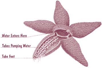
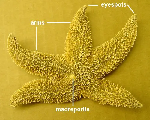
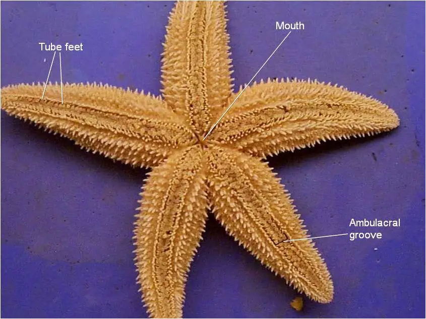
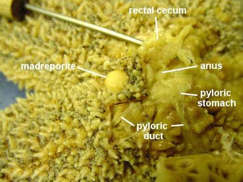
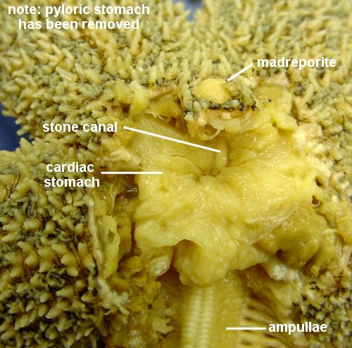
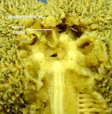
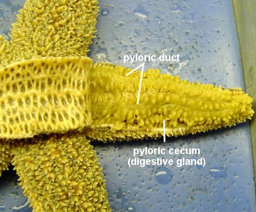
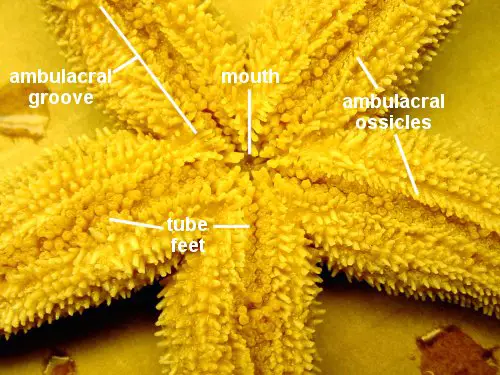
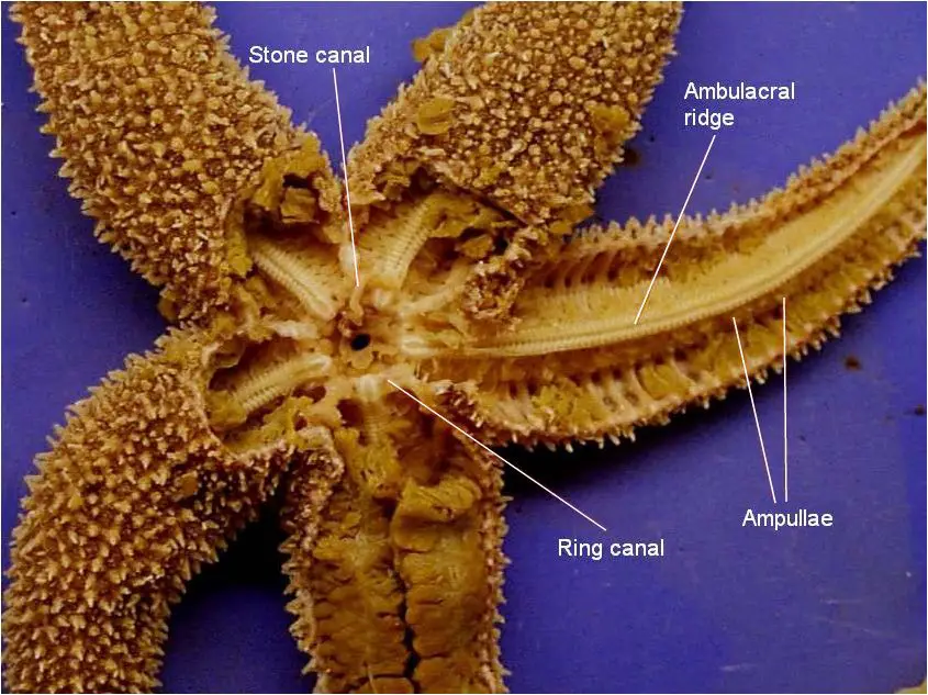
 Sea stars (group name Stelleroidea) are sometimes called starfish, though they are not real fish (they lack both vertebrae and fins). There are two sub-types of sea stars: