
| ENZYME RATE OF REACTION FOR CATALASE |  |
Introduction:
Chemical reactions make life possible. Hundred of chemical reactions are involved in the process of digesting a candy bar. If these reactions proceeded too slowly, not only would the candy bar remain in the stomach for long time, but the ordinary activities of life would come to a halt as well. Since this is not the case, something in the body must be responsible for speeding up the process. Four things that can speed up chemical reactions are: (1) heat; (2) increasing the concentration of reactants; (3) decreasing the concentration of products; and (4) enzymes, which speed up reaction without themselves being used up.
Enzymes are important in regulating chemical pathways, synthesizing materials needed by cells, releasing energy, and transferring information. Enzymes are involved in digestion, respiration, reproduction, vision, movement, thought, and even in the production of other enzymes. With few exceptions, enzymes are proteins. Simple cells may have as many as 2000 different enzymes, each one catalyzing a different reaction. An enzyme may accelerate a reaction by a factor of 1010 making it happen 10,000,000,000 times faster. Thus, a reaction that might take place as long as 1500 years without an enzyme can be completed in just 5 seconds with an enzyme.
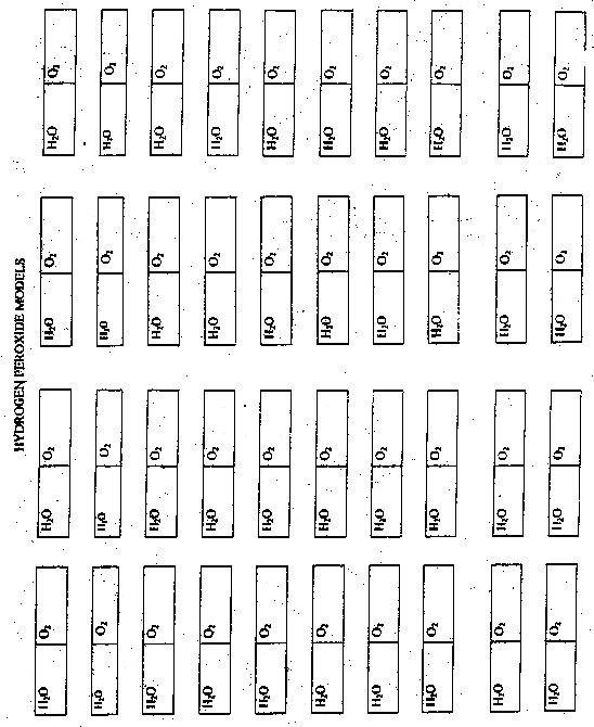
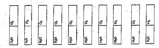
In this lab, your hands are the enzyme Catalase. This enzyme will split H2O2 (a poisonous waste product made by cells) into H20 and O2. You will split the molecule by rippling the paper model down the middle.
Pre Lab Questions:
1. What is an enzyme? What is its functions in living things?
2. What are things that can affect the function of an enzyme?
3 Write the chemical equation for the breakdown of hydrogen peroxide by the enzyme catalase.
4 An enzyme’s efficiency increases with greater substrate concentration, but only up to a point. Why?
Procedure:
1 Cut out 100 hydrogen peroxide molecules from the paper template.
2. Place 100 paper hydrogen peroxide molecules into a paper bag.
3. One member of your group will do the following:
a) When told to, you will grab one hydrogen peroxide molecule and rip it down the middle. Only rip one hydrogen peroxide molecule at a time.
b) Place the pieces back into the paper bag and grab another hydrogen peroxide molecule.
e) Repeat steps a and b, as fast as you can for 10 seconds. A member of group will be timing you for 10 seconds.
d) Empty your container and count the number of ripped hydrogen peroxide molecules.
e) Record the data in the following table.
| Time in seconds | Ripped Hydrogen Peroxide Molecules | Rate of Reaction |
| 0-10 | A | A |
| 10-30 | A | A |
| 30-60 | A | A |
| 60-120 | A | A |
| 120-180 | A | A |
4. Repeat step a – step e for 30, 60, 120, 180 seconds.
5. Graph the results.
6. Determine the rate of reaction for the following times.
The rate of reaction can be calculated by using the following equation:
| Rate = | M2-M1 |
| t2– t1 |
a. 0-10 seconds
b. 10-30 seconds
c. 30-60seconds
d. 60-120 seconds
e. 120-180 seconds
7. Record the above rates in a data table.
8. Graph the results.
Graph Title: ______________________________________________

Post Lab Questions:
1. If you were allowed to continue this lab and rip hydrogen peroxide molecules for 240 and 300 seconds. What would happen to the rate of reaction and why will this happen?
2. What can you say about the length of time and the rate of the reaction?
3. What would happen to the rate of reaction if you remove the H2O and O2 molecules as soon as they are produced?
Paper Molecules:.

The Hardy-Weinberg formulas allow scientists to determine whether evolution has occurred. Any changes in the gene frequencies in the population over time can be detected. The law essentially states that if no evolution is occurring, then an equilibrium of allele frequencies will remain in effect in each succeeding generation of sexually reproducing individuals. In order for equilibrium to remain in effect (i.e. that no evolution is occurring) then the following five conditions must be met:
Obviously, the Hardy-Weinberg equilibrium cannot exist in real life. Some or all of these types of forces all act on living populations at various times and evolution at some level occurs in all living organisms. The Hardy-Weinberg formulas allow us to detect some allele frequencies that change from generation to generation, thus allowing a simplified method of determining that evolution is occurring. There are two formulas that must be memorized:
p = frequency of the dominant allele in the population
q = frequency of the recessive allele in the population
p2 = percentage of homozygous dominant individuals
q2 = percentage of homozygous recessive individuals
2pq = percentage of heterozygous individuals
Individuals that have aptitude for math find that working with the above formulas is ridiculously easy. However, for individuals who are unfamiliar with algebra, it takes some practice working problems before you get the hang of it. Below I have provided a series of practice problems that you may wish to try out. Note that I have rounded off some of the numbers in some problems to the second decimal place.
PROBLEM #1 You have sampled a population in which you know that the percentage of the homozygous recessive genotype (aa) is 36%. Using that 36%, calculate the following:
PROBLEM #2. Sickle-cell anemia is an interesting genetic disease. Normal homozygous individuals (SS) have normal blood cells that are easily infected with the malarial parasite. Thus, many of these individuals become very ill from the parasite and many die. Individuals homozygous for the sickle-cell trait (ss) have red blood cells that readily collapse when deoxygenated. Although malaria cannot grow in these red blood cells, individuals often die because of the genetic defect. However, individuals with the heterozygous condition (Ss) have some sickling of red blood cells, but generally not enough to cause mortality. In addition, malaria cannot survive well within these “partially defective” red blood cells. Thus, heterozygotes tend to survive better than either of the homozygous conditions. If 9% of an African population is born with a severe form of sickle-cell anemia (ss), what percentage of the population will be more resistant to malaria because they are heterozygous (Ss) for the sickle-cell gene?
PROBLEM #3. There are 100 students in a class. Ninety-six did well in the course whereas four blew it totally and received a grade of F. Sorry. In the highly unlikely event that these traits are genetic rather than environmental, if these traits involve dominant and recessive alleles, and if the four (4%) represent the frequency of the homozygous recessive condition, please calculate the following:
PROBLEM #4. Within a population of butterflies, the color brown (B) is dominant over the color white (b). And, 40% of all butterflies are white. Given this simple information, which is something that is very likely to be on an exam, calculate the following:
PROBLEM #5. A rather large population of Biology instructors have 396 red-sided individuals and 557 tan-sided individuals. Assume that red is totally recessive. Please calculate the following:
PROBLEM #6. A very large population of randomly-mating laboratory mice contains 35% white mice. White coloring is caused by the double recessive genotype, “aa”. Calculate allelic and genotypic frequencies for this population.
PROBLEM #7. After graduation, you and 19 of your closest friends (lets say 10 males and 10 females) charter a plane to go on a round-the-world tour. Unfortunately, you all crash land (safely) on a deserted island. No one finds you and you start a new population totally isolated from the rest of the world. Two of your friends carry (i.e. are heterozygous for) the recessive cystic fibrosis allele (c). Assuming that the frequency of this allele does not change as the population grows, what will be the incidence of cystic fibrosis on your island?
PROBLEM #8. You sample 1,000 individuals from a large population for the MN blood group, which can easily be measured since co-dominance is involved (i.e., you can detect the heterozygotes). They are typed accordingly:
| BLOOD TYPE | GENOTYPE | NUMBER OF INDIVIDUALS | RESULTING FREQUENCY |
|---|---|---|---|
| M | MM | 490 | 0.49 |
| MN | MN | 420 | 0.42 |
| N | NN | 90 | 0.09 |
Using the data provide above, calculate the following:
PROBLEM #9. Cystic fibrosis is a recessive condition that affects about 1 in 2,500 babies in the Caucasian population of the United States. Please calculate the following:
PROBLEM #10. In a given population, only the “A” and “B” alleles are present in the ABO system; there are no individuals with type “O” blood or with O alleles in this particular population. If 200 people have type A blood, 75 have type AB blood, and 25 have type B blood, what are the allelic frequencies of this population (i.e., what are p and q)?
PROBLEM #11. The ability to taste PTC is due to a single dominate allele “T”. You sampled 215 individuals in biology, and determined that 150 could detect the bitter taste of PTC and 65 could not. Calculate all of the potential frequencies.
| Heart Dissection |
Introduction
Mammals have four-chambered hearts and double circulation. The heart of a bird or mammal has two atria and two completely separated ventricles. The double-loop circulation is similar to amphibians and reptiles, but the oxygen-rich blood is completely separated from oxygen-poor blood. The left side of the heart handles only oxygenated blood, and the right side receives and pumps only deoxygenated blood. With no mixing of the two kinds of blood, and with a double circulation that restores pressure after blood has passed through the lung capillaries, delivery of oxygen to all parts of the body for cellular respiration is enhanced. As endotherms, which use heat released from metabolism to warm the body, mammals require more oxygen per gram of body weight than other vertebrates of equal size. Birds and mammals descended from different reptilian ancestors, and their four-chambered hearts evolved independently – an example of convergent evolution.
Objective
Using a pig heart, students will observe the major chambers, valves, and vessels of the heart and be able to describe the circulation of blood through the heart to the lungs and back and out to the rest of the body. (The pig heart is used because it is very similar to the human heart in structure, size, & function.)
Materials
Dissecting pan, dissecting kit, safety glasses, lab apron, pig heart, & gloves
Procedure – External Structure

Front or Ventral Side of the Heart

Procedure – Internal Anatomy:
Tricuspid Valve
When you have finished dissecting the heart, dispose of the heart as your teacher advises and clean, dry, and return all dissecting equipment to the lab cart. Wash your hands thoroughly with soap.
| BACK |
Name___________________________ Date__________ Period___________
Heart Dissection
1. Why are pig hearts used to study the anatomy of the human heart?
2. How can you tell which side of the heart is the ventral surface?
3. How many chambers are found in the mammalian heart? What other group of organisms would have this same number of chambers?
4. What is the advantage in having this number of chambers compared to organisms with fewer number of chambers?
5. Which chambers are the pumping chambers of the heart?
6. Which chambers are the receiving chambers of the heart?
7. How do the walls of the atria compare with the walls of the ventricles and why are they different?
8. What is the purpose of heart valves?
9. Name & compare the heart valves found between the upper & lower chambers of the right and left sides of the heart.
10. Vessels that carry blood away from the heart are called __________, while __________ carry blood toward the heart.
11. Which artery is the largest and why?
12. What is the purpose of the coronary artery and what results if there is blockage in this vessel?
13. Use the diagram of the heart below to trace blood flow through the heart:
| BACK |
