
Use this pig heart dissection guide to identify chambers, valves, and major vessels and trace blood through the pulmonary and systemic circuits. The flow diagram supports the specimen observations; it shows connections rather than the physical arrangement of an opened heart.
Student resource: Heart Dissection Questions. Question 13 refers to a heart diagram that is missing from that page; use the flow schematic here as its reference before assigning it. Pig and human hearts share the four-chambered mammalian plan, but their vessel branching and orientation are not identical.
Learning goals and materials
- Identify the right and left atria and ventricles using connections and wall structure.
- Trace blood through all four valves, the lungs, and the body.
- Explain how wall thickness and valves relate to the work of each chamber.
Prerequisites: tissues, diffusion and gas exchange, and the difference between a vessel and a chamber. Materials: commercially supplied preserved pig heart, tray, blunt probe, forceps, approved scissors, PPE, and an observation sheet. The teacher should inspect the specimen first: short or missing vessel stumps can make identification difficult.
Labeled pig heart blood-flow diagram
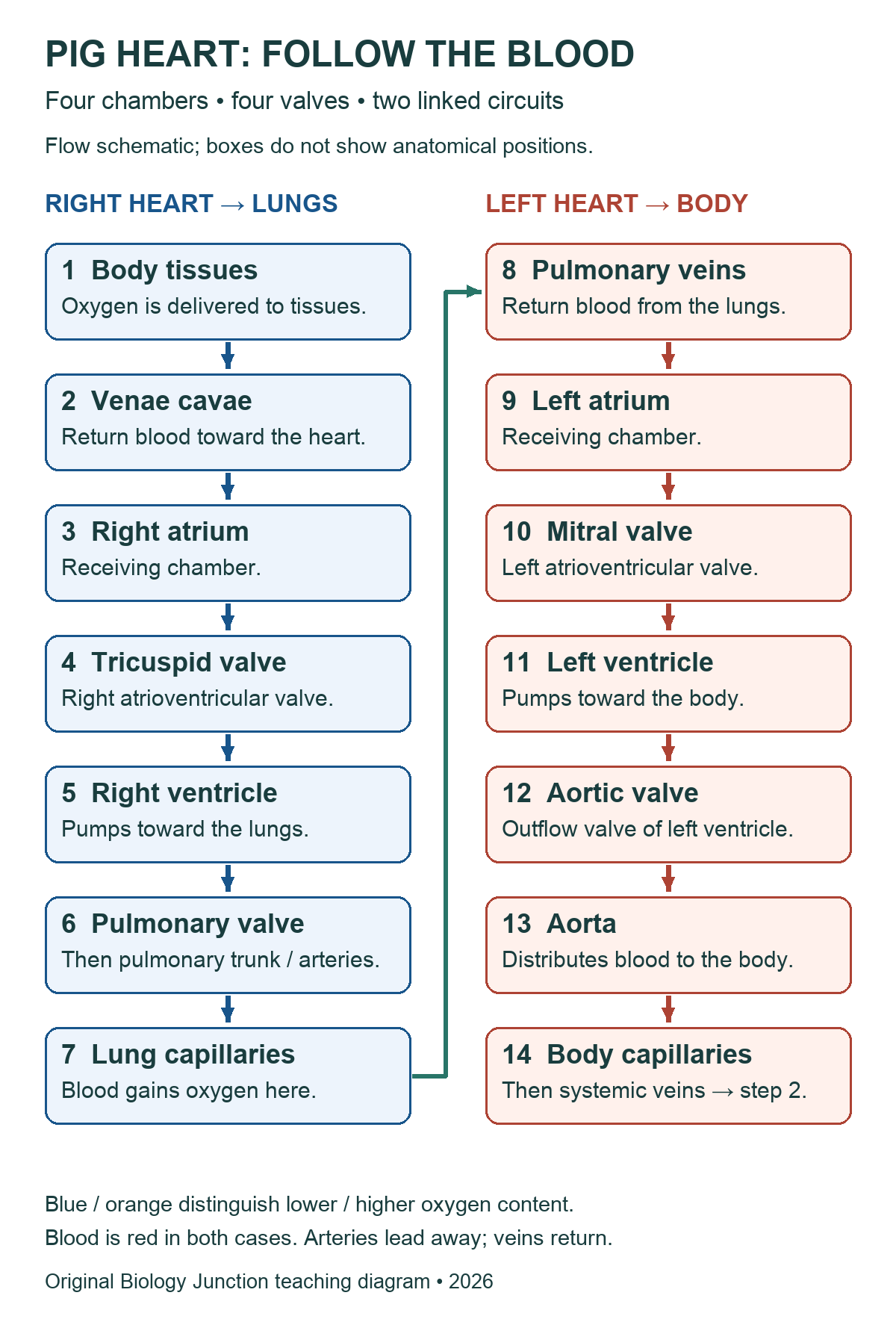
Arteries carry blood away from the heart; veins carry blood toward it. In the adult pulmonary circuit, arteries carry blood with less oxygen and veins return blood with more oxygen. Blood remains red in both cases; diagram colors are teaching conventions.
Teacher preparation and safety
Use commercially supplied preserved specimens in a supervised school lab. Before class, review the specimen label, supplier’s safety data sheet (SDS), school risk assessment, ventilation requirements, and storage and disposal procedures. Demonstrate handling and cutting with the actual tools students will use. Require appropriate splash goggles, preservative-compatible gloves, and protective clothing. Keep hands behind the cutting edge and work in a tray. No food or drink.
Rinse or drain a specimen only if the product instructions and school procedure permit it; do not assume preservative belongs in a sink. Report a cut, splash, or spill promptly and follow the lab’s response procedure. Collect tissue, liquids, and sharps as directed, clean the station, and wash hands. A diagram, model, or teacher demonstration can support an alternative activity.
Review the biology lab safety expectations, NSTA’s dissection safety guidance, and the supplier’s specimen FAQs. These general notes do not replace the product-specific procedure.
Orient a preserved heart before cutting
The broad base contains the major vessel attachments; the apex is the pointed end. In an anterior view, the specimen’s anatomical right is on the viewer’s left. Rotating the specimen changes that screen relationship, so confirm sides by tracing vessels and comparing walls rather than memorizing a left-right picture.
The pulmonary trunk leaves the right ventricle and divides toward the lungs. The aorta leaves the left ventricle. Thin-walled veins may be collapsed or trimmed. An auricle is an appendage of an atrium, not a fifth chamber. Locate surface coronary vessels and the interventricular groove without assuming every visible opening is intact.
The University of Washington pig-heart photographs provide external and internal comparison views. Those photographs are linked for reference, not reproduced here.
Teacher-led observation sequence
- Record the intact specimen: mark base, apex, and the proposed anterior surface. Note missing or damaged tissue.
- Identify external attachments: follow the teacher’s demonstration to distinguish aorta, pulmonary trunk, venae cavae, and pulmonary veins where present. Use a blunt probe gently; never force it through a valve.
- Confirm the right-side route: trace a vena cava toward the right atrium and the pulmonary outflow toward the right ventricle. Surface position alone is not enough.
- Open under supervision: the teacher chooses cuts that expose each chamber while preserving its valves, septum, and chordae. Lift and cut the chamber wall with approved scissors; do not follow a generic incision line through unidentified tissue.
- Inspect atrioventricular valves: locate tricuspid and mitral leaflets, chordae tendineae, and papillary muscles. Distinguish supporting cords from muscular ridges in the wall.
- Compare the ventricles: compare wall thickness at similar locations, excluding the septum and papillary muscles from the measurement. The left ventricle generally has the thicker wall because it develops the pressure needed for systemic circulation.
- Inspect outflow valves: locate pulmonary and aortic semilunar valves at ventricular outlets. If assigned, examine coronary artery openings near the aortic root. Stop if attachments or cut surfaces are unclear.
- Explain and finish: trace a complete blood route on the diagram, mark what was actually observed, and complete the class questions. Follow the approved cleanup, storage, and disposal procedure and wash hands.
Chambers, valves, and supporting structures
| Structure | Location or role |
|---|---|
| Right atrium → right ventricle | Blood passes through the tricuspid atrioventricular valve. |
| Right ventricle → pulmonary trunk | Blood leaves through the pulmonary semilunar valve and travels toward the lungs. |
| Left atrium → left ventricle | Blood passes through the mitral, or bicuspid, atrioventricular valve. |
| Left ventricle → aorta | Blood leaves through the aortic semilunar valve and enters systemic circulation. |
| Chordae tendineae and papillary muscles | Support atrioventricular leaflets against prolapse during ventricular contraction. They do not pull the valves open. Semilunar valves do not have chordae. |
| Interventricular septum | The muscular partition between the ventricles; distinguish it from an outer ventricular wall. |
| Myocardium and endocardium | Myocardium is cardiac muscle; endocardium lines internal surfaces. The pericardial sac surrounds the heart and is not the ventricular chamber wall. |
| Coronary arteries and coronary sinus | Coronary arteries supply myocardium and originate near the aortic root. The coronary sinus is a venous channel draining into the right atrium; it is not a hole in the aorta. |
Valve opening and closing follow pressure differences. During ventricular systole, atrioventricular valves close; outflow valves open when ventricular pressure exceeds arterial pressure. During ventricular relaxation, outflow valves close and filling resumes when pressure conditions allow. “Systole” means contraction and “diastole” relaxation; specify whether discussing atria or ventricles.
Existing valve photographs

Teaching notes and original checks
- Use atrium for one receiving chamber and atria for two. The right ventricle is a lower pumping chamber, not an upper chamber.
- Pulmonary veins enter the left atrium; the two semilunar valves are at the ventricular outlets, not the pulmonary-vein entrances.
- Blood entering the aorta goes into systemic circulation, not out of the body. Coronary circulation supplies the heart muscle itself.
- A preserved specimen cannot demonstrate pressure-driven valve motion or a heartbeat. Distinguish an observed leaflet from an explanation of its function.
Check 1: A student calls a pulmonary artery a vein because its blood contains less oxygen. What decides the name? Direction relative to the heart: the artery carries blood away from the right ventricle.
Check 2: Why compare ventricular walls at similar locations? The septum, papillary muscles, and angled cuts can distort a thickness comparison. Record the sampling location and specimen condition.
Check 3: Would a coronary artery opening in the aortic root be the coronary sinus? No. An artery takes blood to myocardium; the coronary sinus returns venous blood to the right atrium.
Related resources and sources
- Animal Dissections directory — return to the existing collection.
- Heart Dissection Questions — existing question set; the checks here are not a complete verified key for it.
- Frog Dissection — compare an adult frog heart with the mammalian plan, accounting for the different circulatory organization.
- OpenStax Biology 2e: mammalian heart and vessels.
- OpenStax Anatomy and Physiology 2e: heart anatomy and coronary circulation.
- University of Washington: pig-heart specimen views.