Your Guide To Your First Earthworm Dissection

META: follow our handy guide to dissecting your first earthworm and learn some interesting things about them too.

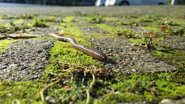
Earthworms play essential roles in many ecosystems. They help introduce oxygen to the soil and mix it up. As they tunnel through the ground, they enrich the soil and push it toward the surface where it’s easier for plants to get to the nutrients. You can see the organs that help these worms do their jobs by dissecting an earthworm.

Safety First

Safety is critical in all aspects of our lives. It may seem trivial in a controlled environment like a school biology lab, but it’s not, and all safety rules should be followed. They are in place to protect you and your classmates, so don’t skip any regulations just because you think it will be ok or those rules don’t seem to apply to your circumstances. The basic common-sense rules are:

  • Wear safety gear when necessary like goggles, gloves, and aprons.
  • Most preserved specimens contain formaldehyde, so wash them first.
  • Do not play with lab equipment or instruments such as scalpels and scissors.
  • Do not eat any parts of your specimen. Yes, there is an apparent reason for this rule.
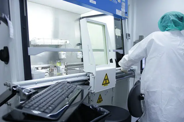
laboratory safety measure

image via Pixabay

Your lab should have the rules and safety measures available plus your instructor will go over them with you. Don’t assume the only rules are the ones we list here. The type of lab and type of specimen determine the rules. Ask for a copy of the rules if you don’t see one posted in the lab. Your teacher should be close by most of the time to help you guide you as well.

Always wear safety goggles and gloves. If you have to carry a sharp instrument, hold it with the pointed end pointing down and away from your body. Don’t rush or run while holding a scalpel or scissors. Never carry a knife or scissors by any part other than the handle. Scalpels are razor sharp, and it only takes a split second for them to cut you open.

Keep your station clean and tend to any spills immediately unless they pose a breathing hazard. Dispose of any blades, gloves, aprons, and specimens according to the established rules in your lab. Your teacher will probably explain all the rules to you, but don’t wait to ask if you aren’t sure what to do. Teachers are there to help educate you and keep you safe.

Earthworm Dissection Guide

Earthworms are great for helping you understand simple organisms and basic anatomy. They’ll help you get a grasp on lab safety before you progress to larger specimens like pigs or frogs. As a bonus, they’re small and soft, so handling them is much more comfortable as well.

The first step is to examine the exterior of the earthworm. Earthworms are segmented works, so they look like a long stack of small rings. They don’t have a head or any limbs, but they do have a fascinating exterior anatomy to study. The anterior end of the earthworm is a little fatter than the posterior. When you locate the anterior end of the work, pin it to the dissecting pan or tray.
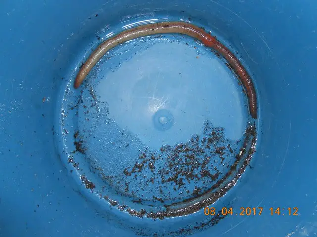
earthworm in laboratory

image via Flickr

Earthworms are annelids which means their bodies are composed of multiple ring-like sections or segments. This part may not be on your teacher’s list, but it’s always interesting to count the segments while you study the exterior anatomy of the earthworm. While you count, notice the small setae on the ventral surface. These little bristles help the worms move through the dirt with ease.

Each segment along the worm’s exterior has small pores. These pores excrete the sticky film you find when you run your finger along a live worm. You may need a magnifying glass or small microscope to see them. It depends on the size of your earthworm specimen and your eyesight as well.

From the anterior end of the worm, count your way down to segment fourteen. Typically, this is where the oviducts are located. The oviducts release the eggs when the worm reproduces. The exciting part is the next segment after the oviducts; it contains the sperm ducts. Earthworms have both male and female reproductive organs.

Further down the worm at segment 31 is the clitellum. It secretes a sticky mucus that binds two earthworms together while the mate. It develops a cocoon to hold the eggs and sperm after mating is finished. Earthworms are simple worms, but fantastic at the same time. Their exterior anatomy is fascinating to study.
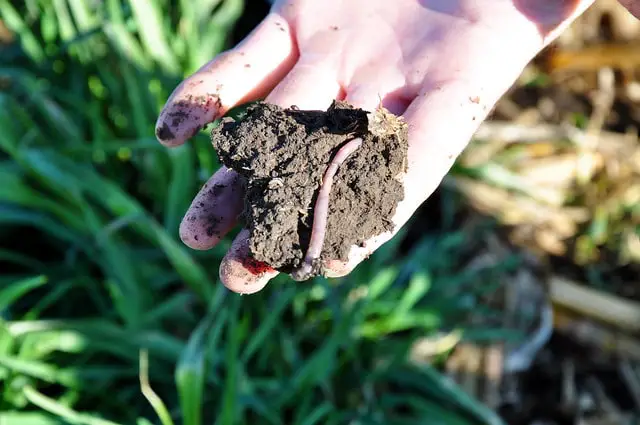
person holding earthwork in hand with soil

image via Flickr

Earthworms are hermaphroditic which means they have both female and male reproductive organs. Eggs come from the ovaries inside segment fourteen, sometimes thirteen. It can be hard to count the segments on small worms. Worms have testes which can form in segments near the oviducts. Study these segments and see if you can find the reproductive organs on your specimen.

When worms mate, they get stuck together briefly to help keep the reproductive organs aligned. Sperm from both worms travels into the other worms seminal receptacle. The clitellum creates the cocoon which moves along the outside of the worm to collect the semen and the eggs. The eggs are fertilized outside the worm in the cocoon.

By now, you should have a good understanding of the exterior anatomy of your earthworm specimen. Remove the pin from the anterior end of the earthworm and place it on its ventral side, then put the pin back in the anterior end of the worm. The ventral side of the worm is a little flatter than the dorsal side, and it may be a lighter color.

Carefully and slowly make a shallow incision using your scalpel from the anterior end of the work to the clitellum. Never cut toward your body or fingers. Be extra careful and keep the incision shallow, so you don’t cut into the worm’s digestive system and internal organs. Use your forceps to spread the worm open and pin the sides of its body to your dissection pan or tray.
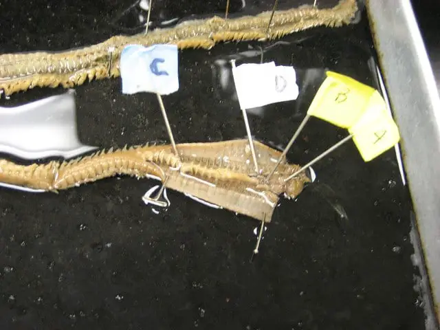
close up photo of earthworm dissection

image via Flickr

The inside of the worm should be exposed now. You may want to lightly sprinkle water over the worm to keep it from drying out while you study the inside of it. The interior part of the walls is called the septa. See if you can tell the difference. If possible, ask your teacher to point them out and help you see the different layers.

Now, the internal digestive organs should be exposed and available for study. Starting with the mount on the anterior end of the worm, locate the organs. The first organ you see is the pharynx. The worm’s esophagus protrudes from the pharynx. About halfway down your incision are the crop and gizzard. Skip the other organs for now and find those two.

The crop is essentially a stomach. It stores food until the food is moved to the gizzard which grinds it up. The food leaves the gizzard and goes into the intestine, much like it does in humans, and travels to the anus. Along the way, the worm’s intestines absorb nutrients from the food the gizzard crushed and ground up. Earthworms don’t eat dirt. The consume organic materials found in the soil.

Make your way back up to the crop. If you look above the crop on the anterior side, you’ll find five pairs of aortic arches. This is the worm’s version of a heart. The hearts are located around the esophagus, and they connect to the dorsal blood vessel. That’s the worm’s version of an artery. Most earthworms can take direct damage to half their aortic arches and live.

Move your attention back to the pharynx at the anterior end of the worm. Locate the cerebral ganglia beneath the pharynx on the dorsal side. You may need to use your forceps to move some organs around to get a good look at it. The ventral nerve starts at the cerebral ganglia and runs the length of the worm. It may be hard to see if it is too small.

They are simple creatures speaking purely on their anatomy, but how their bodies and mating works are truly amazing. If you have time, go back over this tutorial again and study the worm longer. When you finish exploring, make sure you clean your workstation and dispose of your specimen correctly. Dispose of your lab gear according to the lab rules. Wash your hand thoroughly with soap and water.

Some Final Notes

Earthworms are vital to the health of our soil. The improve drainage, help stabilize the land, and add nutrients to the ground. Worms feed on organic materials they find in the dirt. Their bodies use the nutrients they need and deposit what’s left back into the soil as waste. Fortunately for plants, that waste is usually nitrogen-rich along with other nutrients plants need to grow.

Their worm tunnels help loosen the soil which aids plants in root development. We could go on and on about the benefits of earthworms. If you follow our guide to dissecting earthworms and read our interesting facts along the way, we’re sure you’ll be able to dissect an earthworm specimen safely. You may even appreciate these simple creatures a little more when you are done.

Questions To Study For A Brain Anatomy Quiz In AP Biology

Questions To Study For A Brain Anatomy Quiz In AP Biology

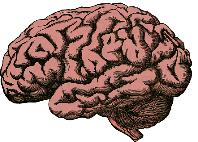
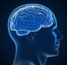
human brain

image via: pxhere.com

Taking AP Biology? Have a brain anatomy quiz coming soon? We’ve got 17 questions to help you study for it, plus some clever tricks and tips for studying smarter, not harder!

Parts Of The Brain

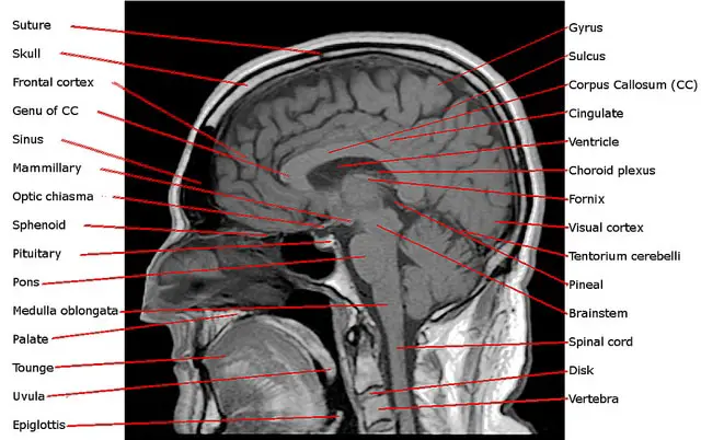
parts of human brain

image via: pixabay.com

One of the first things you should have to ace a brain anatomy quiz is a thorough grasp of the parts of the brain and each part’s function. Here are some of the questions you might expect:

1. Where Is The Cerebellum Located And What Does It Do?

human intelligence

image via: pixabay.com

The cerebellum is the part of the brain situated at the back of the head. It receives sensory information and regulates your motor movements. The cerebellum also controls balance and coordination, helping you to enjoy smooth movements.

2. Which Part Of The Brain Processes Visual Information?

The occipital lobe lies underneath the occipital bone. It is part of the forebrain (you have two, technically; one at the back of each cortex) and is responsible for processing visual information. Here’s a helpful memory device: the “o” in occipital can remind you of the “o” in optometrist or ophthalmologist.

3. If A Person’s Frontal Lobe Is Injured, What Functions Might He Or She Lose?

person in front of paper works

image via: pixabay.com

The frontal lobe can be found in the front of the brain, in each cerebral hemisphere. A deep groove called the central sulcus separates it from the parietal lobe, and another groove called the lateral sulcus separates it from the temporal lobe. A part of the frontal lobe known as the precentral gyrus contains the primary motor cortex, which controls specific body parts’ voluntary movements.

The frontal lobe is responsible for reasoning, higher order thinking, and creativity, so if somebody’s frontal lobe is damaged, he or she could have difficulty making decisions and reasoning.

4. What Are The Gyrus And Sulcus And How Do They Help The Brain?

Gyrus are the ridges on the brain and sulcus are the grooves (also seen as furrows or depressions). Together, their up and down “motion” are responsible for the folded, “spaghetti” appearance of the brain.

They are, in fact, an extremely clever way of making the most of very limited space. The brain is limited to the area inside your cranium, but the folding of the brain tissue allows a much greater surface area for cortical tissue, allowing additional cognitive function even in a relatively small space.

The human brain begins as a smooth surface, but as the embryo develops, the brain begins to form the deep indentations and ridges we see in the adult brain.

5. What Part Of The Brain Controls The Primitive Parts Of Our Body?

human body with light bulb head

image via: pixabay.com 

Pons is the Latin word for bridge, and that’s exactly what the pons appears to do in the brain, as its physically connected to the brainstem. Like any good bridge, the pons contains neural pathways to move signals to the medulla, cerebellum, and thalamus.

Many of the nuclei contained inside the pons are responsible for relaying signals, as we’ve already described, but other nuclei play roles in primitive functions that we don’t normally consider being within our control, such as respiration, sleep, bladder control, and others.

6. What Is The Corpus Callosum?

The corpus callosum sits underneath the cerebral cortex. It’s about 10cm long and is a thick, tough bundle of fibers that connects the cerebral hemispheres (right and left), enabling them to communicate with each other.

It has over 200 million axonal projections, making it the largest white matter structure.

7. Which Part Of The Brain Is The Newest From An Evolutionary Perspective?

orange and black question marks on a black ground

image via: pixabay.com

The cerebrum is the part of the brain that is outermost. In it, the brain can store memories, call upon senses, and establish self-awareness. High order functioning can also take place here and its known for being larger in musicians and left-handed individuals. It is also considered to be the most recent brain development.

8. How Many Lobes Is The Brain Comprised Of, And What Are Their Names And Functions?

Inside the brain is found the occipital lobe (see question #2), the frontal lobe (see question #3), the parietal lobe, and the temporal lobe. The parietal lobe sits behind the frontal lobe and above the temporal lobe. It is where the body becomes self-aware and plays an important role in language processing.

The temporal lobe plays a role in the processing of sensory input, helping the brain to translate these inputs into meaning. If, for example, you smell apple pie and think of your grandmother, you have your temporal lobe to thank!

9. Which Part Of Your Brain Acts Like A Supercomputer?

human brain as supercomputer

image via: pixabay.com

The thalamus is the small organ at the very center of your brain that acts as a supercomputer or switchboard, relaying signals throughout the brain. It is one of the most important parts of the brain and regulates motor signals, sleep, and consciousness.

Closely related to the thalamus is the hypothalamus, which sits just underneath the thalamus and regulates the pituitary gland and homeostasis.

10. Which Part Of The Brain Helps You Sneeze?

The medulla oblongata (medulla is Latin for “middle”), and the medulla oblongata is located on the brainstem close to the cerebellum. It is responsible for involuntary or autonomic processes, which include vomiting and sneezing. It also helps with breathing, cardiac functions such as heart rate, and blood pressure.

 The Central Nervous System

light bulb in a wooden surface

image via: pixabay.com

The central nervous system is another important subject likely to show up on a brain anatomy quiz. The questions below will help you better prepare.

11. What Is The Central Nervous System (CNS) Comprised Of?

The brain and the spinal cord make up the CNS, which is protected by the skull and the spine’s vertebral canal. It is the command center of the entire body, regulating all activity and processing all sensory inputs.

 12. What Role Does The Midbrain Play In The CNS?

smiling woman

image via: pixabay.com

The midbrain controls visual reflexes (including automatic eye movements, such as blinking and focusing). It also contains nuclei that link parts of the body’s motor system, including both cerebral hemispheres.

13. What Is A Neurotransmitter?

A neurotransmitter is a chemical that a nerve fiber releases when a nerve impulse arrives. It diffuses across the junction or synapse so that the impulse may pass to the next nerve fiber, muscle fiber, or other structure. Both neurotransmitters and inhibitory neurotransmitters are found in the brain.

14. What Is The Difference Between Dopamine And Serotonin?

person head with white question mark

image via: pixabay.com

Dopamine and serotonin are both powerful neurotransmitters. Serotonin impacts your sleep, arousal, hunger, and mood, while dopamine impacts your brain’s pleasure and reward system, your learning and attention, and movement.

15. What Is Glutamate And Why Is It Important?

Glutamate is the most abundant neurotransmitter found in the CNS; in fact, it accounts for more than 90% off all the synaptic connections in your brain! Some parts of the brain, including granule cells found in the cerebellum, rely on glutamate almost exclusively. Glutamate also plays a vital role in memory and learning.

16. Can You Name The Most Common Inhibitory Neurotransmitter In The Brain?

boy with different books

image via: pxhere.com

GABA (gamma-Aminobutyric acid) is the most common inhibitory neurotransmitter found in the brain. It is considered inhibitory because it helps to calm or reduce neuron excitability. This means it plays an important role in calming anxiety. It also is responsible for the regulation of muscle tone.

17. What Is The Neurotransmitter That Triggers Our Fight Or Flight Response?

The fight or flight response is also called the acute stress response or hyperarousal; it is a physiological reaction that occurs when the brain perceives an imminent threat. Epinephrine (also known as adrenaline) is the neurotransmitter most responsible for this response. It can signal an increase in blood flow to muscles and greater blood flow through the heart, among other things (this is why your heart starts to beat quickly when you’re afraid).

The Quick Guide To Studying Smarter

man imaginations

image via: maxpixel.net

If you’re reading this article, you’re already well on your way to preparing for your brain anatomy quiz, but here are a few more tips to help you get the most out of your time studying:

Get Lots of Rest

Sleeping instead of studying sounds counterintuitive, but without sleep, your brain will have a hard time committing what you’ve learned to memory. In fact, one of the best things you can do to prepare for a test or quiz is to get a good night’s sleep the night before!

Use Memory Devices

We’ve already hinted at a few tricks for helping your brain remember facts (did you notice them in the questions above?), but mnemonic devices and facts set to music help those boring facts stick much better than just rote memorization.

Setting the major parts of the brain to your favorite song, for example, can help pique your brain’s interest and increase emotional arousal, increasing your odds of remembering the information!

Finally, make it real. Drawing the brain, using models of the brain, or reading stories about people who have injured certain parts of the brain are all ways to make abstract concepts seem real–and make you more likely to remember them. Good luck!

Protein Synthesis Worksheet: Definition, Examples & Practice

Meta: Need to learn how protein synthesis works? We’ve got your complete guide to the process on our protein synthesis worksheet, including the difference between DNA and RNA, important misconceptions about mutations, and an explanation of the central dogma of biology. Plus, get practice exercises and quiz questions. 


What is Protein Synthesis?


Protein synthesis is the construction of proteins within living cells. The process consists of two parts; transcription and translation.

Proteins are an important organic compound that exists in every living organism. They are an essential part of the majority of cell functions. Specific proteins are needed for particular functions. Proteins are made up of long chains of amino acids which can be arranged in either a linear pattern or can be folded to form a more complex structure.

Proteins can be complex in structure and so are filtered into four categories – primary secondary, tertiary and quaternary.

Protein synthesis is a biological procedure which living cells perform to create new proteins. When studied in detail, the chemical synthesis of proteins process is extremely complex. The process begins with the production of new and different amino acids, some of which are collected from food sources.

The process requires ribonucleic acid (RNA), deoxyribonucleic acid (DNA), and a specific set of enzymes. All the different types of ribonucleic acids are needed for protein synthesis to work effectively. These are messenger ribonucleic acid (mRNA), transfer ribonucleic acid (tRNA), and ribosomal ribonucleic acid (rRNA).


Protein Synthesis: Definition, Examples, and Practice

Let’s check out a couple of important definitions to better understand protein synthesis.

Most protein synthesis worksheets will require a working understanding of the following definitions:

Central Dogma of Biology

A polypeptide encoded in a gene is expressed in a directional relationship called the central dogma of biology. It recognizes that information moves from the DNA to the RNA to the protein.

DNA

Deoxyribonucleic acid (otherwise known as DNA), is the carrier of genetic info found in almost every found living organism to date. It is present in the nucleus of cells and is self-replicating, meaning it’s integral to protein synthesis.

RNA

RNA is ribonucleic acid, and it’s present in every living cell discovered to date. It is a messenger and vitally involved in translating genetic code from DNA to the ribosomes so that amino acids can be created.

There are three kinds of RNA: messenger RNA (mRNA) transfers the genetic code from the DNA in the nucleus out to the ribosomes in the cytoplasm. Ribosomal RNA (rRNA) provides the structure for the ribosomes. Finally, transfer RNA (tRNA) works during translation to bring the amino acids to the ribosome so that a polypeptide (an amino acid chain) can be built.

Transcription

Transcription is the stage of manufacturing in which the DNA gene sequence is copied so that an RNA molecule can be made. We’ll explain more shortly.

Translation

The second stage of protein synthesis is translation. At this point in the process, a mRNA (messenger RNA) molecule is “read” and the information is used by the ribosome to build a polypeptide.

Polypeptide

A polypeptide is a chain made up of amino acids.

Codon

Three nucleotides form a codon. This codon is then used to create amino acids.

RNA vs. DNA

It’s tempting to confuse RNA with DNA, but they’re very different, and it’s important to understand these differences. They are both made up of nucleotides, which are the basic units of nucleic acids (like DNA and RNA). These nucleotides contain a phosphate group, a nitrogenous base, and a 5-carbon sugar ribose.

Instead of DNA’s ribose, however, RNA uses deoxyribose, a different kind of sugar. Also, RNA is most often a single strand, while DNA is famously double-stranded. Finally, DNA contains thymine, while RNA uses uracil instead.

Chromosomes

DNA is found by the meter inside even minuscule cells. During replication, the masses of coiled DNA called chromatin (shaped thanks to proteins called histones) organize into what are called chromosomes.

Different types of cells (eukaryotes) have chromosomes in varying amounts. Humans, as you probably know, have 46 chromosomes, while dogs, for example, have 78.

Transcription and Translation

To best understand your protein synthesis worksheet, let’s cover the complete protein synthesis process. It starts with transcription. Special enzymes in the nucleus arrive to gently pull apart the DNA code needed, and RNA begins to transcribe or rewrite the genetic material.

During translation, the mRNA connects with the ribosome and its information is decoded again so that the correct sequence of amino acids will connect to form a polypeptide. It’s important to note here that the ribosome doesn’t make protein nor does it make amino acids. It simply instructs already-made amino acids to form the correct sequence.

The amino acids’ sequence determines its protein’s shape, function, and properties and it can do so thanks to the RNA’s four bases (all of which are nucleotides): adenine (A), cytosine (C), guanine (G), and uracil (U). A codon, as we explained earlier, is a combination of three of these bases in a specific order: UUC, for example.

Some codons tell the ribosome to start or stop (UAA, UAG, and UGA indicate stop) and the rest indicate specific amino acids.

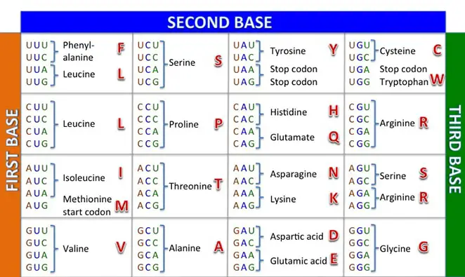
Understanding the Codon Table

codon table by cabal edu, protein synthesis worksheet

Image Source: sabal.uscb.edu

The heart of protein synthesis (and what you’ll most likely see on a protein synthesis worksheet) is the codon table. It helps us work through translation to understand the amino acids the mRNA is prescribing. For example, if you want to know what the codon CAA translates to, you’ll use the first letter of the codon (C) to locate the corresponding row on the left side of the chart.

Next, use the second letter of the codon (A) to identify the corresponding column on the top of the chart. The box indicated includes four codons that began with C and A; if you’d like, you can simply identify your codon there, or you can use the right side of the chart to identify the corresponding order of the third letter in the codon (A).

Either way, the single amino acid for CAA is Gln (glutamine).

Mutations

Mutations sound scary, but don’t worry–we’re not talking about superheroes with latent power and plans for world domination. Instead, we’re talking about what happens when there’s a mistake in the transcription or translation process.

Mutations come in three forms: silent, missense, and nonsense. A mutation that is silent means that the amino acid will not be impacted during translation. Missense mutations mean that the single amino acid has been changed and a nonsense mutation ends prematurely.

How are Mutations Caused?

There are several different reasons a mutation may occur. If at least one base is added to a DNA sequence, this is referred to as an insertion. A deletion, however, occurs when at least one base has been removed from the DNA sequence.

Similarly, when a change is made to the codons so that the reading frame of the sequence is changed, the resulting mutation is called a frameshift mutation. For example, a mRNA codon that reads AUG-AUA-CGG-AAU might experience an insertion of a T in the DNA sequence.

This frameshift mutation leads to a new codon: AUG-UAC-GGA-AU.

If we utilize the codon chart, we find that the polypeptide mutates from Met-Ile-Arg-Asn to Met-Tyr-Gly.

Common Misconceptions About Mutations

Something important to note is that sometimes the DNA sequence experiences an insertion or deletion of three nucleotides in a row. This doesn’t cause a frameshift mutation. Instead, it will just impact whether or not the deleted or inserted amino acids are added or not.

This can cause a dramatic change in the outcome of the polypeptide.

Another common misconception is that a mutation is always dramatic. While this is sometimes the case, mutations are common and provide the genetic variation we so appreciate in life. Many mutations have little to no impact on life, and some mutations even create good changes.

It’s a very limited number of mutations that survive to be problematic.


What Exactly Are Genes?


A gene is a short section of DNA that acts as an instruction manual for our bodies. DNA is found inside almost every cell in the body.

Genes contain the instructions that tell cells to create new proteins via protein synthesis. Every gene carries certain instructions which make up who you are such as eye color, height, and hair color. Genes come in many different types and versions for each feature. For example, one variant of a gene may contain instructions for blue eyes whereas another contains instructions for brown eyes. Genes are so small that there are around 20,000 inside each cell in the body. The entire sequence of your genes is named the genome.


How Do Genes Work?


Genes are responsible for telling each of your cells what to do and when to do it. They do this by making proteins. Why are proteins important? Well, our bodies are made up of proteins. Around 50% of a cell is some form of protein. Proteins are also responsible for many bodily functions such as digestion, immunity, circulation, motion, and communication between cells. These are made possible by the estimated 100,000 different proteins that are produced in the body.

Genes within your DNA don’t make proteins directly. Instead, enzymes read and copy the DNA code. The section of DNA that is to be copied gets unzipped by an enzyme which then uses that segment of DNA as a template to build a single-stranded molecule of ribonucleic acid. This ribonucleic acid then leaves the nucleus of the cell and enters the cytoplasm where ribosomes then translate the code to create the specific protein.

In certain genes, not all of the DNA sequence is used to make a protein. The section of DNA that is non-coding is known as introns. The coding sections of DNA are called exons.


The Structure of DNA


DNA is made up of pairs of nucleotides on a phosphate and sugar backbone. There are four different nucleotides: thymine, cytosine, guanine, and adenine. Each of the types of nucleotides only pairs with one other type. Hydrogen bonds connect to those nucleotide pairs. The sugar and phosphate backbone, along with the nucleotide pairs form a ladder-like structure that twists to form the double helix structure of DNA. Each side of this ladder shape is known as a strand of DNA.


Nucleotides consist of a base, a phosphate group, and five carbon atoms. Each of the different types of nucleotide has a base with a different structure, however, all the bases contain nitrogen. The four bases can be split into two groups. These are pyrimidine bases and purine bases. Pyrimidine bases are small and have one six-atom ring. Purine bases are larger and are made up of a six-atom ring plus a five-atom ring which are joined by two shared atoms. Thymine and cytosine are pyrimidine bases and adenine and guanine are purine bases.


Pyrimidine bases bond to purine bases because the shapes of these bases allow hydrogen bonds to form between them. The base pairing rules states that guanine pairs only with cytosine and adenine pairs only with thymine. This rule is known as complementary base pairing. Three hydrogen bonds form between a guanine and cytosine pair whereas only two hydrogen bonds form between an adenine and thymine base pair.


Protein Synthesis Worksheet Practice

It’s helpful to utilize practice protein synthesis worksheets. To help you, here’s a list of questions–and their answers–that you’re likely to find on tests, worksheets, and protein synthesis projects:

  1. During translation, which RNA carries amino acids to the ribosome? (transfer RNA or tRNA)
  2. Is DNA made with uracil or thymine? (thymine)
  3. In which part of the cell does transcription happen? (in the nucleus)
  4. Which RNA carries the genetic code to the ribosomes from the DNA? (messenger RNA or mRNA)
  5. What is the central dogma of biology? (DNA → RNA → protein)
  6. What are the building blocks of proteins? (amino acids)
  7. What are the three causes of mutations? (insertion, deletion, and frameshift)
  8. What is a codon? (three nucleotides)
  9. What are the three differences between DNA and RNA? (RNA uses deoxyribose instead of ribose, is single-stranded instead of double-stranded, and contains uracil instead of thymine)
  10. In what phase is tRNA molecules used? (translation)
  11. Does protein synthesis build protein? (no; protein synthesis builds amino acids)
  12. What are polypeptides? (chains of amino acids)
  13. What do codons do? (indicate the specific amino acid and in what order, and indicate when to stop and start the amino acid chain)
  14. Which leaves the nucleus: DNA or RNA? (RNA)
  15. What are the three kinds of mutations? (silent, missense, and nonsense)
  16. Which codons indicate stop? (refer to the codon chart for the answer; UAA, UAG, and UGA)
  17. What does chromatin organize into during replication? (chromosomes)

Practice with the Codon Chart

Another great way to increase your knowledge of protein synthesis and better prepare for protein synthesis worksheets is to practice with the codon chart. You can find the solutions in parenthesis after the example:

  1. CUU-CGU-AAU-UGG-AAG (leu-arg-asn-trp-lys)
  2. ACU-ACA-AGU-UGC-UUU (thr-thr-ser-cys-phe)
  3. AAC-AAG-GUC-GUC-AGG (asn-lys-val-ile-arg)

Protein synthesis is a complex, highly tuned process that enables life to flourish. Understanding it, from the DNA to the RNA to the amino acids, gives us a better appreciation for life itself. Use our protein synthesis worksheet practice questions to help you learn the ins and outs of protein synthesis and remember the informaion.

17 Questions To Study For A Brain Anatomy Quiz In AP Biology

Ready to ace your next brain anatomy quiz? We’ve got you covered! Review our 17 practice questions to improve your understanding of the parts of the brain and the central nervous system. Plus, brush up on our tips for studying smarter, not harder.

Taking AP Biology? Have a brain anatomy quiz coming soon? We’ve got 17 questions to help you study for it, plus some clever tricks and tips for studying smarter, not harder!

brain icon

Parts of the Brain

One of the first things you should have to ace a brain anatomy quiz is a thorough grasp of the parts of the brain and each part’s function. Here are some of the questions you might expect:

brain cartoon

1

Where is the cerebellum located and what does it do?

The cerebellum is the part of the brain situated at the back of the head. It receives sensory information and regulates your motor movements. The cerebellum also controls balance and coordination, helping you to enjoy smooth movements.

2

Which part of the brain processes visual information?

The occipital lobe lies underneath the occipital bone. It is part of the forebrain (you have two, technically; one at the back of each cortex) and is responsible for processing visual information. Here’s a helpful memory device: the “o” in occipital can remind you of the “o” in optometrist or ophthalmologist.

3

If a person’s frontal lobe is injured, what functions might he or she lose?

The frontal lobe can be found in the front of the brain, in each cerebral hemisphere. A deep groove called the central sulcus separates it from the parietal lobe, and another groove called the lateral sulcus separates it from the temporal lobe. A part of the frontal lobe known as the precentral gyrus contains the primary motor cortex, which controls specific body parts’ voluntary movements.

The frontal lobe is responsible for reasoning, higher order thinking, and creativity, so if somebody’s frontal lobe is damaged, he or she could have difficulty making decisions and reasoning.

4

What are the gyrus and sulcus and how do they help the brain?

Gyrus are the ridges on the brain and sulcus are the grooves (also seen as furrows or depressions). Together, their up and down “motion” are responsible for the folded, “spaghetti” appearance of the brain.

They are, in fact, an extremely clever way of making the most of very limited space. The brain is limited to the area inside your cranium, but the folding of the brain tissue allows a much greater surface area for cortical tissue, allowing additional cognitive function even in a relatively small space.

The human brain begins as a smooth surface, but as the embryo develops, the brain begins to form the deep indentations and ridges we see in the adult brain.

5

What part of the brain controls the primitive parts of our body?

brain illustration

Pons is the Latin word for bridge, and that’s exactly what the pons appears to do in the brain, as its physically connected to the brainstem. Like any good bridge, the pons contains neural pathways to move signals to the medulla, cerebellum, and thalamus.

Many of the nuclei contained inside the pons are responsible for relaying signals, as we’ve already described, but other nuclei play roles in primitive functions that we don’t normally consider being within our control, such as respiration, sleep, bladder control, and others.

6

What is the corpus callosum?

The corpus callosum sits underneath the cerebral cortex. It’s about 10cm long and is a thick, tough bundle of fibers that connects the cerebral hemispheres (right and left), enabling them to communicate with each other.

It has over 200 million axonal projections, making it the largest white matter structure.

7

Which part of the brain is the newest from an evolutionary perspective?

The cerebrum is the part of the brain that is outermost. In it, the brain can store memories, call upon senses, and establish self-awareness. High order functioning can also take place here and its known for being larger in musicians and left-handed individuals. It is also considered to be the most recent brain development.

parts of the brain

8

How many lobes is the brain comprised of, and what are their names and functions?

Inside the brain is found the occipital lobe (see question #2), the frontal lobe (see question #3), the parietal lobe, and the temporal lobe. The parietal lobe sits behind the frontal lobe and above the temporal lobe. It is where the body becomes self-aware and plays an important role in language processing.

The temporal lobe plays a role in the processing of sensory input, helping the brain to translate these inputs into meaning. If, for example, you smell apple pie and think of your grandmother, you have your temporal lobe to thank!

9

Which part of your brain acts like a supercomputer?

The thalamus is the small organ at the very center of your brain that acts as a supercomputer or switchboard, relaying signals throughout the brain. It is one of the most important parts of the brain and regulates motor signals, sleep, and consciousness.

Closely related to the thalamus is the hypothalamus, which sits just underneath the thalamus and regulates the pituitary gland and homeostasis.

10

Which part of the brain helps you sneeze?

The medulla oblongata (medulla is Latin for “middle”), and the medulla oblongata is located on the brainstem close to the cerebellum. It is responsible for involuntary or autonomic processes, which include vomiting and sneezing. It also helps with breathing, cardiac functions such as heart rate, and blood pressure.

brain icon

The Central Nervous System

nervous system

The central nervous system is another important subject likely to show up on a brain anatomy quiz. The questions below will help you better prepare.

11

What is the central nervous system (CNS) comprised of?

The brain and the spinal cord make up the CNS, which is protected by the skull and the spine’s vertebral canal. It is the command center of the entire body, regulating all activity and processing all sensory inputs.

12

What role does the midbrain play in the CNS?

The midbrain controls visual reflexes (including automatic eye movements, such as blinking and focusing). It also contains nuclei that link parts of the body’s motor system, including both cerebral hemispheres.

13

What is a neurotransmitter?

A neurotransmitter is a chemical that a nerve fiber releases when a nerve impulse arrives. It diffuses across the junction or synapse so that the impulse may pass to the next nerve fiber, muscle fiber, or other structure. Both neurotransmitters and inhibitory neurotransmitters are found in the brain.

14

What is the difference between dopamine and serotonin?

Dopamine and serotonin are both powerful neurotransmitters. Serotonin impacts your sleep, arousal, hunger, and mood, while dopamine impacts your brain’s pleasure and reward system, your learning and attention, and movement.

15

What is glutamate and why is it important?

Glutamate is the most abundant neurotransmitter found in the CNS; in fact, it accounts for more than 90% off all the synaptic connections in your brain! Some parts of the brain, including granule cells found in the cerebellum, rely on glutamate almost exclusively. Glutamate also plays a vital role in memory and learning.

16

Can you name the most common inhibitory neurotransmitter in the brain?

GABA (gamma-Aminobutyric acid) is the most common inhibitory neurotransmitter found in the brain. It is considered inhibitory because it helps to calm or reduce neuron excitability. This means it plays an important role in calming anxiety. It also is responsible for the regulation of muscle tone.

17

What is the neurotransmitter that triggers our fight or flight response?

The fight or flight response is also called the acute stress response or hyperarousal; it is a physiological reaction that occurs when the brain perceives an imminent threat. Epinephrine (also known as adrenaline) is the neurotransmitter most responsible for this response. It can signal an increase in blood flow to muscles and greater blood flow through the heart, among other things (this is why your heart starts to beat quickly when you’re afraid).

brain icon

The Quick Guide to Studying Smarter

If you’re reading this article, you’re already well on your way to preparing for your brain anatomy quiz, but here are a few more tips to help you get the most out of your time studying:

Get Lots of Rest

Sleeping instead of studying sounds counterintuitive, but without sleep, your brain will have a hard time committing what you’ve learned to memory. In fact, one of the best things you can do to prepare for a test or quiz is to get a good night’s sleep the night before!

Use Memory Devices

plugging earphone on a device

We’ve already hinted at a few tricks for helping your brain remember facts (did you notice them in the questions above?), but mnemonic devices and facts set to music help those boring facts stick much better than just rote memorization.

Setting the major parts of the brain to your favorite song, for example, can help pique your brain’s interest and increase emotional arousal, increasing your odds of remembering the information!

Finally, make it real. Drawing the brain, using models of the brain, or reading stories about people who have injured certain parts of the brain are all ways to make abstract concepts seem real–and make you more likely to remember them. Good luck!

Sheep Heart Dissection Lab Report


Sheep Heart Dissection


In this investigation, the external and internal heart structure valves of a sheep’s heart organ were examined and identified by dissection. The heart is a muscular organ that pumps oxygenated blood and nutrients throughout the body. A sheep’s heart has four chambers like most mammals including humans. Two of those chambers are receiving chambers called the right and left atrium. The other two chambers are pumping chambers called the right and left ventricle. A sheep heart dissection can help to identify each of these different areas of the heart.

The efficiency in the cycle of blood depends on the sequential contraction of the atriums and ventricles. Whenever the atriums contract this is called the systolic phase and whenever the ventricles contract this is called the diastolic phase. These contractions ensure the regular flow of blood through the heart. The contractions occur one after another to make a heartbeat. The many heart valves such as the tricuspid and mitral heart valves control the flow of blood from each chamber.

Blood flow through the heart starts when the right atrium takes the blood that flows in through the superior or inferior vena cava. The right atrium then fills with blood and pressure causes the tricuspid valve to open. The blood then goes into the right ventricle where it contracts the blood into the pulmonary arteries. These arteries lead to the lungs where blood is then oxygenated. The oxygenated blood then flows from the lungs to the left atrium through the pulmonary veins. Due to pressure the mitral valve, which leads to the left ventricle, opens up and pushes the blood into the left ventricle. The left ventricle then contracts and forces the blood through the aorta, which provides the rest of the body with blood.


Objectives of a sheep heart dissection in a lab:

  • Describe the appearance of the external and internal structures of the animal’s heart organ
  • Name the structure and function of the animal’s heart organ
  • Understand the anatomy and physiology of a sheep’s heart

What is a Sheep Heart Dissection?


A sheep heart dissection involves cutting into particular areas of a sheep’s heart so that we can see each of the different sections and learn more about what each part of a heart looks and feels like. A sheep’s heart and sheep internal anatomy are very similar to a human, so it gives us an opportunity to learn more about what a human heart might look like on the inside.

By dissecting into a heart, we can see each different section in detail and can learn how each section helps in pumping blood around the body. Following is a full explanation and sheep heart dissection guide so that you can easily and safely complete a sheep heart dissection yourself in a lab setting.


How to do a Heart Dissection


Materials


The materials needed in this dissection include sheep’s heart, a dissecting tray, a blunt metal probe, a pair of scissors, a scalpel, and a pair of tweezers. The safety equipment needed for this dissection is safety goggles, lab aprons, and gloves. The procedure must be completed according to the safety elements of the lab manual.


Sheep Heart Dissection Guide


Most diagrams of a heart show the left atrium and ventricle on the right-hand side of the picture. This is to show the heart in a way as if it is facing you. If a human was facing you, then the left-hand side of their heart would be on your right, and this is how diagrams usually portray a heart.


Observing the External Anatomy and Areas of the Heart


  1. Start by identifying the left and right sides of the heart. If you look closely, you will see a diagonal line of blood vessels on one side which divide the heart. The half of the heart that includes all of the apex is the left side. This can be confirmed by gently squeezing each side of the heart. The left side of the heart will feel much firmer than the right. This is due to all of the muscles that are required to pump blood to the entire body. The right side of the heart is less firm and weaker as this side only pumps blood to the lungs.
  2. Place the heart down so that the right side is on your right. Take a moment to examine the darker flaps that are located at the top of the heart. These flaps are named auricles. There should be a large opening at the top of the heart right next to the auricles. This is the opening to the superior vena cava – the area which brings blood from the top half of the body to the right atrium. If you stick a probe into this opening, you should feel it go right through into the right atrium. Slightly down and to the left of the superior vena cava lies another opening. Inserting a probe into this opening will also lead to the inside of the right atrium. This other opening is the inferior vena cava, which brings blood up to the heart from the lower tissues. Another blood vessel will be visible next to the left auricle. This is the pulmonary vein, which brings blood up from the lungs and into the left atrium.
  3. Right in the center of the heart, you will see the largest blood vessel. This is the aorta, which is responsible for taking oxygenated blood from the left ventricle to the rest of the body. The aorta branches out when it leaves the heart into more than one artery so it may have more than one opening on the heart that you are examining. If you look closely at the openings, you will see that they are connected to each other.
  4. To the left side of the back of the aorta, you will find another large vessel. This vessel is the pulmonary artery which is responsible for taking blood from the right ventricle to the lungs.

The Dissecting Process and Observing the Internal Anatomy and Areas of the Heart


  1. Insert your scalpal into superior vena cava and make an incision through the wall of the right-hand side atrium and ventricle. The area that we cut is called the pericardium. This is the sac that surrounds the internal areas of the heart. Pull the two sides apart and you should notice three flaps of membrane. These form the tricuspid valve between the right-hand atrium and ventricle. These are connected to flaps of muscle which are named the papillary muscles. They are connected via tendons called chordae tendinae, and these tendons are what are known as the “heartstrings”. This valve allows blood flow from the atrium into the ventricle and prevents blood from backflowing in the opposite direction.
  2. If you insert your probe into the pulmonary artery, you will see it appear in the right ventricle. Make an incision into this artery and look on the inside of it and you should see three small membrane pockets. These pockets form the pulmonary semilunar valve, which is responsible for preventing blood from flowing back into the right ventricle.
  3. Insert you scalpal into the base of the left auricle of the aorta and continue the incision down the left ventricular wall. Between the left atrium and ventricle, you will find the mitral valve. This will have two flaps of membrane connect via tendons to the papillary muscles.
  4. Insert your probe into the aorta and see where it emerges in the left ventricle. Proceed to make an incision in the aorta and observe the inside for three small membrane pockets. These are the aortic semilunar valve and are responsible for preventing blood from flowing backwards into the left ventricle.


Results

Internal Anatomy and Physiology of the Heart & Blood Flow
sheep heart dissection


Conclusion


1) Trace the path of blood from the right atrium to the aorta. The path of blood starts from the superior or inferior vena cava to the right atrium. Then it goes from there to the right ventricle to the pulmonary arteries. The blood flows to the lungs and comes back to the heart through pulmonary veins to the left atrium. The blood then flows down to the left ventricle. The blood then travels from there to the aorta and leaves the body.


2) Pulmonary circulation carries blood between the heart and the lungs. Systemic circulation carries blood to the rest of the body. In what chambers of the heart does pulmonary circulation begin and end? In what chambers does systemic circulation begin and end? Pulmonary circulation begins in the right ventricle and ends in the left atrium. Systemic circulation begins in the left ventricle and ends in the right atrium.


3) What is the function of the septum separating the left and right ventricles? The septum is sort of like a barrier between the two chambers.


4) What is the function of the mitral and tricuspid valves? These valves control the flow of blood into and out of each chamber in the heart. They also prevent blood from flowing backwards.


5) Why are the walls of the left ventricle thicker than the walls of the right ventricle? The left ventricle has thicker walls because it uses this extra muscle to propel blood to and through the aorta to the rest of the body.


Following the steps in the sheep heart dissection guide will give you the tools and knowledge to write a complete essay on the internal and external anatomy of a sheep’s heart.