Fetal Pig Dissection and Fetal Pig Anatomy


Fetal Pig Dissection


Fetal Pig Dissection Background:

Mammals are vertebrates having hair on their body and mammary glands to nourish their young. The majority are placental mammals in which the developing young, or fetus, grows inside the female’s uterus while attached to a membrane called the placenta. The placenta is the source of food and oxygen for the fetus, and it also serves to get rid of fetal wastes. The dissection of the fetal pig in the laboratory is important because pigs and humans have the same level of metabolism and have similar organs and systems. Also, fetal pigs are a byproduct of the pork food industry so they aren’t raised for dissection purposes, and they are relatively inexpensive.


Objectives of fetal pig dissection:


  • Identify important external structures of the fetal pig anatomy.
  • Identify major structures associated with a fetal pig’s digestive, respiratory, circulatory, urogenital, & nervous systems.
  • Compare the functions of certain organs in a fetal mammal with those of an adult mammal.

Materials:
preserved fetal pig, dissecting pan, dissecting kit, dissecting pins, string, plastic bag, metric ruler,  paper towels


Pre-lab:
Before observing internal or external structures of the fetal pig, use your dissection manual, textbook, and dissection notebook to answer the pre-lab questions on the fetal pig. You may have to refer to more than one dissection manual to answer all the questions so trade and share with other dissection groups.

Click here for Prelab worksheet


***Wear your lab apron and eye cover at all times. Watch your time and be sure to clean up all equipment and working area each day before leaving.

Day 1 – External Anatomy


  1. Obtain a fetal pig and rinse off the excess preservative by holding it under running water. Lay the pig on its side in the dissecting pan and locate dorsal, ventral,& lateral surfaces. Also locate the anterior and posterior ends.
  2. A fetal pig has not been born yet, but its approximate age since conception can be estimated by measuring its length. Measure your pig’s length from the tip of its snout to the base of its tail and record this on your hand-in. Use the length/age chart on this sheet or the inside cover of your dissection manual to determine the age of your fetal pig & record this.
  3. Examine the pig’s head. Locate the eyelids and the external ears or pinnae. Find the external nostrils.
  4. Study the pig’s appendages and examine the pig’s toes. Count and record the number of toes and the type of hoof the pig has.
  5. Locate the umbilical cord. With scissors, cut across the cord about 1 cm from the body. Examine the 3 openings in the umbilical cord. The largest is the umbilical vein, which carries blood from the placenta to the fetus. The two smaller openings are the umbilical arteries which carry blood from the fetus to the placenta.
  6. Lift the pig’s tail to find the anus. Study the ventral surface of the pig and note the tiny bumps called mammary papillary. These are present in both sexes. In the female these structures connect to the mammary glands.
  7. Determine the sex of your pig by locating the urogenital opening through which liquid wastes and reproductive cells pass. In the male, the opening is on the ventral surface of the pig just posterior to the umbilical cord. In the female, the opening is ventral to the anus. Record the sex of your pig.
  8. Carefully lay the pig on one side in your dissecting pan and cut away the skin from the side of the face and upper neck to expose the masseter muscle that works the jaw, lymph nodes, and salivary glands. Label these on your hand-in.
  9. With scissors, make a 3-cm incision in each corner of the pig’s mouth. Your incision should extend posteriorly through the jaw.
  10. Spread the jaws open and examine the tongue.
  11. Observe the palate on the roof of the mouth. The anterior part of the palate is the hard palate, while the posterior part is the soft palate.
  12. Locate the epiglottis, a cone-shaped structure at the back of the mouth. Above the epiglottis, find the round opening of the nasopharynx. This cavity carries air from the nostrils to the trachea, a large tube in the thoracic which supplies air to the lungs.
  13. Dorsal to the glottis, find the opening to the esophagus. Examine the tongue and note tiny projections called sensory papillae.
  14. Examine the teeth of the pig. Canine teeth are longer for tearing food, while incisor are shorter and used for biting. Pigs are omnivores, eating plants and animals.
  15. Label the drawing of the inside of the pig’s mouth.
  16. Clean up your materials and work area. Wrap the pig in damp paper towels and put it in a zip-lock plastic bag. Obtain a piece of masking tape and label your bag with your names. Return your lab equipment and pig to the supply cart and then thoroughly wash your hands with soap.

Click here for Day 1 Worksheet

Day 2      Part A: The Incision

  1. Be sure to wear your lab apron and eye cover. Obtain your dissecting equipment and pig from the supply cart.
  2. Place the fetal pig ventral side up in the dissecting tray.
  3. Tie a string securely around a front limb. Run the string under the tray, pull it tight, and tie it to the other front limb. Repeat this procedure with the hind limbs to hold the legs apart so you can examine internal structures.
  4. Study the diagram below. The dashed lines numbered 1-5 show the first set of incisions that you will make. To find the exact location for the incision marked 2, press along the thorax with your fingers to find the lower edge of the ribs. This is where you will make incision 2.
  5. With scissors, make the incisions in order, beginning with 1. Be sure to keep the tips of your scissors pointed upward because a deep cut will destroy the organs below. Also, remember to cut away from yourself.
  6. After you have made your incisions through the body wall, you will see the peritoneum, a thin layer of tissue that lines the body cavity. Cut through the peritoneum along the incision lines.
  7. Spread the flaps of the body wall apart. Cut the umbilical vein which extends through the liver.
  8. Once the vein is cut, carefully pull the flap of skin, including the end of the umbilical cord between the hind legs. Your are now able to see the organs of the abdominal cavity.


If time remains continue with part B, the digestive tract. Otherwise, clean up and return your materials and pig as you did on day 1.

Click here for day 2 worksheet

Part B: Digestive System


  1. Be sure you are wearing your lab apron and eye cover.
  2. Locate the diaphragm, a sheet of muscle that separates the abdominal cavity from the thoracic cavity. Find the most obvious structure in the abdominal cavity, the brownish-colored liver. Count the number of lobes.
  3. Find the tube-like esophagus which joins the mouth and the stomach. Food moves down the esophagus by muscular contractions after being softened by saliva in the mouth. Follow the esophagus and locate the soft, sac-like stomach beneath the liver.
  4. With scissors, cut along the outer curve of the stomach. Open the stomach and note the texture of its inner walls. These ridges inside the stomach are called rugae and increase the area for the release of digestive enzymes. The stomach may not be empty because fetal pigs swallow amniotic fluid.
  5. The pig has a digestive system which is classified as monogastric or nonruminant. Humans also have this type of digestive system. They have one stomach (mono=one, gastric=stomach). Locate the entrance to the stomach or esophageal area, the cardiac region which is largest, and the pyloric region where the stomach narrows to join to the small intestine.
  6. At the end of the stomach, there is a sphincter, or ring-shaped muscle to control food leaving the stomach and entering the duodenum. Locate the cardiac sphincter at the junction of the stomach and esophagus, and the pyloric sphincter at the junction of the stomach and small intestine. Fetal pigs receive their nourishment from their mother through the umbilical cord.
  7. Identify the first part of the small intestine, the U-shaped duodenum, which connects to the lower end of the stomach. Pancreatic juice, made by the pancreas, and bile, made by the liver and stored in the gall bladder, are add to food here to continue digestion.
  8. Study the rest of the small intestine. Notice that it is a coiled, narrow tube, held together by tissue called mesentery. The soupy, partly digested food that enters the small intestine from the stomach is called chyme.
  9. Carefully cut through the mesentery and uncoil the small intestine. Note and record its length in centimeters. The mid-section is called the jejunum, while the last section is called the ileum.
  10. With scissors, remove a 3-cm piece of the lower small intestine. Cut it open and rinse it out.
  11. Observe the inner surface of the small intestine. Run your finger along it and note its texture. Using a magnifying glass, examine the villi, the tiny projections that line the small intestine and increase the surface area for absorption.
  12. Follow the small intestine until it reaches the wider, looped large intestine. Cut the mesentery and unwind the large intestine or colon. Measure and record its length.
  13. At the junction of the large and small intestine, locate a blind pouch called the caecum. The caecum has no known function in the pig.
  14. Notice that the large intestine leads into the rectum, a tube that runs posteriorly along the dorsal body wall. The rectum carries wastes to the opening called the anus where they are eliminated.
  15. Locate the thin, white pancreas beneath the stomach and duodenum. Pancreatic juice flows through pancreatic ducts to the duodenum.
  16. Between the lobes of the liver, find the small, greenish-brown gall bladder. Locate the hepatic duct which carries bile from the liver to the gall bladder.
  17. Find the spleen, a long, reddish-brown organ wrapped around the stomach. The spleen filters out old red blood cells and produces new ones for the fetus.
  18. On the diagram on the back of day 2 hand-in, label the pig’s body organs.


Clean up your materials and work area. Wrap the pig in damp paper towels and put it in a zip-lock plastic bag. Return your lab equipment and pig to the supply cart and then thoroughly wash your hands with soap.


Day 3     Respiratory System


  1. Be sure to wear your lab apron and eye cover.
  2. Examine the diaphragm, a sheet of muscle that stretches across the abdominal cavity and separates it from the thoracic cavity where the lungs are located. The diaphragm isn’t used by the fetal pig because gas exchange occurs through the umbilical cord. The diaphragm in adult pigs moves up and down changing air pressure in the chest cavity causing air to move into and out of the lungs.
  3. In order to see the upper part of the respiratory system, you will need to extend cut #1 up under the pig’s throat and make to more lateral incisions in order to fold back the flaps of shin covering the throat.
  4. In the thoracic cavity, carefully separate the pericardium or sac surrounding the heart and the diaphragm from the body wall.
  5. Locate the two, spongy lungs that surround the heart. The tissue that covers and protects the lungs is called pleura. The lungs haven’t been used by the fetus so they have never contained air.
  6. Find the trachea, a large air tube that lies anterior to the lungs. The trachea is easy to identify because of the cartilaginous rings that help keep it form collapsing as the animal inhales and exhales.
  7. Notice that the trachea branches into each lung. These two tubes are called bronchial tubes. Inside the lungs these branch into smaller bronchioles that end with a grape-like cluster of air sacs or alveoli where oxygen and carbon dioxide are exchanged with capillaries.
  8. Lying ventral to the trachea or windpipe, locate the pinkish-brown, V-shaped structure called the thyroid gland. This gland secretes hormones that control metabolism.
  9. At the top, anterior end of the trachea, find the hard, light-colored larynx or voice box. This organ contains the vocal cords that enable the animal to produce sound.
  10. Locate the epiglottis at the top of the trachea. This flap of skin closes over the trachea whenever you swallow. Find the area called the pharynx at the back of the nasal cavity. Air enters an adult pig through the mouth or nose before passing through the pharynx and down the trachea to the lungs.
  11. Label the diagram of the respiratory system on your day 3 hand-in.


Clean up your materials and work area. Wrap the pig in damp paper towels and put it in a zip-lock plastic bag. Return your lab equipment and pig to the supply cart and then thoroughly wash your hands with soap.

Click here for day 3 worksheet


Day 4     Circulatory System


  1. Be sure to wear your lab apron and eye cover.
  2. Locate the heart. It is covered by a thin tissue called the pericardium. Remove this membrane to study the heart.
  3. Pigs, like all mammals, have four-chambered hearts. The right side of the heart pumps blood to the lungs, while the left side of the heart pumps blood to all other parts of the body. Locate the right and left sides of the heart.
  4. Each side of the heart has an upper and a lower chamber. Upper chambers are called atria and receive blood, while lower chambers are called ventricles and pump blood out of the heart. Locate the right and left atria and ventricle.
  5. Notice that the surface of the heart is covered with blood vessels. These are part of the coronary circulation, a set of arteries and veins whose only job is to nourish the heart tissue. Blockage in these vessels causes heart attacks.
  6. Anterior to the heart, locate another large vein that enters the right atrium. This vein, the anterior vena cava, brings blood to the right atrium from the anterior part of the body.
  7. Now lift the heart to view its dorsal surface. Observe the posterior vena cava that carries blood from the posterior part of the body and empties it into the right atrium.
  8. Find the pulmonary artery which leaves the right ventricle. After birth, this vessel carries blood to the lungs. However, in a fetus, a shunt called the ductus arteriosus allows fetal blood to bypass the lungs and go directly to the aorta, the largest artery of the body.
  9. Locate the pulmonary veins that enter the left atrium. After birth, these vessels carry oxygenated blood from the lungs to the heart.
  10. Identify the aorta, a large artery that transports blood from the left ventricle. Many arteries that carry blood throughout the body branch off of the
  11. Remove the heart by severing the blood vessels attached to it.
  12. Hold the dorsal and ventral surfaces of the heart with your thumb and forefinger and rest the ventricles on your dissecting tray. With a scalpel, cut the heart into dorsal and ventral halves. Caution: The scalpel is very sharp. Use it carefully and always cut away from yourself.
  13. Remove any material inside the heart and expose the walls of the atria and the ventricles.
  14. Study the internal features of these chambers and note where vessels leave or enter each chamber. Locate the valves between each atrium and ventricle. These structures prevent blood from flowing backward in the heart.
  15. Label the fetal pig heart diagram on your day 4 hand-in.


Clean up your materials and work area. Wrap the pig in damp paper towels and put it in a zip-lock plastic bag. Return your lab equipment and pig to the supply cart and then thoroughly wash your hands with soap.

Click here for day 4 worksheet

Day 5     Urogenital System


  1. Be sure to wear your lab apron and eye cover.
  2. Remove the digestive organs to study the excretory and reproductive organs that make up the urogenital system.
  3. Locate the large, bean-shaped kidneys lying against the dorsal body wall. Notice that they are covered by the peritoneum. Kidneys filter wastes from blood.
  4. Find the ureters, tubes which extend from the kidneys to the bag-like urinary bladder. The urinary bladder lies between the umbilical arteries and temporarily stores liquid wastes filtered from the blood.
  5. Lift the urinary bladder to find the urethra, the tube which carries urine out of the body. Follow the urethra to the urogenital opening on the outside of the pig’s body.
  6. Make sure that incision #6 extends all the way to the anus but be careful to not cut too deep and damage the internal organs.
  7. Follow the directions below for locating the excretory and reproductive organs in either a male or female pig. When you finish observing the organs in a pig of one sex, exchange specimens with another classmate to view the organs in a pig of the opposite sex.


Male System


  1. In the male pig, locate the two scrotal sacs at the posterior end of the pig. If the pig is in the later stages of development, you will find a testis in each sac. If the pig is in an early stage of development, the oval-shaped testes will be in the abdominal cavity. These testes have not yet descended into the scrotal sacs.
  2. On each testis, find the coiled epididymis. Sperm cells produced in the testis pass through the epididymis and into a tube called the vas deferens. This tube crosses over a ureter and enters the urethra.
  3. Follow the urethra to the penis, a muscular tube lying just below the skin posterior to the umbilical cord. In mammals, the penis is the organ that transfers sperm.
  4. Label the diagram of the male urogenital system on your day 5 hand-in.

Female System 

  1. In the female pig, find the two bean-shaped ovaries at the posterior end of the abdominal cavity. Observe the coiled Fallopian tube attached to each ovary, which carries eggs from the ovary.
  2. Follow the Fallopian tube to the uterus. The uterus is dorsal to the urinary bladder and the urethra.
  3. Trace the uterus to a muscular tube called the vagina. The vagina will appear as a continuation of the uterus. Sperm from the male are deposited into this organ during mating. The vagina and the urethra open into a common area called the urogenital sinus. This cavity opens to the outside at the urogenital opening.
  4. Label the diagram of the female urogenital system on your day 5 hand-in.

When you have completed your study of the urogenital system of both sexes, then clean up your materials and work area. Wrap the pig in damp paper towels and put it in a zip-lock plastic bag. Return your lab equipment and pig to the supply cart and then thoroughly wash your hands with soap.

Click here for day 5 worksheet


Day 6     Nervous System


  1. Be sure to wear your lab apron and eye cover.
  2. With the pig dorsal side down, open both thoracic and abdominal flaps and locate the spinal column.
  3. Select a site along the spine and remove any organs blocking your view of the spine. Using a scalpel, expose the spine and locate any emerging nerves. Trace one as far as you can into the body.
  4. Place the pig dorsal side up in your dissecting tray. In the thoracic region, remove the skin and muscle to expose 10mm of the vertebral column.
  5. Using forceps to grip the spine and scissors to cut, open the vertebral canal by cutting off the vertebral arch. Note the dura mater or outermost covering of the brain & spinal cord.
  6. Make a second cut on the other side of this vertebrae, and fold the spine section upward so you can view the cross-section. Locate the white and gray matter, dorsal and ventral root, central canal, and a dorsal root ganglion.
  7. With the dorsal side of the pig up, remove the skin from the entire skull.
  8. Cut through the skull near the center being careful not to break the meninges or membranes covering and protecting the brain.
  9. After the skull is open, chip away the pieces but do not use the scalpel blade for chipping.
  10. When the brain is completely exposed, locate the 2 large hemispheres called the cerebrum. Fissures indenting the surface of the cerebrum are called sulci (sulcus, singular). Gyri (gyrus, singular) are ridges projecting outward from the surface.
  11. Locate the longitudinal fissure or indention that runs laterally between the right and left cerebral hemispheres. The olfactory lobes that control smell are at the front of the cerebrum. The cerebrum controls thinking, senses, etc.
  12. Posterior to the cerebrum is the cerebellum. Locate the cerebellum and the transverse fissure that separates it from the cerebrum. The cerebellum consists of 2 lateral hemispheres and is involved with the control of muscles and coordination.
  13. Find the fissure between the right and left cerebellum hemispheres called the vermis.
  14. Carefully remove the brain from the skull in order to locate the hind section of the brain known as the medulla oblongata. The medulla connects the brain to the spinal cord and controls all vital functions of the body such as heart beat and breathing.
  15. Label the diagrams of the brain and spinal cord on your day 6 hand-in.

Clean up your materials and work area. Wrap the pig in damp paper towels and put it in a zip-lock plastic bag. Return your lab equipment and pig to the supply cart and then thoroughly wash your hands with soap.

Click here for day 6 worksheet

Click here for Online Test

Fish & Amphibian Study Guide

Fish & Amphibian Study Guide

Ø    List several characteristics found in all vertebrates.

Ø    What is the function of the kidney in fish?

Ø    What type of fish has skin covered by overlapping scales?

Ø    What type of fish feeds parasitically on other fish?

Ø    What type of fish has small scales embedded in the skin?

Ø    What does the word “Agnatha” mean?

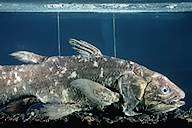
Ø    Name 2 fish that retain their notochord throughout their life cycle.

Ø    What does the word “Chondrichthyes” mean?

Ø    Give 2 examples of agnathans.

Ø    Which fin propels bony fish through the water?

Ø    The word “amphibian” means  ___________________.

Ø    Name the 2 major groups of bony fish.

Ø    What is the function of the swim bladder in bony fish?

Ø    What structure covers the gills of bony fish?

Ø    Describe several characteristics of lungfish.

Ø    What makes up the skeleton of fish in the group Osteichthyes?

Ø    What structure helps draw water into the mouth of bony fish?

Ø    Give 2 ways amphibians breathe.

Ø    In what order are amphibians without tails found?

Ø    Describe the feeding habits of adult frogs.

Ø    Describe metamorphosis in frogs.

Ø    Give several reasons why frogs & toads return to water to reproduce.

Ø    Which order of amphibians is legless?

Chapter 43 AP Obj Body Defenses


Chapter 43    Body’s Defenses
Objectives
Nonspecific Defenses Against Infection
1.Explain what is meant by nonspecific defense and list the nonspecific lines of defense in the vertebrate body.
2.Distinguish between:
a. innate and acquired immunity
b. humoral and cell mediated response
3.Explain how the physical barrier of skin is reinforced by chemical defenses.
4.Define phagocytosis. Name four types of phagocytic leukocytes.
5.Explain how interferon limits cell-to-cell spread of viruses.
6.Describe the inflammation response, including how it is triggered.
7.Describe the factors that influence phagocytosis during the inflammation response.
8.Explain how the action of natural killer cells differs from the action of phagocytes.
9.Explain what occurs during the condition known as septic shock.
10.Describe the roles of antimicrobial proteins in innate immunity.
How Specific Immunity Arises
11.Distinguish between antigens and antibodies.
12.Distinguish between antigen and epitope.
13.Explain how B lymphocytes and T lymphocytes recognize specific antigens
14.Explain how the particular structure of a lymphocyte’s antigen binding site forms during development. Explain the role of recombinase in generating the staggering variability of lymphocytes.
15.Explain why the antigen receptors of lymphocytes are tested for self-reactivity during development. Predict the consequences that would occur if such testing did not take place.
16.Describe the mechanism of clonal selection. Distinguish between effector cells and memory cells.
17.Distinguish between primary and secondary immune responses.
18.Describe the cellular basis for immunological memory.
19.Describe the variation found in the major histocompatibility complex (MHC) and its role in the rejection of tissue transplants. Explain the adaptive advantage of this variation.
20.Compare the structures and functions of cytotoxic T cells and helper T cells.
21.Compare the production and functions of class I MHC and class II MHC molecules.
Immune Responses
22.Distinguish between humoral immunity and cell-mediated immunity.
23.Describe the roles of helper T lymphocytes in both humoral and cell-mediated immunity.
24.Describe the functions of the proteins CD4 and CD8.
25.Explain how cytotoxic T cells and natural killer cells defend against tumors.
26.Distinguish between T-dependent antigens and T-independent antigens.
27.Explain why macrophages are regarded as the main antigen-presenting cells in the primary response but memory B cells are the main antigen-presenting cells in the secondary response.
28.Explain how antibodies interact with antigens.
29.Diagram and label the structure of an antibody and explain how this structure allows antibodies to (a) recognize and bind to antigens, and (b) assist in the destruction and elimination of antigens.
30.Distinguish between the variable (V) and constant (C) regions of an antibody molecule.
31.Describe the production and uses of monoclonal antibodies.
32.Compare the processes of neutralization, opsonization, and agglutination.
Immunity in Health and Disease
33.Distinguish between active and passive immunity and describe examples of each.
34.Explain how the immune response to Rh factor differs from the response to A and B blood antigens.
35.Describe the potential problem of Rh incompatibility between a mother and her unborn fetus and explain what precautionary measures may be taken.
36.Explain what is done medically to reduce the risk of tissue transplant rejection due to differences in the MHC. Explain what is unique about the source of potential immune rejection in bone marrow grafts.
37.Describe an allergic reaction, including the roles of IgE, mast cells, and histamine.
38.Explain what causes anaphylactic shock and how it can be treated.
39.List three autoimmune disorders and describe possible mechanisms of autoimmunity.
40.Distinguish between inborn and acquired immunodeficiency.
41.Explain how general health and mental well-being might affect the immune system.
42.Describe the infectious agent that causes AIDS and explain how it enters a susceptible cell.
43.Explain how HIV is transmitted and describe its incidence throughout the world. Note strategies that can reduce a person’s risk of infection.

.

BACK


Chapter 34 AP Objectives


Chapter 34     Vertebrate Evolution and Diversity
Objectives
Invertebrate Chordates and the Origin of Vertebrates
1.Distinguish between the phyla of deuterostomes.
2.Describe the four derived traits that define the phylum Chordata.
3.Distinguish among the three subphyla of the phylum Chordata and give examples of each.
4.Discuss the evidence for and against Garstang’s hypothesis that vertebrates had a tunicate-like ancestor.
5.Explain what lancelets suggest about the evolution of the chordate brain.
Craniates Are Chordates with a Head
6.Discuss the importance of genetic duplication in chordate evolution.
7.Explain the fate of the neural crest cells in craniate development.
8.Explain what Haikouella and Haikouichthys tell us about craniate evolution.
Vertebrates Are Craniates with a Backbone
9.Describe the way of life and unique characters of the lamprey.
10.Describe conodonts, and explain why they are considered vertebrates.
11.Describe the trends in mineralized structures in early vertebrates.
Gnathostomes Are Vertebrates with Jaws
12.Explain one hypothesis for the evolution of the jaws of gnathostomes.
13.List the shared, derived characters that characterize gnathostomes.
14.Describe the evidence that suggests that the loss of bone in Chondrichthyes is a derived feature.
15.Describe the features of sharks that are adaptive for their active, predatory lifestyle.
16.Describe and distinguish between Chondrichthyes and Osteichthyes, noting the main traits of each group.
17.Identify and describe the main subgroups of Osteichthyes.
18.Name the three living lineages of lobe-fins.
Tetrapods Are Gnathostomes with Limbs and Feet
19.Define and distinguish between gnathostomes, tetrapods, and amniotes.
20.Explain what Acanthostega suggests about the origin of tetrapods.
21.Describe the common traits of amphibians and distinguish among the three orders of living amphibians.
Amniotes Have Amniotic Eggs
22.Describe an amniotic egg and explain its significance in the evolution of reptiles and mammals.
23.Explain why the reptile clade includes birds.
24.Describe a number of reptile features that are adaptive for life on land.
25.Explain why non-bird reptiles should be called “ectothermic” rather than “cold-blooded.”
26.Define and describe the parareptiles.
27.Distinguish between the lepidosaurs and the archosaurs.
28.Compare the interpretations of dinosaurs as ectotherms or endotherms.
29.Describe the specialized adaptations of snakes that make them successful predators.
30.List the modifications of birds that are adaptive for flight.
31.Summarize the evidence supporting the hypothesis that birds evolved from theropod dinosaur ancestors.
32.Explain the significance of Archaeopteryx.
33.Describe the characteristic derived characters of mammals.
34.Describe the evolutionary origin of mammals.
35.Distinguish among monotreme, marsupial, and eutherian mammals.
36.Describe the adaptive radiation of mammals during the Cretaceous and early Tertiary periods.
37.Compare and contrast the four main evolutionary clades of eutherian mammals.
Primates and the Evolution of Homo sapiens
38.Describe the general characteristics of primates. Note in particular the features associated with an arboreal existence.
39.Distinguish between the two subgroups of primates and describe their early evolutionary relationship.
40.Distinguish between hominoid and hominid.
41.Explain what Sahelanthropus tells us about hominid evolution.
42.Describe the evolution of Homo sapiens from australopith ancestors. Clarify the order in which distinctive human traits aro
BACK

Bird


Birds
All Materials © Cmassengale


Birds:

Well adapted to marine, freshwater, & terrestrial habitats
Bodies adapted for flight
Endothermic – body temperature controlled by metabolism

Evolution:

  • Evolved from reptiles
  • Few fossils due to lack of preservation of feathers or thin, hollow bones
  • Archaeopteryx:
    1. Possible link between birds & reptiles
    2. Lived during Jurassic period
    3. Large skull with reptile like teeth
    4. Bones not hollow
    5. Claws on forelimbs
    6. Long tail
    7. Strong legs & rounded wings for gliding
    8. Feathers
    9. Furculum – fused collarbone or wishbone

Archaeopteryx
Archaeopteryx Fossil

  • Hesperonis:
    1. Bird fossils from Cretaceous period
    2. Large, flightless bird
    3. Had teeth like reptiles

kish-02.jpg (71663 bytes)
Hesperonis

  • Ichthyornis:
    1. Smaller, tern like bird
    2. Lived during Cretaceous period
    3. Had large flight wings


Section 1 Review


Characteristics of Birds:

  • Body covered with feathers made of protein called keratin
  • Thin, hollow bones
  • Some bones fused for extra strength
  • Forelimbs modified into wings for  flight
  • Two hind limbs with claws to support upright body
  • Scales on legs
  • Toothless, horny beak
  • Additional air sacs  with lungs for more oxygen
  • Endotherms (40 to 41 degrees Celsius body temperature)
  • Four chambered heart with single, right aortic arch
  • Amniote egg with calcium carbonate shell
  • Oviparity with both parents often caring for eggs
  • Eggs usually incubated within a nest

Feathers:

  • Modified scales
  • Function to provide lift for flight & help conserve body heat
  • Five kinds of feathers —– down, contour, flight, filoplume, & bristles


Types of Feathers

  • Down feathers:
    1. Soft & fluffy
    2. Cover the body of nestlings
    3. Provide an undercoat insulating adult birds
  • Contour Feathers:
    1. Give streamline shape to body
    2. Provide coloration to adult birds
    3. Give additional insulation to body
  • Flight Feathers:
    1. Specialized contour feathers
    2. Found on wings & tail

  • Filoplumes:
    1. Called pin feathers
    2. Hairlike feathers under contour feathers on body

Parts of a feather:

  • Develop from tiny pits in the skin called follicles
  • Shaft emerges from the follicle
  • Two vanes develop on either side of shaft
  • Barbs branch off of each vane & have projections called barbules
  • Barbules have microscopic hooks to hold barbules together


Parts of a Flight Feather


Microscopic Hooks on Barbules

  • Birds preen their feathers to clean them & coat them with oil
  • Preen glands – oil glands located at the base of the tail
  • Birds shed or molt feathers periodically:
    1. Molting usually in late summer between breeding & migration
    2. Flight feathers replaced
    3. Some birds molt before courtship

Beaks and Feet:

  • Adapted to habitat & feeding
  • Hawks & eagles have hooked beaks & talons for tearing meat


Gentoo Penguin
TalonsHooked BeakPenguin Flippers


  • Swifts have tiny beaks that open wide to catch insects in midair
  • Flightless birds like ostriches have legs & feet modified for running & walking
  • Penguins have wings modified into flippers for swimming
  • Ducks & geese with webbed feet


Running Legs of OstrichWebbed Feet on Duck


  • Legs of some birds such as herons &  egrets turn vivid colors to attract mates; caused by hormones

Skeleton and Muscles:

  • Pelvic & pectoral girdles fused for strength
  • Bones thin & hollow so bird lighter

A birdbone(notice the honey combed shape)
Hollow Bones

  • Furculum or wishbone is a fused collarbone that stabilizes bird in flight
  • Lighter beak replaces heavy teeth & jaws
  • Lower vertebrae fused so no heavy ligaments needed
  • Enlarged eye sockets reduce skull weight
  • Keeled sternum for attachment of large flight muscles
  • Pygostyle – terminal vertebrae support tail & aids in flight (lift, steering, & braking)
  • Two digits in forelimbs lost & other three digits fused to form wings
  • Wings shaped like air foils (thicker in front & tapering to back) so air moves faster on top causing lift

  • Powerful muscles make up 50% of body weight
  • each wing movement uses different set of muscles
  • Flight muscles called pectorals & are attached to wing & keeled sternum
  • When large pectorals contract, wings move down
  • When large pectorals relax & small pectorals contract, wings move upward

Body Temperature:

  • Metabolism generates body heat (endothermic)
  • Enables birds to survive in warm & cold environments
  • Rapid breathing & increased air sacs in lungs bring in more oxygen

Diagram of a bird's lung and air sac system, and countercurrent exchange
Air Sacs in Bird Lungs

  • Ingest large amounts of food for energy
  • Fluff out feathers to trap air for insulation
  • Aquatic birds have thin layer of fat for insulation

Digestive System:

  • Fast & efficient digestion (mouse digested in 3 hours)
  • No chewing
  • Crop for temporary food storage
  • Two part stomach — proventriculus & gizzard
  • Proventriculus is 1st chamber where digestive juices added
  • Gizzard is 2nd part for crushing food
  • Small stones & gravel eaten by birds aids grinding in gizzard
  • Pyloric sphincter valve at lower end of gizzard controls food movement into intestines
  • Duodenum – beginning of small intestine where bile (digests fats) & pancreatic juice are added & digested food is absorbed

birdanat.gif (87464 bytes)

Excretory System:

  • Paired kidneys filter nitrogen wastes (uric acid) from blood
  • No urinary bladder to store liquid wastes
  • Uric acid travels down ureters to cloaca where intestinal wastes & reproductive products added
  • Uric acid secreted in white, semi solid mass
  • Shorebirds have salt secreting glands above the eyes & secrete excess salt through their nostrils

Respiratory System:

  • Fly at high altitudes where there is less oxygen so need efficient respiratory system
  • High metabolic rate requires large amount of oxygen
  • Nine air sacs associated with lungs increase oxygen level & decrease density
  • Air sacs connected to air spaces in hollow bones
  • One way flow of air in lungs & air sacs so more oxygen is removed
  • Air pathway:
    air enters body through nostrils on beak  trachea (windpipe) syrinx (voice box) 2 primary bronchi 75% of air into two posterior air sacs and 25% of air into lungs air from lungs into other seven air sacs
  • When carbon dioxide exhaled, oxygen from posterior air sacs moves into lungs to always keep fresh oxygen supply

Circulatory System:

  • Four chambered heart
  • Right side of heart pumps deoxygenated blood from body cells to lungs
  • Left side of heart receives oxygenated blood from lungs & pumps it to the body cells
  • Single aortic arch
  • Rapid heartbeat (hummingbird 600X/minute & chickadee 1000X/minute)
  • Less active birds such as ostrich have slower heart rates (70X/minute)

Nervous System:

  • Large brains relative to size of bird
  • Most highly developed brain areas control flight
  • Cerebellum coordinates movement
  • Cerebrum controls navigation, mating, nest building, & care of young
  • Optic lobes receive & interpret visual stimuli
  • Keen vision
  • Have color vision for locating food
  • Large eyes located on side of head for wide field of vision in most birds
  • Some birds such as owls with eyes on front of head for binocular vision (depth perception)
  • No external ears, but have feathers around ear openings to direct sounds into ear canals
  • Tympanic membrane or eardrum for picking up sound vibrations
  • Semicircular canals in inner ear regulate balance
  • Poorly developed sense of smell except in ducks & flightless birds
  • Sense of taste helps avoid bitter tasting or toxic foods

Reproductive System:

  • Testes in males produces sperm that travels by the vas deferens to cloaca
  • Females have single ovary that makes eggs
  • Eggs are fertilized in the oviducts
  • Shell added by shell gland & then egg moves into
  • In mating, male presses cloaca to female to transfer sperm (internal fertilization)
  • Lay an amniote egg:
    1. Embryo suspended in fluid called albumen (white of egg)
    2. Chalaza – rope like strands suspending embryo in albumen
    3. Chorion is membrane inside of shell
    4. Yolk is stored food surrounded by yolk sac


Bird Egg

Incubation & development of Egg:

  • Eggs incubated by one or both parents
  • Brood patch – thickened, featherless patch of skin on abdomen of bird used to warm eggs
  • Membranes grow out of embryo’s digestive tract & surround yolk
  • Membranes make digestive enzymes to dissolve proteins & lipids in yolk
  • Yolk sac has blood vessels to carry food to embryo
  •  Wastes from embryo collect in membrane called allantois
  • Chorion membrane lines the shell & allows gas exchange
  • Young birds may be precocial or altricial
  • Precocial young:
    1. Have longer incubations
    2. More eggs laid
    3. Active as soon as hatch
    4. Nestlings can swim, walk, & feed themselves
    5. Need some parental care
    6. Includes ducks, geese, & swans
  • Altricial young:
    1.Lay fewer eggs
    2. Hatch quickly
    3. Hatchlings are blind, naked, & helpless
    4. Depend on parents for warmth & food for several weeks
    5. Includes songbirds, woodpeckers, hawks, pigeons, doves, raptors


Dunnock & Cuckoo
Altricial YoungPrecocial Young


Behavior:

  • Longer parental care allows more complex learning (courtship, nesting, migration, etc.)
  • Territoriality allows males to establish & defend breeding areas
  • Courtship behaviors are used by males to attract mates:
    1. Brightly colored feathers
    2. Flight displays
    3. Songs


Male Scarlet Tanager Breeding Plumage

  • Nest building holds eggs, conceals & shelters young birds, may help attract mates
  • Nests are built in sheltered, well-hidden spots in trees, on the ground, etc. & are made of twigs, mud, grass, feathers…

  • Migration to new areas is triggered by dropping temperatures & dwindling food supplies
  • Birds use migration clues including:
    1. Position of sun & stars
    2. Topographical landmarks
    3. Magnetic clues
    4.Air pressure changes
    5. Low frequency sounds


Section 2 Review

Classification:

  • Class Aves
  • 27 orders
  • Gaviiformes – loons
  • Pelecaniformes – pelicans & cormorants
  • Ciconiiformes – wading birds like ibises & herons
  • Anseriformes – ducks, geese, & swans
  • Falconiformes – falcons, eagles, hawks, vultures
  • Galliformes – turkey, quail, pheasants
  • Gruiformes – cranes, coots, & rails
  • Charadriiformes – snipes, sandpipers, gulls, terns
  • Columbiformes – pigeons & doves
  • Psittaciformes – parrots, parakeets, & macaws
  • Cucluiformes – cuckoos & roadrunners
  • Strigiformes – owls
  • Caprimulgiformes – whippoorwill & nighthawk
  • Apodiformes – hummingbird & swifts
  • Coraciiformes – kingfishers
  • Piciformes – woodpeckers, sapsuckers, & flickers
  • Passeriformes – perching birds like robins, cardinals, blue jays


Pelican at Oranjestad waterfront
Pygmy OwlBrown Pelican
photograph of macawFemale Northern Cardinal Photograph
MacawFemale Cardinal


Food & Habitat Adaptations:

  • Anseriformes (ducks, geese, & swans) have webbed feet for swimming & flattened bills; young are precocial but need some parental care
  • Strigiformes (owls) have sharp, hooked beaks & talons (claws) for meat eating, keen hearing & eyesight, & forward facing eyes
  • Apodiformes (hummingbirds) are small, fast-flying birds with tiny feet & long tongues for drinking nectar; found only in western hemisphere
  • Psittaciformes (parrots, cockatoos, parakeets…) have a strong, hooked beak for seed opening & two forward & two rear facing toes for perching & climbing
  • Piciformes (woodpeckers, toucans, & flickers) have two rear facing toes for dwelling in tree cavities & sharp, chisel like bills for drilling into trees
  • Falconiformes or raptors ( hawks, eagles, vultures) have hooked beaks & talons & keen vision for seeing prey
  • Passeriformes or songbirds (blue jays, cardinals, sparrows, robins …) have enlarged rear facing toe to grip branches, a syrinx or voice box in males to produce songs, & a variety of beak shapes to feed on seeds, nectar, fruits, & insects; known as passerines or perching birds
  • Columbiformes (pigeons & doves) have small heads & bills, a crop that makes “pigeon’s milk” for feeding young, short incubation period (2 weeks)
  •  Ciconiiformes (herons, ibises, & egrets) have long legs for wading & sharp pointed bills for piercing frogs & fish
  • Galliformes (turkeys, quail, pheasants, & chickens)  have plump bodies with limited flying &a large gizzard for grinding grains
  • Sphenisciformes (penguins) have wings modified into flippers, an extra layer of body fat for insulation, & webbed feet for swimming
  • Struthioniformes (ostrich) are the largest birds that can’t fly but have long legs with only two toes adapted for fast running
Section 3 Review