|
Diffusion and Osmosis |
Introduction:
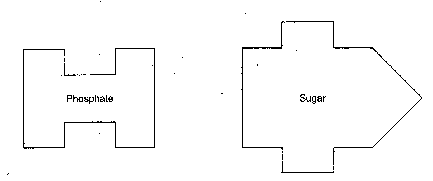
In this exercise you will measure diffusion of small molecules through dialysis tubing, an example of a semi permeable membrane. The movement of a solute through a semi permeable membrane is called dialysis. The size of the minute pores in the dialysis tubing determines which substance can pass through the membrane. A solution of glucose and starch will be placed inside a bag of dialysis tubing. Distilled water will be placed in a beaker, outside the dialysis bag. After 30 minutes have passed, the solution inside the dialysis tubing and the solution in the beaker will be tested for glucose and starch. The presence of reducing sugars like glucose, fructose, and sucrose will be tested with Benedict’s Solution. The presence of starch will be tested with Lugol’s solution (iodine-potassium-iodide).
Procedure:
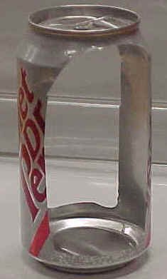
- Obtain a 30 -cm piece of 2.5-cm dialysis tubing that has been soaking in water. Tie off one end of the tubing to form a bag. To open the other end of the bag, rub the end between your fingers until the edges separate.
- Place 15 mL of the 15% glucose/ 1% starch solution in the bag. Tie off the other end of the bag, leaving sufficient space for the expansion of the bag’s contents. Record the color of the solution in Table 1.1.
- Test the 15% glucose / 1% starch solution in the bag for the presence of glucose. Your teacher may have you do a Benedict’s test. Record the results in Table1.1.
- Fill a 250 mL beaker or cup 2/3 full with distilled water. Add approximately 4 mL of Lugol’s solution to the distilled water and record the color in Table 1.1. Test the solution for glucose and record the results in Table 1.1.
- Immerse the bag in the beaker of solution.
- Allow your set up to stand for approximately 30 minutes or you see a distinct color change in the bag or the beaker. Record the final color of the solution in the bag, and of the solution in the beaker, in Table 1.1.
- Test the liquid in the beaker and in the bag for the presence of glucose. Record the results in Table 1.1.
Table 1.1
|
Initial Contents |
Initial Solution Color |
Final Solution Color |
Initial Presence of Glucose |
Final Presence of Glucose |
| Bag |
15% Glucose & 1% starch |
|
|
|
|
| Beaker |
H2O + IKI |
|
|
|
|
Analysis of Results:
1. Which substance(s) are entering the bag and which are leaving the bag? What experimental evidence supports your answer?
_______________________________________________________________________
_______________________________________________________________________
_______________________________________________________________________
_______________________________________________________________________
2. Explain the results you obtained. Include the concentration differences and membrane pore size in your discussion.
________________________________________________________________________
________________________________________________________________________
________________________________________________________________________
________________________________________________________________________
3. Quantitative data uses numbers to measure observed changes. How could this experiment be modified so that quantitative data could be collected to show that water diffused into the dialysis bag?
________________________________________________________________________
________________________________________________________________________
________________________________________________________________________
________________________________________________________________________
________________________________________________________________________
4. Based on your observations, rank the following by relative size, beginning with the smallest : glucose molecules, water molecules, IKI molecules, membrane pores, starch molecules.
_______________________________________________________________________
_______________________________________________________________________
5. What results would you expect if the experiment started with glucose and IKI solution inside the bag and only starch and water outside? Why?
________________________________________________________________________
________________________________________________________________________
________________________________________________________________________
________________________________________________________________________
Osmosis:
In this experiment you will use dialysis tubing to investigate the relationship between solute concentration and the movement of water through a semi permeable membrane by the process of osmosis. When two solutions have the same concentration of solutes, they are said to be isotonic to each other. If the two solutions are separated by a semi permeable membrane, water will move between the two solutions, but there will be no net change in the amount of water in either solution. If two solutions differ in the concentration of solutes that each has, the one with more solute hypertonic to the one with the less solute. The solution that has less solute is hypotonic to the one with more solute. These words can only be used to compare solutions.
Procedure:
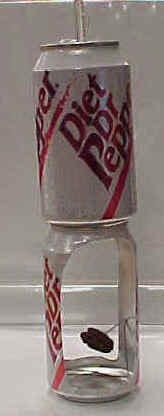
1. Obtain six 30-cm strips of presoaked dialysis tubing.
2. Tie a knot in one end of each piece of dialysis tubing to form six bags. Pour approximately 25 mL of each of the following solutions into separate bags:
- Distilled water
- 0.2 M sucrose
- 0.4 M sucrose
- 0.6 M sucrose
- 0.8 M sucrose
- 1.0 m sucrose
Remove most of the air from the bags by drawing the dialysis bag between two fingers. Tie off the other end of the bag. Leave sufficient space for the expansion of the contents in the bag.
3. Rinse each bag gently with distilled water to remove any sucrose spilled during filling.
4. Carefully blot the outside of each bag and record in Table 1.2 the initial mass of each bag.
5. Fill six 250 mL beakers 2/3 full with distilled water.
6. Immerse each bag in one of the beakers of distilled water and label the beaker to indicate the molarity of the solution in the dialysis bag. Be sure to completely submerge each bag.
7. Let them stand for 30 minutes.
8. At the end of 30 minutes remove the bags from the water. Carefully blot and determine the mass of each bag.
9. Record your group’s results in Table 1.2. Obtain data from the other lab groups in your class to complete Table 1.3: Class Data.
Table 1.2 Dialysis Bag Results: Individual Data
| Contents in Dialysis Bag |
Initial Mass |
Final Mass |
Mass Difference |
% Change in Mass |
| a). Distilled Water |
|
|
|
|
| b). 0.2 M |
|
|
|
|
| c). 0.4 M |
|
|
|
|
| d). 0.6 M |
|
|
|
|
| e). 0.8 M |
|
|
|
|
| f). 1.0 M |
|
|
|
|
To Calculate:
| % change in mass |
= |
Final Mass-Initial Mass |
X |
100 |
|
|
———————–
Initial Mass |
|
|
Table 1.3 Dialysis Bag Results: Class Data
percent change in Mass of Dialysis Bags
| Bag Contents |
Group 1 |
Group 2 |
Group 3 |
Group 4 |
Group 5 |
Group 6 |
Total |
Class Average |
| Distilled Water |
|
|
|
|
|
|
|
|
| 0.2 M |
|
|
|
|
|
|
|
|
| 0.4 M |
|
|
|
|
|
|
|
|
| 0.6 m |
|
|
|
|
|
|
|
|
| 0.8 M |
|
|
|
|
|
|
|
|
| 1.0 M |
|
|
|
|
|
|
|
|
10. Graph the results for both your individual data and class average on the following graph. For this graph you will need to determine the following:
a). the independent variable. __________________________________
b). the dependent variable. ___________________________________
Graph Title ______________________________________________

Analysis of Results:
1. Explain the relationship between the change in mass and the molarity of sucrose within the dialysis bag.
________________________________________________________________________
________________________________________________________________________
________________________________________________________________________
________________________________________________________________________
________________________________________________________________________
2. Predict what would happen to the mass of each bag in this experiment if all the bags were placed in a 0.4 M sucrose solution instead of distilled water. Explain your response.
________________________________________________________________________
________________________________________________________________________
________________________________________________________________________
________________________________________________________________________
________________________________________________________________________
3. Why did you calculate the per cent change in mass rather than using the change in mass?
________________________________________________________________________
________________________________________________________________________
________________________________________________________________________
________________________________________________________________________
________________________________________________________________________
4. A dialysis bag is filled with distilled water and then placed in a sucrose solution. The bag’s initial mass is 20 g. and its final mass is 18 g. Calculate the percent change of mass, showing your calculations in the space below.
5. The sucrose solution in the beaker would have been ___________________ to the distilled water in the bag.
 Introduction: