AR Wildflowers

Arkansas Wildflowers


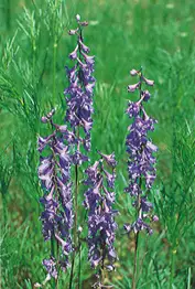
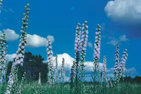
Carolina Larkspur (Delphinum carolinium) – 4′ tall.
Blooms May – July.  These spurred flowers may be deep blue, reddish – blue, or white.  Native perennial.  OZ, OU, CP.


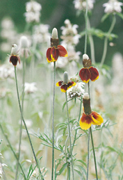
Mexican Hat  (Ratibida columnifera) ― 2 – 3′ tall.
Blooms June – October.  A widely planted form of a native perennial.  Statewide.


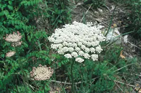
Queen Ann’s Lace (Daucus carota)  ― 1 – 4′ tall

Blooms May – frost.  This is the ancestor of the cultivated carrot.  Introduced biennial. Statewide.


Black-eyed Susan  (Rudbeckia hirta) ― 2 – 3′ tall with one 2″ flower head on each hairy stem.

Blooms May – October.  Native Biennial or short-lived perennial.  Statewide.


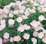
Showy Evening Primrose  (Oenothera speciosa) ― 1 – 2′ tall.

Blooms April – July.  White or pink flowers.  Native perennial.  Statewide.


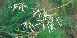
Pale Purple Coneflower (Echinacea pallida) – 3′ tall.

Blooms May – July.  Native perennial.  OZ, OU, CP.


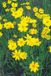
Lance-leaved Coreopsis (Coreopsis lanceolata) – 3′ tall.

Blooms April – June.  Native perennial.  Statewide.


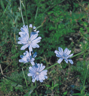
Chicory  (Coreopsis intybus) – 4′ tall.

Blooms May – October.

This European native’s roots are sometimes used as a coffee substitute or additive.  Perennial.  OZ, OU.


Rough Blazing Star  (Liatrus aspera)  ― 3 – 4′ tall.
Blooms July – October.  The unopened flower buds resemble small cabbages.  Native perennial.  Statewide.


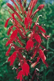
Cardinal Flower (Lobelia cardinalis) – 3′ tall.
Blooms August – October. This flower attracts hummingbirds.  Native perennial. Statewide.


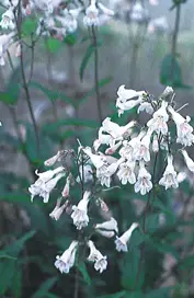
Arkansas Beard Tongue (Penstemon arkansanus) – Less than 2′ tall.
Blooms April – June.  The 3/4″ whitish flowers have lavender streaking.  Native perennial. OZ, OU.


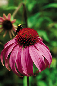
Purple Coneflower  (Echinacea purpurea) – Up to 4′ tall.
Blooms from June – October.
The ray flowers are more purple than those of pale purple coneflower. Native perennial. OZ, OU.
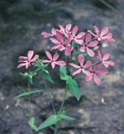
Downy Phlox (Phlox pilosa) – 2′ tall.

Blooms April – July.

Flowers can be pink, pale pink, or sometimes white with purple centers.  Native perennial.  OZ, OU, CP.


Spider Lily (Hymenocallis caroliniana) – 3′ tall.

Blooms May – August.  These large white flowers have a distinctive spider-like shape. Native perennial. OU, GP, AP.


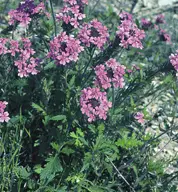
Rose Vervain (Glandularia canadensis) – Plants less than 2′ tall.

Blooms March – September.  The source of many garden hybrids.  Native perennial.  OZ, OU, CP, AP.


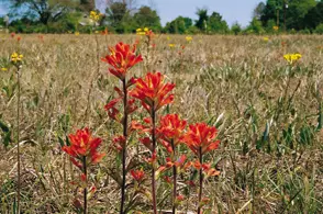
Indian Paintbrush (Castilleja coccinea) ― 1 – 2′ tall.  The bracts that surround the small flowers displays brilliant colors.

Blooms April – June.  Native annual.  Found on prairies in the OZ, CP, AP.


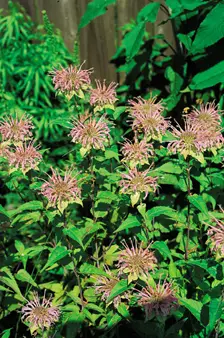
Wild (Monarda fistulosa) ― 2 – 4′ tall.
Blooms June – September.  Also called Bee Balm.  Flowers pinkish, lavender, or lilac.  Statewide.


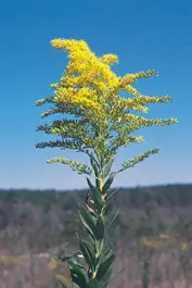
Goldenrod (Solidago canadensis) ― 4 – 6′ tall.

Blooms July – September.  Native perennial.  Statewide.


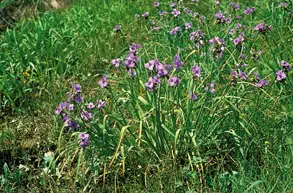
Ohio Spiderwort (Tradescantia ohiensis) – Stems 3′ tall.
Blooms May – July.
So named because the internal jellylike substance resembles a spider’s web.  Native perennial.  OZ, OU, CP.
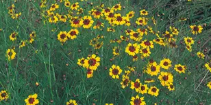
Plains Coreopsis (Coreopsis tinctoria) – 3′ tall.

Blooms June – September.  Native annual.  Statewide.

Bird’s Foot Violet (Viola pedata) – 6″ tall.

Blooms April – May.  This violet occurs in several different colors:  light violet, dark violet, or dark violet with 2 dark purple petals. Native perennial.  OZ, OU, CP.


Butterfly Weed (Asclepias tuberosa) ― 1 – 2′ tall.

Blooms May – September.  Flower’s nectar attractive to butterflies.  Native perennial.  Statewide.


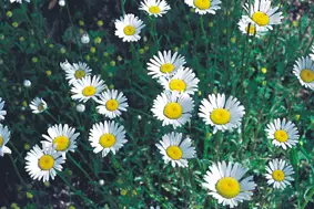
Ox-eyed Daisy (Chrysanthemum leucanthemum) – 2″ flower heads.

Blooms May – July.  Introduced perennial.  OZ, OU, CP.


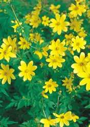
Tickseed (Bidens aristosa) ― 1 – 6′ tall.

Blooms August – November.  This late bloomer is often found in large stands.  Native perennial.  Statewide.


Arthropod

Arthropods
Non-Insects

Characteristics

  • Makes up 3/4’s of all animal species
  • Includes insects, spiders, scorpions, millipedes, centipedes, crabs, lobsters, & crayfish
  • Arthropod means “jointed foot”
  • Jointed appendages (legs, antenna, mouthparts)
  • Segmented body with paired appendages on each segment)
  • External exoskeleton made of chitin (carbohydrate) & protein for protection & support
  • Exoskeleton has 3 layers — outer waxy layer repels water, middle layer has calcium for extra strength, & inner layer has flexible joints for movement
  • Protostomes (blastopore develops into mouth)
  • Coelomate (mesoderm-lined body cavity)
  • Ventral nervous system
  • Open circulatory system
  • Specialized sensory receptors & high degree of cephalization
  • Have simple or compound eyes & segmented antenna

Movement & Growth

  • Muscles occur in bundles & are attached to inside of exoskeleton on each side of joints
  • Exoskeleton must be periodically molted (shed) for organism to grow
  • Molting called ecdysis
  • Molting hormone released & causes epidermal cells to secrete enzymes that digest & loosen inner exoskeleton
  • New exoskeleton secreted by epidermal cells flexible at first & must harden so arthropod not vulnerable to predators so often stay in hiding after molting
  • Arthropods go through numerous molts


Butterfly Molting Pupal Case

Evolution & Taxonomy

  • Evolved from ancestral arthropod with many body segments each with appendages
  • Modern arthropod segments fused into larger, specialized structures called tagmata
  • Four subphyla
    * Trilobita – extinct trilobites
    * Crustacea – shrimps, lobsters, crayfish, & barnacles
    * Chelicerata – spiders, scorpions, & ticks
    * Uniramia -centipedes, millipedes, & insects

Subphylum Trilobita
Characteristics

  • Includes extinct trilobite
  • Marine
  • Have a head & segmented trunk with one pair of legs on each segment
  • Breathe through gills
  • Single pair of antenna


TRILOBITE

Subphylum Chelicerata
Characteristics

  • Includes 2 classes — Xiphosura (horseshoe crab) and Arachnida (spiders, ticks, scorpions, & mites)
  • Have a cephalothorax (fused head& thorax) and abdomen
  • No antenna
  • Simple eyes or ocelli
  • Have 6 pairs of jointed appendages:
    * Chelicerae – claws or fangs (1 pair)
    * Pedipalps – used for feeding, walking, sensing, transferring sperm (1 pair)
    * Walking legs – movement (4 pairs)
  • Horseshoe crab
    * Marine
    * Not true crabs
    * Fanglike pincers or chelicerae
    * Use book gills to breathe


HORSESHOE CRAB

  • Arachnids
    * Terrestrial
    * Eight legs
    * Chelicerae or fangs with venom
    * Ocelli
    * No antenna
    * Breathe by book lungs &/or tracheal tubes
  • Spiders
    * Arachnid that feeds on insects (carnivores)
    *  Have oval shaped, unsegmented abdomen
    * Cephalothorax connected by narrow waist to abdomen
    * Have 8 simple eyes or ocelli
    * Fangs pierce prey, inject poison, & suck out body fluids
    * Pedipalps on head help sense prey & move it to the mouth
    * Open circulatory system
    * Ostia are openings in heart where blood reenters
    * Body cavity called hemocoel
    * Hemocycanin is oxygen-carrying pigment in blood
    * Have silk glands to make silk & spinnerets to release silk for webs
    * Breathe by book lungs & tracheal tubes
    * Malpighian tubules filter wastes & reabsorb water


GARDEN SPIDER

  • Ticks & Mites
    * Parasitic arachnid
    * Fused cephalothorax & abdomen
    * Most abundant arachnid
    * Need blood meal to molt
    * Mites can damage fruit & feed on dead skin at base of hair follicle
    * Ticks carry Lyme disease & Rocky Mountain Spotted Fever


MITE

TICK


  • Scorpions
    * Have a cephalothorax & long segmented abdomen curled over body
    * Prefer dry regions
    * Poisonous stinger on end of abdomen
    * Breathe through book lungs
    * Pedipalps modified into claws
    * Nocturnal predators


SCORPION

Subphylum Crustacea
Characteristics

  • Marine members include shrimp, lobster, copepods, barnacles, & crabs


CRAB SHRIMP


  • Terrestrial crustaceans called isopods include pillbugs & sowbugs


PILLBUG

  • Freshwater members include crayfish & Daphnia (water fleas)


DAPHNIA

  • All have jaws are mandibles for chewing or tearing
  • Known as mandibulates
  • Have cephalothorax & abdomen
  • Have 10 pairs of jointed appendages
  • Breathe through gills
  • Barnacles
    * Marine
    * Sessile crustaceans that live in limestone case
    * Filter plankton with 12 appendages called cirri


BARNACLE

  • Isopods (pillbugs & sowbugs)
    * Live on land in dark places
    * Have 7 pairs of legs on a segmented body
    * Can roll into a ball for protection
  • Crayfish
    * Cephalothorax made of 13 fused segments & covered by protective carapace
    * Antennules located on head help in balance, touch, & taste
    * Statocysts – balancing organs at the base of antennules
    * Antenna on head used for touch & taste
    * Maxillae – paired mouthparts that move side to side to tear food
    * Maxillipeds – help hold food
    * Chelipeds – claws used to capture food & for protection
    * Mandibles – jaws that move up & down to crush  food
    * Walking legs – 8 pairs used for movement
    * Swimmerets – under abdomen to swim, gas exchange, & protect eggs/young
    * Abdomen ends in flat segment called telson with flat uropods on each side


CRAYFISH

               * Compound eyes on stalks
* Chitinous teeth in stomach grind food
* Wastes leave through anus
*  Green glands filter wastes from blood & help with salt balance
*  Open circulatory system with heart to pump blood to gills & body cells
* Ostia – one way valves allowing blood from dorsal sinus to reenter heart
* Gills attached to walking legs
* Separate sexes that mate in fall & sperm stored in seminal receptacle
*  Eggs attach to swimmerets of female & hatch in several weeks

  • Copepods
    * Largest group of crustaceans
    *  Make up most of the marine plankton
    *  Serve as food for many marine animals
    *  Found in freshwater, marine, & moist terrestrial environments


COPEPOD

Subphylum Uniramia
Characteristics

  • All have antenna, mandibles (jaws), & unbranched appendages
  • Includes 3 classes — Chilopoda (centipedes), Diplopoda (millipedes), & Insecta
  • Known as myriapods
  • Most are terrestrial
  • Exoskeleton prevents desiccation (water loss)

Class Chilopoda

  • Terrestrial centipedes
  • Flattened body with longer legs for fast movement
  • Have 1 pair of legs per body segment
  • Predators
  • Mandibles & maxilla for chewing prey (insects & earthworms)
  • Claw-like appendages or pincers on 1st body segment that can inject venom
  • Can coil up for defense


CENTIPEDE

Class Diplopoda

  • Terrestrial millipedes
  • Have 2 pairs of legs per body segment
  • Rounded body
  • Scavengers on decaying vegetation as they burrow through soil
  • Roll into ball when threatened & spray noxious chemical containing cyanide


MILLIPEDE

BACK


Bacteria

KINGDOMS ARCHAEBACTERIA & EUBACTERIA


All Materials © Cmassengale

Bacterial Evolution & Classification 

  • Most numerous organisms on earth
  • Earliest life forms (fossils date 2.5 billion years old)
  • Microscopic prokaryotes (no nucleus nor membrane-bound organelles)
  • Contain ribosomes
  • Infoldings of the cell membrane carry on photosynthesis & respiration
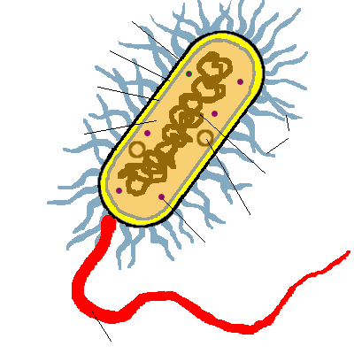
  • Surrounded by protective cell wall containing peptidoglycan (protein-carbohydrate)
  • Many are surrounded by a sticky, protective coating of sugars called the capsule or glycocalyx (can attach to other bacteria or host)
  • Have only one circular chromosome
  • Have small rings of DNA called plasmids
  • May have short, hairlike projections called pili on cell wall to attach to host or another bacteria when transferring genetic material
  • Most are unicellular

  • Found in most habitats
  • Most bacteria grow best at a pH of 6.5 to 7.0
  • Main decomposers of dead organisms so recycle nutrients
  • Some bacteria breakdown chemical & oil spills
  • Some cause disease 
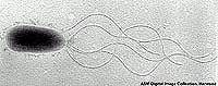
  • Move by flagella, gliding over slime they secrete ( e.g. Myxobacteria)
  • Some can form protective endospores around the DNA when conditions become unfavorable; may stay inactive several years & then re-activate when conditions favorable
  • Classified by their structure, motility (ability to move), molecular composition, & reaction to stains (Gram stain)
  • Grouped into 2 kingdoms — Eubacteria (true bacteria) & Archaebacteria (ancient bacteria)
  • Once grouped together in the kingdom Monera


STRUCTURE FUNCTION
Cell Wall protects the cell and gives shape
Outer Membrane protects the cell against some antibiotics (only present in Gram negative cells)
Cell Membrane regulates movement of materials into and out of the cell; contains enzymes important to cellular respiration
Cytoplasm contains DNA, ribosomes, and organic compounds required to carry out life processes
Chromosome carries genetic information inherited from past generations
Plasmid contains some genes obtain through genetic recombination
Capsule, and slime layer protects the cell and assist in attaching the cell to other surfaces
Endospore protects the cell against harsh environmental conditions, such as heat or drought
Pilus (Pili) assist the cell in attaching to other surfaces, which is important for genetic recombination
Flagellum moves the cell


Kingdom Archaebacteria

  •   Found in harsh environments (undersea volcanic vents, acidic hot springs, salty water)
  • Cell walls without peptidoglycan
  • Subdivided into 3 groups based on their habitat — methanogens, thermoacidophiles, & extreme halophiles

Methanogens

  • Live in anaerobic environments (no oxygen)
  • Obtain energy by changing H2 and CO2 gas into methane gas
  • Found in swamps, marshes, sewage treatment plants, digestive tracts of animals
  • Break down cellulose for herbivores (cows)
  • Produce marsh gas or intestinal gas (methane)

Extreme Halophiles

  •   Live in very salty water
  •   Found in the Dead Sea, Great Salt Lake, etc.
  • Use salt to help generate ATP (energy)

Thermoacidophiles (Thermophiles)

  • Live in extremely hot  (1100C) and acidic (pH 2) water
  • Found in hot springs in Yellowstone National Park, in volcanic vents on land, & in cracks on the ocean floor that leak scalding acidic water

Kingdom Eubacteria (true bacteria)

  • Most bacteria in this kingdom
  • Come in 3 basic shapes — cocci (spheres), bacilli (rod shaped), spirilla (corkscrew shape)

  • Bacteria can occur in pairs ( diplo– bacilli or cocci)
  •   Bacteria occurring in chains are called strepto- bacilli or cocci
  • Bacteria in grapelike clusters are called staphylococci
  • Most are heterotrophic (can’t make their own food)
  • Can be aerobic (require oxygen) or anaerobic (don’t need oxygen)
  • Subdivided into 4 phyla — Cyanobacteria (blue-green bacteria), Spirochetes, Gram-positive, & Proteobacteria
  • Can be identified by Gram staining (gram positive or gram negative)  

Gram Staining

  • Developed in 1884 by Danish microbiologist, Hans Gram
  •   Bacteria are stained purple with Crystal Violet & iodine; rinsed with alcohol to decolorize; then restained with Safranin (red dye)

  • Bacterial cell walls either stain purple or reddish-pink

Gram-positive bacteria (Gram +)

  • Thick layer of peptidoglycan (protein-sugar) complex in cell walls & single layer of lipids
  • Stain purple

  • Lactobacilli are used to make yogurt, buttermilk ….
  • Actinomycetes make antibiotics like tetracycline & streptomycin
  • Disease-causing gram + bacteria produce poisons called toxins
  • Clostridium causes tetanus or lockjaw
  • Streptococcus cause infections such as “strep” throat

  • Staphylococci cause “staph” infections

  • Also cause toxic shock, bacterial pneumonia, botulism (food poisoning), & scarlet fever
  • Can be treated with penicillin (antibiotics) & sulfa drugs

Gram-negative bacteria (Gram -)

  • Cell walls have a thin layer of peptidoglycan & an extra layer of lipids on the outside
  • Stain pink or reddish 

  • Lipid layer prevents the purple stain & antibiotics from entering (antibiotic resistant)
  • Some are photosynthetic but make sulfur, not oxygen
  • Rhizobacteria grow in root nodules of legumes (soybeans, peanuts…) & fix nitrogen form the air for plants
  • Rickettsiae are parasitic bacteria carried by ticks that cause Rocky Mountain spotted fever
  • Spirochetes can cause syphilis & Lyme disease

Phylum Cyanobacteria

  • Gram negative
  •   Carry on photosynthesis & make oxygen
  • Called blue-green bacteria
  • Contain pigments called phycocyanin (red & blue) & chlorophyll a (green)
  •    May be red, yellow, green, brown, black, or blue-green
  • Some grow in chains (e.g. Oscillatoria)  & have specialized cells called heterocysts that fix nitrogen


OSCILLATORIA

  •  First bacteria to re-enter devastated areas
  • Anabaena that live on nitrates & phosphates in water can overpopulate & cause “population blooms” or eutrophication
  •   After eutrophication, the cyanobacteria die, decompose, & use up all the oxygen for fish

Phylum Spirochetes

  •   Gram positive
  • Have flagella at each end so move in a corkscrew motion
  •   Some are aerobic (require oxygen); others are anaerobic
  • May be free-living, parasitic, or live symbiotically with another organism  

Phylum Gram Positive bacteria

  • Most are Gram +, but some are Gram –
  • Lactobacilli grow in milk & make lactic acid (forms yogurt, cottage cheese, buttermilk) & also found on teeth & cause tooth decay
  • Actinomycetes grow in the soil & make antibiotics
  • Gram + members are found in the oral & intestinal cavities & slow the growth of disease-causing bacteria

Phylum Proteobacteria

  • Largest & most diverse bacterial group
  • Subdivided into Enteric bacteria, Chemoautotrophic bacteria, & Nitrogen-fixing bacteria  

Enteric bacteria

  • Gram negative heterotrophs
  • Can live in aerobic & anaerobic environments
  • Includes E. coli that lives in the intestinal tract making vitamin K & helping break down food
  • Salmonella causes food poisoning

Chemoautotrophs

  • Gram negative bacteria that obtain energy from minerals  
  • Iron-oxidizing bacteria found in freshwater ponds use iron salts for energy

Nitrogen-Fixing bacteria

  • Rhizobium are Gram negative & live in legume root nodules

  • 80% of atmosphere is N2, but plants can’t use nitrogen gas
  • Nitrogen-fixing bacteria change N2 into usable ammonia (NH3)
  • Important part of the Earth’s nitrogen cycle

Methods of Nutrition

  •  Saprobes feed on dead organic matter
  •  Parasites feed on a host cell
  •  Photoautotrophs use sunlight for energy, but get carbon from organic compounds (not CO2) to make their own food  
  • Chemoautotrophs obtain food by oxidizing inorganic substances like sulfur, instead of using sunlight

Methods of Respiration

  •   Obligate aerobic bacteria can’t live without oxygen; (tuberculosis bacteria)
  •  Obligate anaerobes die if oxygen is present; (tetanus bacteria that causes lockjaw)
  • Facultative anaerobes do not need oxygen, but don’t die if oxygen is present; (E. coli)
  • Anaerobes carry on fermentation, while aerobes carry on cellular respiration 

Bacterial Reproduction & Genetic Recombination

  • Most bacteria reproduce asexually by binary fission (chromosome replicates & then the cell divides)  
  •   Bacteria replicate (double in number) every 20 minutes under ideal conditions  
  • Bacteria contain much less DNA than eukaryotes
  • Bacterial plasmids are used in genetic engineering to carry new genes into other organisms  
  • Bacteria recombine genetic material in 3 ways — transformation, conjugation, & transduction

Conjugation

  • Sexual reproductive method
  • Two bacteria form a conjugation bridge or tube between them

  •   Pili hold the bacteria together
  •   DNA is transferred from one bacteria to the other       

Transformation

  • Bacteria pick up pieces of DNA from other dead bacterial cells
  • New bacterium is genetically different from original

Transduction

  • A bacteriophages (virus) carries a piece of DNA from one bacteria to another

  • Human insulin is produced in the lab by this method

Pathogenic bacteria

  •   Known as germs or pathogens
  • Cause disease
  • Can produce poisonous toxins
  • Endotoxins are made of lipids & carbohydrates by Gram – bacteria & released after the bacteria die (cause high fever, circulatory vessel damage…)
  • E. coli  produce endotoxins
  • Exotoxins are made of protein by Gram + bacteria 
  • Clostridium tetani produce exotoxins
  • Antibiotics interfere with cellular functions (Penicillin interferes with synthesis of the cell wall; tetracycline interferes with protein synthesis)
  • Some antibiotics are made by bacteria or fungi
  • Broad-spectrum antibiotics affect a wide variety of organisms
  • Bacteria can mutate and become antibiotic resistant (often results from overuse of antibiotics)
BACK


Ap Lab 1 Sample 5


Osmosis & Diffusion – Lab 1 

Introduction:

All molecules have kinetic energy and are constantly in motion.  This motion causes the molecules to bump into each other and move in different directions.  The result is diffusion.  Diffusion is the random movement of molecules from an area of high concentration to an area of low concentration. This will continue until dynamic equilibrium is reached; no net movement will occur.  Osmosis is a special kind of diffusion.  It is the diffusion of water through a selectively permeable membrane. A selectively permeable membrane means that the membrane will only allow certain molecules through such as water, small solutes, oxygen, carbon dioxide, and glucose, because no additional ATP is required. The membrane will not let ions, nonpolar molecules, or large molecules through because extra ATP is needed for them to travel across the membrane.  Active transport is how molecules (such as ions) move against the concentration gradient.  Additional ATP is required to perform this process.

Water will travel from an area of high water potential to an area of low water potential.  Water potential is the measure of free energy of water in a certain solution.  It is measured by using the Greek letter psi (ψ).  The formula for figuring water potential is:

ψ          =             ψp             +           ψs

Water Potential   =   Pressure Potential   +  Solute Potential

Water potential is affected by 2 different factors.  They are the addition of a solute and the pressure potential.  If a solute is added to the water, then the water potential is lowered.  If more pressure is placed on the water, then the potential is raised. The addition of a solute and water potential are inversely proportional.  Pressure being placed onto the water and the potential of the water are directly proportional.

Solutions can have three relationships with each other; isotonic, hypertonic, or hypotonic.  When the solutions have the same concentration of solutes, they are isotonic.  There is no net change in the amount of water on each side of the membrane.  If the solutions differ in their solute concentrations, the solution that has the most solute is hypertonic to the other solution.  The solution with the smaller amount of solute is hypotonic to the other solution. The net movement of water will be from the hypertonic solution to the hypotonic solution. Net movement will occur until dynamic equilibrium is reached, then there will be no net movement of water.

Hypothesis:

In this lab, osmosis and diffusion will occur between the solutions of different concentration until dynamic equilibrium is reached and there is no net movement of water.

Materials:

Exercise 1A:

The materials used include a 30cm piece of 2.5cm dialysis tubing, string, scissors, 15mL of 15% glucose/1% starch solution, 250mL beaker, distilled water, and 4mL of Lugol’s solution (Iodine Potassium-Iodine or IKI).

Exercise 1B:

This exercise required six 30cm strips of presoaked dialysis tuning, six 250mL cups or beakers, string, scissors, a balance, and 25mL of  these solutions: distilled water, 0.2M sucrose, 0.4M sucrose, 0.6M sucrose, 0.8M sucrose, and 1.0M sucrose.

Exercise 1C:

The materials that were required include 100mL of these solutions: distilled water, 0.2M sucrose, 0.4M sucrose, 0.6M sucrose, 0.8M sucrose, and 1.0M sucrose, six 250mL beakers or cups, a potato, a cork borer, a balance, paper towel, and plastic wrap.

Exercise 1D:

The materials used include a calculator, and a pencil.

Procedure:

Exercise 1A:

Soak the dialysis tubing in water.  Tie off one end of the tubing to form a bag.  Open the bag and place the glucose/starch solution in it.  Tie off the other end of the bag, leaving enough room for expansion of the contents in the bag.  Record the color of the solution in Table 1.1.  Next, test the glucose/starch solution for the presence of glucose.  Record the results in Table 1.1.  Fill a 250mL beaker or cup with 2/3 full with distilled water.  Add 4mL of Lugol’s solution to the distilled water and record the color of the solution in Table 1.1.  Test the solution for glucose and record the results in Table 1.1.  Immerse the bag in the beaker of solution.  Allow the beaker and bag to stand for approximately 30 minutes or until you see a distinct color change in the bag and the beaker.  Record the final color of the solution in the bag, and the solution in the beaker, in Table 1.1.  Test the liquid in the beaker and in the bag for the presence of glucose.  Record the results in Table 1.1.

Exercise 1B:

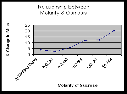
Obtain the six strips of presoaked dialysis tubing and create a bag out of each one by tying off one end.  Pour 25mL of the 6 solutions into separate bags. Tie off the other end of the 6 bags.  Rinse each bag gently with distilled water and blot dry.  Determine the mass of each bag and record it in Table 1.2.  Immerse each bag in one beaker filled will distilled water and label the beaker to indicate the molarity of the solution in the bag.  Let the setups stand for 30 minutes.  Remove the bags from the water.  Carefully blot them dry and determine their masses.  Record them in Table 1.2.  Obtain the other lab groups data to complete Table 1.3.

Exercise 1C:

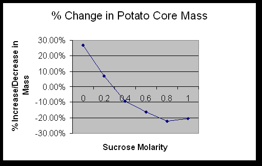
Pour 100mL of the solutions into a labeled 250mL beaker.  Use a cork borer to cut potato cylinders.  You need 4 cylinders for each cup.  Determine the mass of the 4 cylinders together and record the amount in Table 1.4.  Place the cylinders into the beaker of sucrose solution.  Cover the beaker with plastic wrap to prevent evaporation.  Let it stand overnight.  Remove the cores from the beaker and blot them gently on a paper towel and determine their total mass.  Record the results in Table 1.4.  Calculate the percentage change.  Do this for the individual and class data.  Graph the class average percentage change in mass.

Exercise 1D:

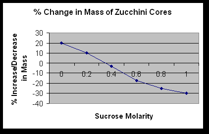
Determine the solute, pressure, and water potential of the sucrose solution.  Then, graph the information that is given about the zucchini cores.

Results:

Exercise 1A:

 Table 1.1


Initial Contents Initial Color Final Color Initial Presence of Glucose Final Presence of Glucose
Bag 15% glucose & 1% starch Cloudy White Purple Yes Yes
Beaker Water & IKI Brown Orange No Yes


  1. Which substances are entering the bag and which are leaving the bag? What evidence supports the answer?  Distilled water and IKI are  leaving and entering.  Glucose is able to leave the bag.
  2. Explain the results that were obtained.  Include the concentration differences and membrane pore size in the discussion.  Glucose and small molecules were able to move through the pores.  Water and IKI moved from high to low concentration.
  3. How could this experiment be modified so that quantitative data could be collected to show that water diffused into the dialysis bag?  You could mass the bag before and after it was placed into the solution.
  4. Based on your observations, rank the following by relative size, beginning with the smallest: glucose molecules, water molecules, IKI molecules, membrane pores, and starch molecules.  Water molecules, IKI molecules, Glucose molecules, Membrane pores, and Starch molecules
  5. What results would you expect if the experiment started with a glucose and IKI solution inside the bag and only starch and water outside?  The glucose and IKI would move out of the bag and turn the starch and water solution purple/blue.  The starch couldn’t move inside the bag because its molecules are too big to pass through the membrane of the tubing.

Exercise 1B:


Table 1.2: Dialysis Bag Results: Individual Data


Contents in dialysis bag Initial mass (g) Final mass (g) Mass difference (g) % Change in mass
Distilled Water 24.7 23.7 1 4.1
0.2M 26.7 27.4 .7 2.62
0.4M 27.4 29 1.6 5.84
0.6M 25.9 29 3.1 12
0.8M 29 32.6 3.6 12.41
1.0M 28 33.7 5.7 20.4


Table 1.3: Dialysis Bag Results: Class Data


Group 1

Group 2

Group 3

Total Class Average
Distilled Water 4.1% .7% 1.6% 6.4% 2.13%
0.2M 2.62% 6.4% 4.1% 13.12% 4.37%
0.4M 5.84% 9.9% 9.5% 25.24% 8.41%
0.6M 12% 13.4% 9.3% 34.37% 11.57%
0.8M 12.41% 14.6% 15.2% 42.21% 14.07%
1.0M 20.4% 19.7% 15.9% 56% 18.67%


  1. Explain the relationship between the change in mass and the molarity of sucrose within the dialysis bags.  The solute is hypertonic and water will move into the bag.  As the molarity increases the water moves into the bag.
  2. Predict what would happen to the mass of each bag in this experiment if all the bags were placed in a 0.4M sucrose solution instead of distilled water.  Explain.  With the 0.2M bag, the water would move out.  With the 0.4M bag, there will be no net movement of water because the solutions reach dynamic equilibrium.  With the 0.6M-1M bags, the water would move into the bag.
  3. Why did you calculate the percent change in mass rather than simply using the change in mass?  This was calculated because each group began with different initial masses and we would have different data.  All the groups needed consistent data.
  4. A dialysis bag is filled with distilled water and then places in a sucrose solution.  The bag’s initial mass is 20g and its final mass is 18g.  Calculate the percent change of mass, showing your calculations.  ((18-20)/20) x 100 = 10%
  5. The sucrose solution in the beaker would have been hypotonic to the distilled water in the bag.

Exercise 1C


Table 1.4: Potato Core: Individual Data


Contents of Beaker Initial Mass (g) Final Mass (g) Difference in Mass % Change in Mass
Distilled Water 2.8 3.7 .9 32.14
0.2M 2.9 3.1 .2 7
0.4M 2.5 2.2 .3 12
0.6M 2.3 1.9 .4 17.39
0.8M 2.5 1.9 .6 24
1.0M 2.3 1.8 .5 21.74


Table 1.5: Potato Core: Class Data


Group 1 Group 2 Total Class Average
Distilled Water 32.14% 21.1% 53.24% 26.62%
0.2M 7% 6.7% 13.7% 6.85%
0.4M -12% -6.5% -18.5% -9.25%
0.6M -17.39% -15.2% -32.59% -16.30%
0.8M -24% -20% -44% -22%
1.0M -21.74% -19% -40.74% -20.37%


Determine the molar concentration of the potato core.  0.3M

Exercise 1D


What is the molar concentration of the zucchini cores? .35M


  1. If a potato core is allowed to dehydrate by sitting in the open air, would the water potential of the potato cells decrease or increase? Why?  It would decrease because the water would leave the cells and cause the water potential to go down.
  2. If a plant cell has a lower water potential than its surrounding environment and if pressure is equal to zero, is the cell hypertonic or hypotonic to its environment? Will the cell gain water or lose water?  It is hypotonic and it will gain water.
  3. The beaker is open to the atmosphere.  What is the pressure potential of the system?  The pressure potential is zero.
  4. Where is the greatest water potential?  In the dialysis bag.
  5. Water will diffuse out of the bag. Why? It is because the water moves from and area of high water potential to an area of lower water potential.
  6. What effect does adding solute have on the solute potential component of that solution? Why?  It makes is more negative.
  7. Consider what would happen to a red blood cell placed in distilled water: a) Which would have the higher concentration of water molecules?  Distilled Water  b) Which would have the higher water potential?  Distilled Water  c)  What would happen to the red blood cell? Why?  It would lyce, because it would take on too much water.

Error Analysis:

Possible errors that could have affected the results of the lab include incorrectly mixing the solutions, ineffectively tying the dialysis tubing, inaccurately measuring , and inaccurately calculating.

Conclusion:

            During Exercise 1A the data that was collected help determine which molecules can and can not move across a cell membrane. Obviously, because of the color change in the bag, the IKI was able to move across the membrane.  It is small enough to fit through the pores in the selectively permeable membrane, along with water.  Starch was too large to move across the membrane. Glucose, as the Benedict’s test proves, was able to move freely along with the water and IKI solution.

In Exercise 1B, it was proven that water moves faster across the cell membrane than sucrose.  The water moved to help reach dynamic equilibrium between the 2 solutions.  The sucrose molecules are too big to move across the membrane as fast as water can.

The data in Exercise 1C showed that the potatoes contained sucrose.  The sucrose in the potato raised the solute potential, which lowered the water potential.  The beaker of distilled water had a high water potential.  Water moves down the concentration gradient, causing the potato cores to take on water.

Exercise 1D helped better understand the lab with simple algebra equations.  It proved that the data that was collected was correct through mathematics.


Ap Unit 6 Heredity Study Guide


Unit 5    Molecular Genetics  Study Guide 


ü     Be able to describe & explain the experiments of the following scientists:
Frederick Griffith
Erwin Chargraff
Alfred Hershey
Martha Chase
Watson & Crick
Meselson & Stahl
Beadle & Tatum
“Dolly” experiment

ü     Know how DNA replicates including steps & the enzymes involved, energy required, nucleotides, leading & lagging strands, proofreading

ü     Be able to describe the ultrastructure of each component of the DNA & RNA molecules

ü     Know the steps of transcription, enzymes involved, etc

ü     Know the steps of translation, enzymes involved, etc.

ü     Be able to describe the structure & function of free and bound ribosomes

ü     Know the processing steps of newly made mRNA

ü     Know the types of mutations and their effects

ü     Know viral structure, reproduction, host recognition, viral genome, etc

ü     Be able to describe the prokaryotic genome, mechanisms for genetic recombination, & defenses against phages

ü     Differentiate between hetero- & euchromatin

ü     Know the function and use of restriction enzymes

ü     Be able to describe genomic differentiation

BACK