 CHAPTER 32, PLANT REPRODUCTION
SECTION 32-1, PLANT LIFE CYCLES
A life cycle includes all of the stages of an organism’s growth and development. A plant’s life cycle involves two alternating multicellular stages – a Diploid (2n) sporophyte stage and a Haploid (1n) gametophyte stage. This type of life cycle is called Alternation of Generations. The size of gametophytes and sporophytes varies among the plant groups.
OBJECTIVES: Describe the life cycle of a moss. Describe the life cycle of a typical fern. Describe the life cycle of a gymnosperm. Compare and contrast homospory and heterospory.
THE LIFE CYCLE OF MOSSES
1. A Moss is a Nonvascular Seedless Plant belonging to the Phylum Bryophyta.
2. Mosses are the best known and most common Bryophytes. The other Bryophytes are Liverworts and Hornworts. There are about 14,000 kinds of mosses.
CHARACTERISTICS OF MOSSES
1. Mosses grow on moist brick walls, in sidewalks, as thick mats on forest floors, and on the Shaded Side of Trees. Some are adapted to the Desert, or can survive periodic dry spells, reviving when Water becomes available.
2. ALL MOSSES NEED WATER TO COMPLETE THEIR LIFE CYCLE.
3. MOSSES SHARE SOME CHARACTERISTICS OF OTHER BRYOPHYTES:
A. They do not have complicated Vascular Systems. – Nonvascular Plants
B. Water passes from cell to cell by osmosis. They are only a few cells thick.
C. They do NOT have True Roots, Leaves, or Stems.
D. They Require Water for Fertilization.
E. They are small land plants.
THE LIFE CYCLE OF MOSSES
1. The Dominant form of a moss is a clump of leafy Green Gametophytes.
2. A typical moss Alternates between a HAPLOID GAMETOPHYTE and DIPLOID SPOROPHYTE Phases. (Figure 32-1)
3. Haploid and Diploid refer to the number of Chromosomes in the Cells of an Organism.
4. A Gametophyte is the Haploid (1n) generation that produces GAMETES.
5. The Sporophyte is the Diploid (2n) that produces SPORES by Meiosis.
6. The Gametophyte of a moss is usually the largest and longest-lived generation of the moss life cycle.
7. Gametophytes of Mosses have RHIZOIDS, slender, Rootlike Structures that Anchors the Moss in place.
8. The Gametophytes are the Photosynthetic Part of a Moss.
9. The Sporophyte of a Moss is usually smaller than the Gametophyte and is attached to and dependent on the Gametophyte.
10. Sporophytes lack Chlorophyll, they Depend on the Photosynthetic Gametophyte for Food.
11. The Sporophyte consists of a Foot that anchors it to the Gametophyte and a Stalk. The Stalk grows up from the Foot and resembles a Street Lamp.
12. Atop the long, slender Stalk is a CAPSULE.
13. A CAPSULE IS THE STRUCTURE OF A MOSS THAT FORMS HAPLOID SPORES.
SEXUAL REPRODUCTION
1. Mosses, like most sexually reproducing organisms, produce TWO Kinds of GAMETES: EGGS AND SPERM.
2. GAMETES OF ALL BRYOPHYTES ARE SURRROUNDED BY A JACKET OF STERILE CELLS. The Sterile Cells are an important adaptation that protects the gametes from drying out and dying.
3. EGGS of Mosses are large, contain much Cytoplasm, and CANNOT Move.
4. SPERM are smaller and have FLAGELLA, enabling them to reach the Egg by swimming through Water.
5. THE EGG AND SPERM OF MOSSES FORM IN DIFFERENT REPRODUCTIVE STRUCTURES.
6. THE EGG-PRODUCING ORGAN OF A MOSS IS CALLED AN ARCHEGONIUM (ar-keh-GOH-nee-um). Each Flask-Shaped Archegonium forms ONE EGG by Mitosis. The Archegonia form on Branches of the Gametophyte.
7. THE SPERM-PRODUCING ORGAN OF A MOSS IS CALLED AN ANTHERIDIUM (an-theh-RIH-dee-um). Each Antheridium produces Many Sperm.
8. BOTH THE ARCHEGONIA AND ANTHERIDIA ARE PART OF THE GAMETOPHYTE.
9. Bryophytes such as Mosses are sometimes called the “Amphibians of the Plant Kingdom”. Mosses are Land Plants but they require Water for Sexual Reproduction.
10. For most Mosses, Fertilization can occur only during or soon after RAIN or after Flooding, when the Gametophyte is COVERED with Water.
11. The Sperm Swim to the Egg by following a Trail of Chemicals released by the Egg in the Water.
12. Fertilization produces a Zygote that undergoes Mitosis and becomes a Sporophyte.
13. When the Sporophyte matures cells inside the Capsule undergoes Meiosis and form Haploid Spores which are all the Same.
14. The production of One type of spores is called HOMOSPORY. The life cycle of Mosses is called HOMOSPOROUS ALTERNATION OF GENERATION.
15. THESE SPORES BEGIN THE GAMETOPHYTE GENERATION.
16. When spores are mature, the Capsule opens and Spores are carried off by Wind. If a spore lands in a Moist place, it Sprouts and forms a New Gametophyte.
ASEXUAL REPRODUCTION
1. Asexual Reproduction of most Mosses can occur in TWO WAYS:
A. FRAGMENTATION – Small pieces broken from a Gametophyte grow into a new plant.
B. GEMMAE – These are tiny pieces of Tissue that can form new Gametophytes.
2. When raindrops splash Gemmae from the Parent Plant, The Gemmae are carried to a new area where they can form Gametophytes.
THE LIFE CYCLE OF FERNS
1. Ferns are by far the LARGEST Group of Living Seedless Vascular Plants.
2. Ferns grow in a variety of places and are diverse in their appearance.
3. Like other Seedless Plants, Ferns usually live in Moist Habitats because they Need Water for Fertilization.
4. A TYPICAL FERN ALTERNATES BETWEEN HAPLOID GAMETOPHYE AND DIPLOID SPOROPHYTE PHASES. (Figure 32-2)
5. The Sporophyte Phase of the Fern’s Life Cycle is the Dominant Phase.
6. Fern Gametophytes are Tiny, Flat Plants that are Anchored to the soil by Rhizoids.
7. Both ANTERIDIA (Male) and ACHEGONIA (Female) form on the lower surface of a Fern Gametophyte.
8. When Water is present, Sperm released by Antheridia Swim to Archegonia.
9. One Sperm Fuses with the Egg in an Archegonium. Forming a Zygote, which is the First Cell of the Sporophyte.
10. In its Sporophyte Stage, a typical Fern has a Stem with True Roots and True Leaves. The Stem, Roots and Leaves are considered TRUE because they have special Water-Carrying Tissues.
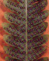 11. The Gametophyte Generation of Ferns begins with Spores. Some Ferns form Spores on specialized stalk-like leaves. Like mosses, most ferns are Homosporous.
12. Other Ferns form Spores in Special Structures on the UNDESIDE OF THE LEAVES. A SORUS (SORI) IS A GROUP OF SPORE-CONTAINING STRUCTURES (SPORANGIA) CLUSTERED ON THE UNDERSIDE OF A FERN LEAF (Figure 32-2).
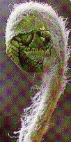 13. The Leaf of a Mature Fern Sporophyte is a Compound Leaf and is divided into smaller Leaflets. A MATURE LEAF OF A FERN IS CALLED A FROND, which grows from an underground stem, or RHIZOME. The new young immature leaf is called a FIDDLEHEAD.
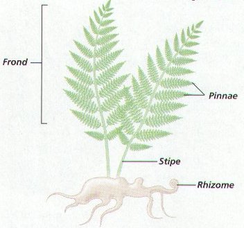 14. Each Frond consists of TWO Parts: a BLADE and PETIOLE (PEH-tee-ohl).
A. The BLADE is the broad, flat, photosynthetic surface of the Frond. The Blade contains the Chloroplast. The Blade also contains Vascular Tissue that brings water and minerals from roots.
B. On most ferns, a Blade does not attach directly to a Stem. Instead, a Stalk attaches the Blade to the Stem. The PETIOLE is the Stalk that attaches the Frond’s Blade to the Stem. The Petiole contains vascular tissue that carries Water and nutrients through the Plant.
LIFE CYCLES OF CONIFERS – GYMNOSPERMS – NAKED SEEDS – CONES
1. The oldest surviving Seed Plants on Earth are Gymnosperms. In Seed Plants the Sporophyte Phase is the Dominant Phase.
2. Gymnosperms are referred to as Naked Seeds, because they develop on the Scales of Female Cones and NOT inside a Fruit.
3. Gymnosperms are adapted to live in cold climates; there are extensive forests of gymnosperms in most of the colder zones of northern temperate regions.
4. There are about 700 species of gymnosperms, such as pine, fir, and spruce, which are also called Evergreen Trees.
5. Gymnosperms include one of the largest and some of the oldest organisms on Earth. The Giant Redwood is one of the Earth’s largest organisms. The Bristlecone Pines is among the oldest, some more than 5000 years old.
6. Unlike mosses, and most Ferns, Gymnosperms produce TWO Types of spores – MALE MICROSPORES AMD FEMALE MEGASPORES.
7. Microspores grow in into Male Gametophytes, while Megaspores grow into Female Gametophytes.
8. The Production of different types of Spores is called HETERSPORY. The Gymnosperm Life Cycle is called HETEROSPOROUS ALTERNATION OF GENERATION.
9. Heterospory ensures that a Sperm will fertilize an Egg from Different Gametophyte and increase the chance that New Combinations of Genes will occur among Offspring.
10. Gymnosperms are Plants (Trees) that reproduce by way of CONES. (Figure 32-3)
11. The Pine Tree is a typical Gymnosperm. The Large, Familiar Cones known as Pinecones are actually the FEMALE Cones of a Pine Tree.
12. Pine trees also have MALE Cones, which are SMALLER than Female Cones. Male and Female Cones have a vital roles in the reproductive cycle of pine trees and other Gymnosperms.
13. THE LIFE CYCLE OF A PINE TAKES TWO OR THREE YEARS FROM THE TIME THE CONES FORM UNTIL SEEDS ARE RELEASED.
14. Male and Female Gametes are made by the Male and Female Cones, which are on the SAME Tree.
15. The Female Cones consist of spirally arranged Scales and Secrete a STICKY RESIN.
16. At the Base of each scale are TWO EGG-CONTAINING OVULES.
17. AN OVULE IS A STRUCTURE, CONSISTING OF AN EGG INSIDE PROTECTIVE CELLS, THAT DEVELOPS INTO A SEED.
 18. Each Male Cone produces huge amounts of POLLEN that are released in Spring. Pollen Grains have WINGLIKE Structures that keep them aloft in the WIND. Pollen Grains can be carried long distances to reach Female Cones FOR POLLINATION.
19. POLLINATION IS THE TRANSFER OF POLLEN FROM THE MALE TO THE FEMALE PART OF A PLANT.
20. When a Pollen Grain reaches a Female Cone, it sticks to the RESIN of the cone. As the Resin dries, the Pollen Grain begins to grow a structure called a POLLEN TUBE that extends to an Ovule near the base of a Scale, it enables the sperm to reach an egg. The Pollen Tube takes about a year to grow and reach the Egg.
21. A Sperm Cell released from the Pollen Tube Fertilizes an Egg in the Ovule forming a Zygote. Pine sperm Do Not have Flagella and they do Not Swim to an Egg.
22. A Zygote forms and grows into an Embryo surrounded by a SEED.
23. As the Embryo Matures, the Pinecone enlarges and the scales Separate releasing the Seed from the Female Cone.
24. If the seed lands in an environment with the proper conditions for growth, it will sprout and form a New Sporophyte Pine Plant.
25. Seed Plants Do Not Require Water for Reproduction, Sexual Reproduction in Seed Plants can therefore take place independent of seasonal rains or other periods of moisture.
SECTION 32-2 SEXUAL REPRODUCTION IN FLOWERING PLANTS – ANGIOSPERMS – FLOWERS & FRUITS
You have probably admired flowers for their bright colors, attractive shapes, and pleasing aromas. These characteristics are adaptations that help ensure sexual reproduction by attracting animal pollinators. But some flowers are not so colorful, large, or fragrant. Such flowers rely on wind or water for pollination.
OBJECTIVES: Identify the four main flower parts, and state the function of each. Describe ovule formation and pollen formation in angiosperms. Relate flower structure to methods of pollination. Describe fertilization in flowering plants. Compare and contrast the gymnosperm and angiosperm life cycles.
ANGIOSPERMS REPRODUCTION (FLOWERS & FRUITS)
1. The importance of a Flower is NOT in the way it LOOKS or SMELLS, but in WHAT IT DOES.
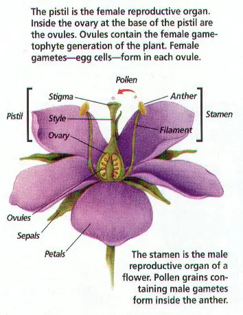 2. A FLOWER IS IMPORTANT BECAUSE IT IS THE REPRODUCTIVE STRUCTURE OF AN ANGIOSPERM.
3. FLOWERS are MODIFIED STEMS with SPECIALIZED LEAVES and other structures for REPRODUCTION. All of these specialized leaves from on the Swollen Tip of a floral “Branch” which is called the RECEPTACLE.
4. FLOWERS HAVE THREE BASIC COMPONENTS: MALE, FEMALE, AND STERILE PARTS.
5. The Male and Female Parts Produce the GAMETES. Sterile Parts ATTRACT POLLINATORS (The Birds and The Bees) and Protect the Female Gametes.
6. Flowers that produce BOTH Male and Female Gametes in the SAME Flower are called PERFECT FLOWERS.
7. IMPERFECT FLOWERS are EITHER a Male or a Female Flower.
8. Some Angiosperms have separate Male and Female Flowers, but BOTH SEXES are on the SAME Plant. Others, the entire plant is Male or Female.
FEMALE STRUCTURES OF FLOWERS
1. The Female Structures of flowers produce EGGS.
2. THE FEMALE, OR EGG-PRODUCING, PART OF A FLOWER IS CALLED THE CARPELS.
3. ONE OR MORE CARPELS FUSED TOGETHER MAKE UP THE STRUCTURE CALLED THE PISTIL. Pistils form at the CENTER of the Flower and usually have THREE PARTS: STIGMA, STYLE, AND OVARY, EACH PART HAS A DIFFERENT FUNCTION:
A. THE STIGMA IS THE STRUCTURE ON WHICH POLLEN LANDS AND GERMINATES. IT IS USUALLY STICKY OR HAS HAIRS TO HOLD POLLEN GRAINS. THE TIP OF THE STYLE.
B. THE STYLE IS THE STALK-LIKE STRUCTURE CONNECTING THE STIGMA TO THE OVARY.
C. THE OVARY IS THE ENLARGED BASE OF A PISTIL, IT IS THE STRUCTURE THAT CONTAINS OVULES AND DEVELOPS INTO A FRUIT. OVULES FORM IN THE OVARY, AND EACH OVULE CONTAINS AN EGG.
MALE STRUCTURES OF FLOWERS
1. THE MALE STRUCTURES OF FLOWERS PRODUCE POLLEN.
2. THE MALE, OR POLLEN-PRODUCING PART OF A FLOWER, IS CALLED THE STAMEN.
3. STAMENS USUALLY HAVE TWO PARTS: ANTHER AND FILAMENT. EACH PART HAS A DIFFERENT FUNCTION:
A. THE ANTHER IS THE STRUCTURE THAT CONTAINS MICROSPORANGIA, WHICH PRODUCE MICROSPORES THAT DEVELOP INTO POLLEN GRAINS. POLLEN GRAINS CONTAIN SPERM CELLS.
B. THE FILAMENT IS THE STRUCTURE THAT HOLDS UP AND SUPPORTS THE ANTHER.
STERILE PARTS OF A FLOWER (ATTRACT/PROTECT)
1. THE STERILE PARTS OF A FLOWER ARE THE PETALS AND SEPALS.
2. PETALS are usually Colorful, Leaflike appendages on a Flower. Their Function is to ATTRACT Pollinators.
3. ALL THE PETALS IN A FLOWER ARE COLLECTIVELY CALLED THE COROLLA.
4. The Protective Leaves at the Base of a Flower are SEPALS. Sepals are often Green, cover the BUD of a Flower and Protect the developing Flower parts as they Grow.
5. ALL THE SEPALS ARE COLLECTIVELY CALLED THE CALYX.
6. Monocots and Dicots can often be distinguished by their Flowers. MONOCOT Floral Parts are arranged in multiples of THREE, The Floral Parts of DICOTS are arranged in multiples of FOUR OR FIVE.
7. FLOWER PARTS ARE USUALLY FOUND IN FOUR CONCENTRIC WHORLS, OR RINGS. (Figure 32-5)
A. OUTERMOST WHORL – THE SEPALS (Calyx) (#1)
B. THE PETALS (Corolla) MAKE UP THE NEXT WHORL. (#2)
C. THE TWO INNERMOST WHORLS OF FLOWER PARTS CONTAIN THE REPODUCTIVE STRUCTURES. FIRST THE MALE (STAMENS, #3) AND THE INNERMOST WHORL CONTAINS THE FEMALE (CARPELS, #4). ONE OR MORE CARPELS FUSED TOGETHER MAKE UP THE PISTIL.
LIFE CYCLE OF ANGIOSPERMS
1. An Angiosperm undergoes Alternation of Generations. The Sporophyte undergoes meiosis to form spores, which then divide mitotically to form Gametophytes.
2. The Gametophytes form the GAMETES: EGG AND SPERM.
3. Sexual Reproduction BEGINS WHEN MICROSOPORE MOTHER CELLS undergo Meiosis in the ANTHER to become Pollen Grains, which is a two-celled or three-celled Male Gametophyte. (Figure 32-7) Notice: Each of the four Microspores will form a Pollen Grain that consists of Two Cells a Tube Cell and a Generative Cell, The Male Gametophyte.
4. During the same time, MEGASPORE MOTHER CELLS undergo Meiosis in Ovules, forming four megaspores in each Ovule, One will become an EGG. (Figure 32-6) Notice: Of the four Megaspores, Three of the Megaspores Degenerate, and the Fourth forms the structures of the Embryo Sac, The Female Gametophyte.
5. Because the Ovule of a flower contains the egg, the ovule contains the Female Gametophyte.
6. The next step is Pollination, the transfer of Pollen from the Anther to the Stigma.
7. When a pollen Grain lands on a Stigma, it sends out a POLLEN TUBE that grows through the Style to the Ovary. Inside the Ovary it enters and Ovule which contains an Egg.
8. Fertilization occurs when a Sperm Nucleus from the Pollen Tube FUSES with the Egg and forms a Zygote.
9. While one sperm fertilizes and Egg, a Second Sperm Nucleus from the pollen tube fertilizes TWO Polar Nuclei.
10. The Second Fertilization forms a Food-Storing Tissue in the Seed called ENDOSPERM.
11. The process in plants that involves TWO Fertilizations is called DOUBLE FERTILIZATION. ONLY ANGIOSPERMS HAVE DOUBLE FERTILIZATION.
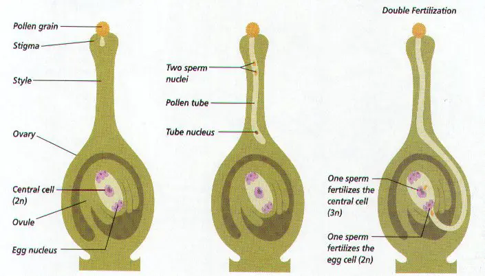
12. After Fertilization, The Zygote develops into an Embryo; The Ovule Becomes a Seed, and Ovary and Surrounding Tissue from a Fruit. A FRUIT IS A MATURE OR RIPPENED OVARY.
13. After a mature seed is planted, it sprouts and begins to develop a plant that is the next Sporophyte Generation.
POLLINATION – THE BRIDS AND THE BEES
1. POLLEN is usually carried from Plant to Plant by WIND, WATER or ANIMALS.
2. Many plants are Pollinated by Animals. Plants may Attract Pollinators with Colorful Flowers, Fragrances, and Sugary Nectar.
3. As Pollinators climb around a Flower searching for and Drinking Nectar, they cover their bodies with Pollen.
4. As a Pollinator moves from one Flower to the Next, Pollen falls from the Pollinators Body as it moves, thus Pollinating the Flowers.
5. BEES, LADYBUGS, MOTHS, BUTTERFLIES, BIRDS, BATS, AND BEETLES ARE SOME ANIMALS THAT POLLINATE PLANTS. (Figure 32-6)
6. WE CAN ALSO POLLINATE PLANTS, AS WE BRUSH UP AGAINST FLOWERS AND INADVERTENLY COLLECT AND TRANSFER POLLEN.
7. In some plants, Pollen Falls from Anther to Stigma on the same flower, thus eliminating the need for a Pollinator. THIS PROCESS IS CALLED SELF-POLLINATION.
8. Self-Pollination is beneficial for plants that are isolated from their own kind. Self-Pollination is usually Undesirable, because it reduces the chances of getting new combination of genes.
9. HYBRIDIZING OR CROSS POLINATION, THE MATING OF TWO INDIVIDUALS WITH DIFFERENT TRAITS, IS MORE DESIRABLE, BECAUSE IT ALLOWS FOR NEW COMBINATIONS OF GENES.
10. Mechanisms for increasing the chances of Hybridizing are common in many types of plants:
A. Producing separate Male and Female Flowers.
B. Having separate Male and Female Plants.
C. Pollen of one plant matures at a different time from the eggs in the ovary.
ALL OF THESE MECHANISMS PROMOTE HYBIRDIZING, THE RECOMBINATION OF
GENES IN THE SPECIES.
11. FERTILIZATION, which is the Union of Gametes, follows Pollination. (Figure 32-9)
12. In order for Fertilization to occur, a Pollen Tube must grow to an Egg, and Sperm must form.
SECTION 32-3, DISPERSAL AND PROPAGATION
Fruits and seeds normally result from sexual reproduction in flowering plants. Fruits are adaptations for dispersing seeds, while seed function in the dispersal and propagation of plants. Many plants also propagate (produce new individuals) through asexual reproduction.
OBJECTIVES: Name different types of fruits. Describe several adaptations for fruit and seed dispersal. Compare and contrast the structure and germination of different types of seeds. Recognize the advantages of asexual reproduction. Describe methods of vegetative propagation.
DISPERSAL OF FRUITS AND SEEDS
1. Seeds are as diverse as the plants they produce. Some seeds, such as peach and apple seeds, grow inside Fleshy Fruit. Others, such as bean seeds, grow in Pods. Seeds of gymnosperms grow on the scales of cones.
2. The main difference between Seed Plants and Seedless Plants is that Seed Plants develop Reproductive Structures called SEEDS instead of Spores.
3. A Seed Protects and Nourishes the Embryo it contains.
4. Seeds may differ in appearance and some structures; ALL SEEDS contain a Plant Embryo and Stored Food in a Protective Coat.
5. It takes more energy for a plant to produce Seeds than to produce Spores. But Seeds have an advantage over Spores, the ability to remain Dormant.
6. Dormant Seeds are inactive while they wait for optimal growing conditions. Some seeds can only remain dormant for a few weeks, others for several or even thousands of years.
7. Spores are light and are easily dispersed by wind to new environments, Most Seeds are too HEAVY to be carried by Wind and require a means of Dispersal.
8. Fruits and Seeds Dispersed by wind or water are adapted to those methods of dispersal. (Figure 32-10) Milkweeds Seeds have “parachutes” that help them drift with the wind.
9. Many plants that grow near Water produce Fruits and Seeds that Contain Air Chambers, which allows them to Float.
10. Some seeds have Sticky or Prickly exteriors that cling to passing animals. The animals carry the seeds away from the parent plant to new locations.
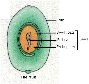 11. Other Seeds are encased in Fleshy Fruit. The Smell, Bright Color, or Flavor of many Fruits Attract Animals.
12. When animals eat the fruit, the Seeds pass unharmed through the Digestive Tracts and are Deposited Elsewhere.
13. Botanists define a FRUIT as a Mature OVARY. Many different types of fruits have evolved among flowering plants. (Figure 32-11)
14. Fertilization usually initiates the development of Fruits. Fruits Protect the Seeds, aid in their dispersal, and often Delay their Sprouting.
15. Fruits are Classified Mainly on the basis of HOW MANY PISTILS OR FLOWERS FORM THE FRUIT AND WHETHER IT IS DRY OR FLESHY. There are Three Basic Types of Fruits: Table 32-1
16. SIMPLE FRUIT – formed from One Pistil of a Single Flower. Can be Dry or Fleshy at Maturity.
17. AGGREGATE FRUIT – formed from Several Pistils of a Single Flower. Can be Dry or Fleshy at Maturity.
18. MULTIPLE FRUIT – Formed from SEVERAL FLOWERS Growing Together. Can be dry or Fleshy at Maturity.
STRUCTURE OF SEEDS
1. ANGIOSPERMS ARE FLOWERING PLANTS. TODAY, ABOUT 270,000 SPECIES OF ANGIOSPERMS HAVE BEEN DISCOVERED AND NAMED.
2. Angiosperms (Flowering Plants) are divided into TWO Groups Monocots and Dicots.
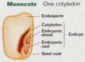 3. Angiosperms with only ONE Cotyledon in their Seeds are called Monocots.
 4. Angiosperms with TWO Cotyledons are called Dicots.
5. Cotyledons are a leaflike structure that is part of the Plant Embryo.
6. Although Cotyledons look like leaves and develop before leaves, they ARE NOT TRUE Foliage Leaves.
7. In Angiosperms Seeds, the THREE Parts of the Embryo are Named according to their relationship with the Cotyledons. (Figure 32-12)
8. THE PART OF THE PLANT EMBRYO ABOVE THE COTYLEDON IS CALLED THE EPICOTYL (EP-ih-kot-ul). The Epicotyl includes most of what will become the Stem and Leaves of the plant.
9. THE AREA OF THE PLANT EMBRYO THAT WILL BECOME THE EMBRYONIC ROOT IS CALLED THE RADICLE.
10. THE AREA OF THE PLANT EMBRYO BETWEEN THE COTYLEDONS AND THE RADICLE IS CALLED THE HYPOCOTYL (Hy-poh-kot-ul).
11. The Epicotyl, along with any Embryonic Leaves, is called the PUMULE.
12. Along the concave edge of the Seed is the HILUM, which is a Scar that marks where the Seed was Attached to the Ovary Wall.
13. Surrounding the Seed is a SEED COAT that Protects the Embryo and its Food Supply (ENDOSPERM).
14. In the Seeds of Monocots, the Sheath that Protects the young plant as it grows out of the soil is the COLEOPTILE. (Figure 32-14)
15. In most Seeds, food is stored as Starch, a Carbohydrate (SPECIAL FOOD STORING TISSUE CALLED ENDOSPERM); some Seeds also contain Proteins and Lipids (Fats).
SEED GERMINATION
1. Many plants are easily grown from seeds. Although its embryo is alive, a Seed will Not Germinate, or Sprout, until it is exposed to Certain Environmental Conditions.
2. Delaying of Germination often assures the survival of the plant. If Seeds that mature in the fall were to sprout immediately, the young plant could be killed by cold weather.
3. If all a plant’s seeds were to sprout at once and all of the New Seeds Died before producing seeds, the species could become Extinct.
4. Many seeds Will Not GERMINATE even when exposed to conditions ideal for Germination. Such seeds exhibit DORMANCY, which is a state of reduced metabolism.
CONDITIONS NEEDED FOR GERMINATION
1. Environmental Factors, such as Water, Oxygen, and Temperature Trigger Seed Germination.
2. Most Seeds are Very DRY and must absorb Water to Germinate.
3. Water Softens the Seed Coat and Activates Enzymes that convert Starch in the Cotyledons or Endosperm into Simple Sugars, which provided energy for the embryo to grow.
4. As the embryo begins to grow, the soften seeds coat cracks open, enabling the Oxygen needed for Cellular Respiration to reach the embryo.
5. Seeds will only Germinate it the Temperature is within a certain Range. Many Seeds need Light for Germination, this prevents the seeds from sprouting it they are buried to deeply.
6. Some Seeds Germinate only after being exposed to Extreme Conditions, After Freezing or passing through a digestive system that breaks down the Seed Coat.
PROCESS OF GERMINATION
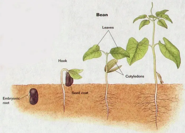
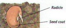 1. The first Visible Sign of Seed Germination is the emergence of the RADICLE (ROOT). (Figure 32-14)
2. Soon after the Radicle Breaks the Seed Coat, the SHOOT begins to Grow.
3. In some Seeds (Dicot, Bean) the Hypocotyl curves and become hooked-shaped. Once the hook breaks through the soil, the Hypocotyl Straightens.
4. The Plumule’s Embryonic Leaves unfold, synthesize Chlorophyll, and begin Photosynthesis. After their Stored Nutrients are used up, the shrunken Cotyledons fall off.
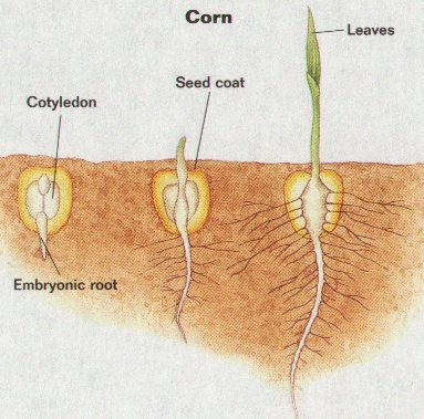 5. In contrast (Monocot, Corn), the Cotyledon of the Corn Seed Remains Underground and transfers Nutrients from the Endosperm to the growing Embryo.
6. The Corn Hypocotyl Does not Hook or Elongate, and the Cotyledons remains Below Ground. The Corn Plumule is protected by a Sheath (Coleoptile) as it passes through the soil.
7. When the Shoot breaks through the soil surface, the Leaves of the Plumule unfold.
ASEXUAL REPRODUCTION IN PLANTS
1. ASEXUAL REPRODUCTION involves NO FERTILIZATION AND PRODUCES OFFSPRING THAT ARE GENETICALLY IDENTICAL TO THE PARENTS -CLONES.
2. Most plants reproduce Asexually at least some of the time, while other plants reproduce Asexually most of the time.
3. In a sable environment with abundant resources, asexually reproduction is FASTER, and produces offspring that are well adapted to the existing environment.
4. ASEXUAL REPRODUCTION THAT OCCURS NATRUALLY IN PLANTS IS CALLED VEGETATIVE REPRODUCTION. Reproduction occurs from Non-Reproductive Parts, such as Leaves, Stems, and Roots. (Figure 32-15)
5. WHEN WE USE ASEXUAL METHODS TO GROW PLANTS WE CALL IT VEGETATIVE (ARTIFICIAL) PROPAGATION.
6. VEGETATIVE PROPAGATION IS A BY-PRODUCT OF A PLANT’S ABILITY TO REGENERATE LOST PARTS.
7. Many species of plants are Vegetative Propagated from Specialized Structures such as Runners, Rhizomes, Bulbs, and Tubers. (Table 32-2)
8. METHODS OF VEGETATIVE PROPAGATION INCLUDE CUTTINGS, GRAFTING, TISSUE CULTURING AND LAYERING.
A. CUTTING – Taking a piece of Stem or Leaf and planting it in soil to grow a new plant.
B. GRAFTING – A way to make TWO Different plants grow as one by fusing their cut ends.
C. TISSUE CULTURING – Growing a new plant from individual cells, or from small pieces of Leaf, Stem or Roots. (Figure 32-16)
D. LAYERING – Roots form on Stems where they make Contact with the Soil. People Stake the Branch Tips to the Soil or Cover the Base of Stems with Soil to Propagate the Plants.



