PLANT EVOLUTION AND CLASSIFICATION
1. There are more than ________________ different plant species.
2. Plants share Four Characteristics:
A._________________________________________________________________
B._________________________________________________________________
C._________________________________________________________________
D._________________________________________________________________
3. In their characteristics plants are most similar to the ________________________.
4. Plants and Green Algae Have these Characteristics in Common:
A.__________________________________________________________________
B.__________________________________________________________________
C.__________________________________________________________________
D.__________________________________________________________________
5. There are also some important Difference:
A.__________________________________________________________________
B.__________________________________________________________________
C.__________________________________________________________________
D.__________________________________________________________________
6. All plants are photosynthetic, multicellular, __________________________ organisms, and can _________________________ _________________________.
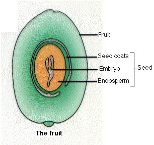
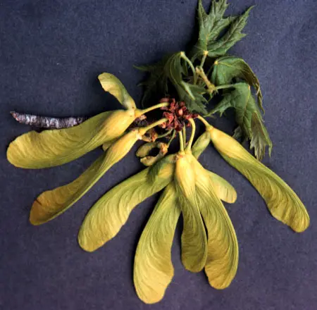
7. A ____________________ is a ripen ovary that surrounds the seeds of angiosperms.
8. All plants probably evolved from ______________________ __________________.
9. One of the greatest problems that encountered by the first land plants was the need for
___________________________.
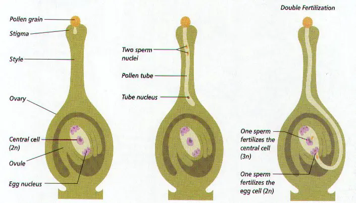
10. How does water aid the fertilization of some organisms? ______________________
____________________________________________________________________
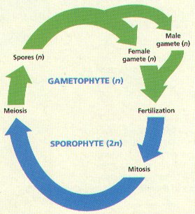
11. _________________________ of _______________________ means that there are TWO
phases in the life cycle of plants:
A. The first phase: ___________________ ______________________ phase that produces ________________________ and _______________________.
B. The second phase: ___________________ _____________________ phase that produces ________________________.
12. Sexual reproduction ensures there will be __________________________ ______________________ in plants.
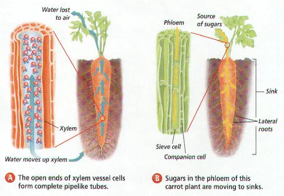
13. The type of vascular tissue that transports organic compounds is ____________________________.
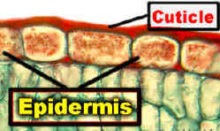
14. The _____________________ is a waxy, waterproof layer that coats the parts of a plant
exposed to air.
15. The earliest plants were probably __________________, and had NO true ___________,
____________________, or ______________________.
16. __________________ is a hard compound that strengthens cell walls, enabling cells to support additional weight.
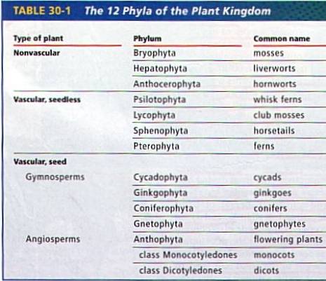
17. The 12 Phyla of plants can be divided into two groups based on the presence of __________________________ ___________________________.
18. One adaptation that help land plants to slow the evaporation of water was a
____________________________.
19. The type of vascular tissue that transports water is _________________________.
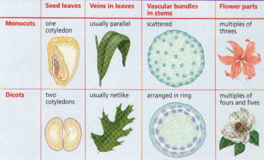
20. This type of angiosperm has parallel leaf venation __________________________.
21. The waxy covering on plant surfaces is called _____________________________.
22. The plant material in peat bogs decomposes very ________________________ because the bogs are ____________________________.
23. How many plant phyla produce seeds? _____________________
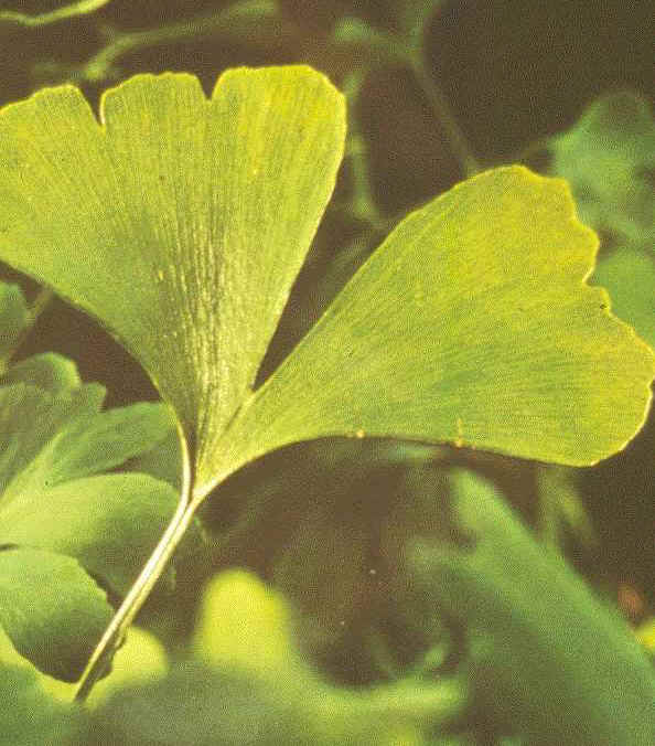
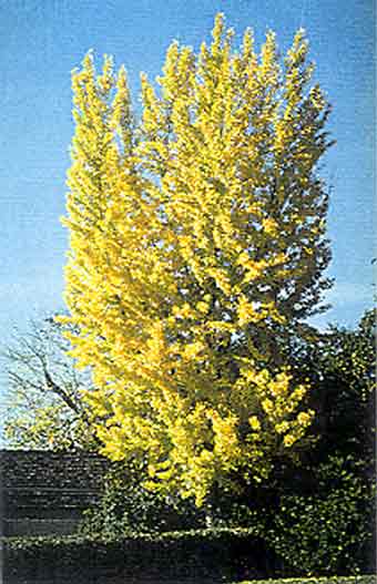
24. What type of gymnosperm produces fleshy seeds? ____________________________
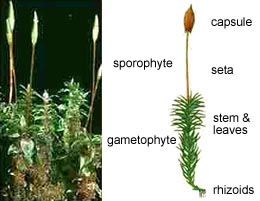
25. What is the photosynthetic phase of a moss called? ______________________________
26. Bryophytes, instead of roots, they have long, thin strands of cells called ____________________ that attach the plant to the soil.
27. Vascular plants absorb water from the soil through underground structures called
_____________________. They also provide a plant with ___________________.
28. Non-woody plants are usually called ___________________________.
29. _____________________ carries organic compounds in any direction depending on the plant’s needs.
30. In order to reproduce, a nonvascular plant must have ________________________.
31. Rhizoids are long, thin strands of cells that resemble ________________________.
32. The roots of vascular plants absorb water and _________________________ _________________________.
33. What is the non-photosynthetic phase of a moss called ____________________________.
34. Gymnosperms produce “_____________________” seeds, while angiosperms produce _______________________ protected inside a _____________________________.
35. This type of angiosperm has net leaf venation __________________________.
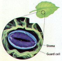
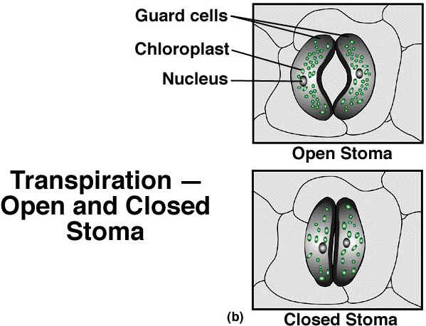
36. The _________________________ allow for the exchange of carbon dioxide and oxygen.
37. Sphagnum is often used to ______________________ soil and help it ____________________ __________________________.
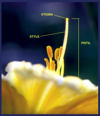
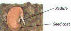
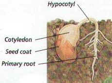
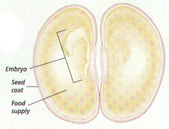
38. A ___________________ is a protective structure that contains a plant
__________________, and _________________ __________________.
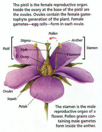
39. A __________________ is a structure that develops in plants with flowers and contains the
____________________.
40. Nonvascular plants are distinguished by the absence of ______________________ and ____________________________.
41. All nonvascular plants are collectively called _______________________________.
42. Vascular plants are classified into one of Two Types: _______________________ or
________________________________ plants.
43. What are the Four Phyla of Seedless Vascular Plants? ________________________,
________________________, ______________________, ________________________.
44. What are the Five Phyla of Seed Vascular Plants? _______________________,
_________________________, _________________________,
________________________, and ______________________________.
45. Vascular seed plants are subdivided into TWO general categories according to the type of seeds they produce: _________________________________ and
____________________________________.
46. A ____________________________ is a special reproductive structure composed of hard scales, that produces seeds without a fruit.
47. ____________________ are vascular plants that produce seeds lacking a protective
_______________________. They are often called _______________ _________.
48. A seed is a _________________________ embryo inside a __________________________ _____________________.
49. The _____________________ are vascular plants that produce seeds enclosed and
__________________ by a __________________.
50. All angiosperms produce _________________ and _________________.
51. The protective structure that contains the seed or seeds of an angiosperm is the
______________________.
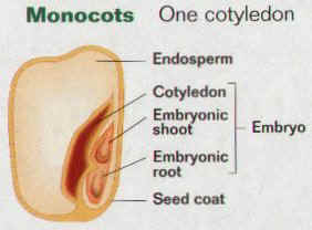
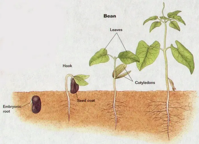
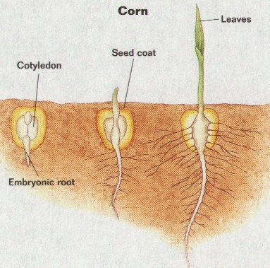
52. One way of distinguishing among the many types of angiosperms is by counting the number of seed leaves or ________________________.
53. Angiosperms with only ONE cotyledon are called _______________________________ or simply _____________________.
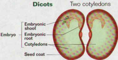
54. An angiosperm whose embryo has TWO cotyledons are called __________________________________ or simply _______________________.
56. Plants that produce seed protected by a fruit are called _______________________________.
57. A dicot is an angiosperm whose embryo has Two _______________________.
58. Plants remove carbon dioxide from the air by the process of ________________________.
59. Bryophytes are _______________-growing plants that live in _____________________ ________________________________.
60. All vascular plants have __________________________ tissues and _____________________________ of _________________________________.
61. True roots, stems, and leaves are characteristics of all ______________________ _________________________.
62. What are the primary functions of spores and seeds?
63. In what ways do green algae differ from plants?
64. Why do nonvascular plants have to live in moist environments?
65. Name three bryophytes, and identify their common characteristics.
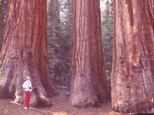
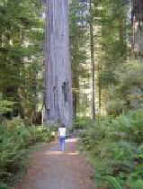
66. Which plant phylum contains the tallest and most massive plants? Is this a phylum of nonvascular, seedless vascular, or seed plants?
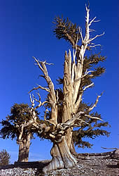
67. Conifers are often found living at high elevations in locations with cold, dry winters. What characteristic enables them to retain their leaves in these conditions?


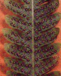
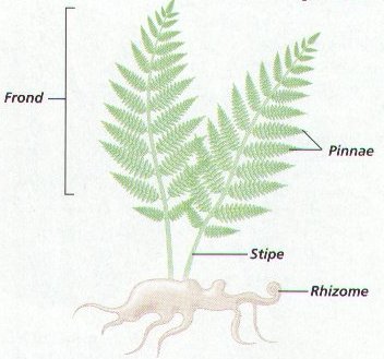
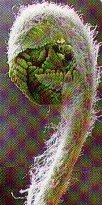
 Fern Characteristics & Life Cycle:
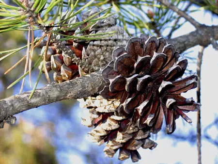
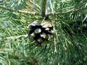
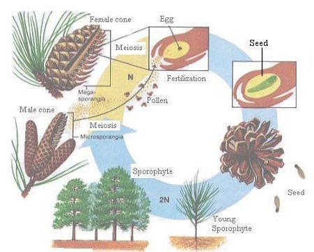
 Characteristics & Life Cycle of Conifers: