PreAP Protozoan Study Guide


Protozoan Review    


1. Protists with animal-like characteristics are called ___________________________.

2. Protozoans are all ______________________ organisms.

3. One convent way to classify protozoans is based on the way they _______________.

4.  _____________________ move by extending their ___________________.  ______________________ propel themselves by ______________________. _____________________ move by hairlike ____________, and _________________________ do ___________ move by themselves at all.

5. Protozoans that move by extending lobes of cytoplasm are called _______________________________.

6. The lobes of cytoplasm that sarcodinians extend are called _____________________ which means “_____________  ________________”.

7. When conditions are unfavorable, many amoebas survive by becoming hard ______________.  The ____________ can withstand drought, heat, or being eaten by other organisms.

8. Not all sarcodinians are soft “Naked”, some have hard shells or  _______________ made of   __________________________________ or _______________________.  They are called ____________________________ and _______________________________.

9. Sarcodinians are protozoans that move by extending lobes of _________________________.

10. ________________________________ are protozoans that move by means of flagella.

11. Some zooflagellates are free-living ________________________ or ____________________ organisms; many can live inside other organisms in _____________________ relationship.  Zooflagellates may have a ________________________ or _________________________________ relationships with other organisms.

12. Which zooflagellate parasite causes African Sleeping Sickness in humans? ____________________________.  The disease is spread by the bite of the __________________  ___________________.

13.  _________________________ are protozoans that have bodies covered with short hairlike projections called _________________.  They beat like ___________ to propel these protists through the water.

14. The ____________________________ is a common freshwater ciliate.

15. Paramecium gathers food with its _____________.  The ____________ sweeps food particles into the ______________  _________________, the Mouth _____________ opens into a ___________________  which pinches off around them to form a ____________________  ______________________.  It ejects wastes through an opening called the ______________________  __________________________.

16. Water is constantly enters the Paramecium cell by _______________________.  They would burst if they did not have a way to get rid of excess water.  ______________________________  _____________________________ collect the excess water and pump it outside.

17. Like all ciliates, Paramecium has ______________ distinct kinds of nuclei, each with a different function.  The ___________________________ controls ongoing functions of the cell and __________________________ reproduction.  Ciliates reproduce _______________________ by cell division.  The _____________________________ is involved in genetic exchange during ___________________________ reproduction.

18. When a Paramecium reproduces sexually, it exchanges genetic information by ________________________________________.

19. The protozoans that have NO structures for movement, and lives by being a parasite in animals are the ______________________________.  They are ____________-__________________ ____________________ protozoans.

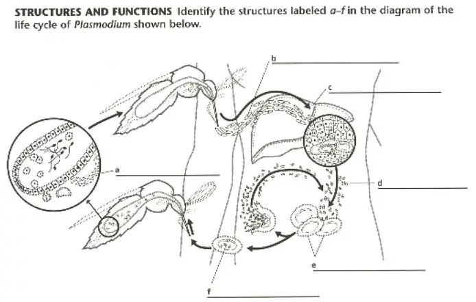
20. The protozoan that causes malaria is named ________________________ and is carried within _________________  ____________________.

21. Protozoans can grow and reproduce only in _____________ environments.

22.   _____________________ is a collection of mostly microscopic organisms that float near the surface of the ocean and lakes.

23. Type of sarcodina that moves by pseudopodia is an _________________________.

24. The sporozoan that causes malaria is _______________________________.

25. The ciliophora that moves by cilia ________________________________.

26. Types of sarcodinians that are covered by a protective test are __________________________ and ______________________________.

27. The insect that transmits malaria to humans is the _____________________.

28. The sporozoan that is found in cats is ________________________  _____________________.

29. The zooflagellate that causes Chaga’s disease is ___________________________  _______________________.

30. The zooflagellate that is known to contaminate stream water in the U.S. is _______________________  __________________________.

31.  ________________________ is a process of sexual reproduction in ciliates.

32. Protozoa are thought to have descended from ________________________ Eukaryotes.

33. Protozoan habitats are characterized by the presence of _______________________.

34. An adaptation to extreme environments is called ________________  _______________________________.

35. Sarcodines use their pseudopodia for ________________________________________, _____________________________________, and ________________________________.

36.  Certain sarcodines affect Earth’s geology by having mineralized shells that form __________________________  _________________ after they die.

37. What do trypanosomiasis, Chaga’s disease, leishmaniasis, and giardiasis have in common?  (Hint) They are all caused by __________________________________.

38. Pseudopodia are extensions of a sarcodine’s ____________________________.

39. In Paramecium, the macronucleus contains _______________________  _______________________ of _____________________.

40. What Two terms best describes members of the Kingdom Protista? (Hint) They are _________________________ – _______________ and ____________________________.

41. Most protists are made up of ______________________________ cell(s).

42. Most protists live in a ________________________________ environment.

43. Some protists undergo sexual reproduction only at times of environmental ________________________________.

44. Some protists have __________________________________ that contain light sensitive pigments.

45. Sleeping sickness is caused by a group of ____________________________ called trypanosomes.

46. _________________________________ has been used since the 1600s to relieve the symptoms of malaria.

47. Disease causing protists are transmitted mainly by insects and contaminated ______________________________.

DIRECTIONS: Answer the questions below as completely and as thoroughly as possible. Answer the question in essay form (not outline form), using complete sentences. You may use diagrams to supplement your answers, but a diagram alone without appropriate discussion is inadequate.

1. What kind of organisms are found in the kingdom Protista? What characteristics do they share?

2. Explain how parasitic zooflagellates infect their hosts. Give two examples.

3. Describe the life cycle of Plasmodium. What features typical of sporozoans does this life cycle exhibit.

4.  Describe the four phyla of protozoa.

5. Would a motile or nonmotile protozoan be more likely to be free-living? Explain your answer.

6. Distinguish between the terms Protist and Protozoan.

7. What is conjugation? How is this process advantageous for ciliates, such as Paramecium?

8. Describe the process of ameboid movement and how it helps with the amoeba’s nutrition.

9. What are pseudopodia? What functions do they serve in sarcodines?

10. Describe Three means of locomotion among protozoa.

11. What is a cyst? Under what conditions might certain protozoa form cysts?

12. Explain how Conjugation in protozoa (Paramecium) differs from conjugation in bacteria?

13. Explain the role of protozoa in aquatic ecosystem food chains?

14. How have sarcodines built geological features of the environment?

15. What adaptive significance does the contractile vacuole have in fresh water sarcodine?

16. What Kinds of diseases can zooflagellates cause in humans?


Properties of Water Labs

Properties of Water

INTRODUCTION:

Water covers about three fourths of the surface of the earth? It is ubiquitous. It is also one of the simplest yet most important molecules in living systems. It makes up from 50 to 95 percent of the weight of living organisms. The cytoplasm of a cell is a water-based solution that contains a variety of ions, salts, and molecules which make life ‘happen.’ Water is literally involved in every facet of life.

Figure 2. Polarity of Water Molecule


The simplicity of the water molecule belies the complexity of its properties. Based on its small size and light weight, one can predict how it should behave, yet it remains liquid at a much higher temperatures than expected. It also boils and freezes at much too high, or low, of a temperature for a molecule of its size. Many of these unexpected properties of water are due to the fact that water molecules are attracted to each other like small magnets (cohesion). This attraction results in turn from the structure of the water molecule and the characteristics of the atoms it contains.

Each molecule of water is made up of two atoms of hydrogen connected to one atom of oxygen, as shown below. This summarized in the familiar formula, H2O.

Figure 3.  Formation of a Water Molecule

Hydrogen in water will take on a partial positive charge and why oxygen will take on a partial negative charge. This causes a water molecule to be polar, having opposite + and – charges on each end of the molecule. These partial charges cause water molecules to ‘stick’ to each other like magnets. The ‘stickiness’ in this particular case is due to ‘hydrogen bonding‘. In this case, hydrogen bonding involves the attraction between the positively charged hydrogen atom of one water molecule and the negatively charged oxygen atom of another water molecule. As no electrons are actually shared however, hydrogen bonds are much weaker than covalent bonds – they easily break and easily form again.

Figure 4. Hydrogen Bonding in Water


Water is everywhere. It’s in the air we breathe. It’s in our sink faucets, and it’s in every cell of our body. Water is an unusual substance with special properties. The properties of water help to answer several questions such as:

  1.  “How does water rise from the roots of a redwood tree to the very top?”
  2.  “How do insects walk on water?”
  3.  “Why does ice float rather than sink?”
  4.  “Why do people become seriously ill, or die, if they go without water for a week or so?”
  5. “How would life in a lake be affected if ice sank and lakes froze from the bottom up? “

In this first lab, we will investigate the properties of water in an attempt to understand how water behaves in relation to both our bodies and the environment. Through a set of experiments, the unique properties of water and its consequent importance to living things will become apparent.

MATERIALS:

chromatography paper strips
detergent
vis-a-vis black ink pens
wax paper
pennies
glue
cooking oil
red food coloring
water
10 ml grad cylinders
50 ml grad. cylinders
beaker
glass slides
stirring rods
medicine droppers
scissors


Objectives


Once you have completed this exercise you should be able to:
1. Describe the polarity of a water molecule and explain how that polarity affects the properties of water.
2. Explain why water climbs the inside of a thin glass capillary but not a thin plastic capillary.
3. Explain why water climbs a paper strip.

PreAP Syllabus


PreAP Biology Syllabus
Cheryl Massengale, Instructor
Collegeboard
Textbook: HRW Modern Biology
All Materials © Cmassengale

Course Overview:
Biology is the study of living organisms, their origins, how they survive, reproduce, change over time, and interact with each other and their environments. The Pre-AP Biology curriculum is an introductory course taught in two semesters of high school.  The primary objective of the course is to provide students with a fundamental understanding of modern biology and scientific processes, building a foundation for success in the college level AP courses to follow. Course material is roughly divided as follows:  35% molecules and cells, 35% evolution and genetics, and 30% organisms and populations.  Nature of science will be taught throughout the year.

Pre-AP Biology is recommended for high-achieving students and for students who have a particular interest in biology and the natural sciences, including students who are traditionally underrepresented in AP courses. Students will be ultimately responsible for their learning; therefore, they should be organized, prepared, and motivated to learn every day.

The Pre-AP Biology curriculum differs from the regular Biology curriculum in meaningful ways. The Pre-AP course places a higher priority on developing critical thinking skills by examining real world problems. The Pre-AP curriculum examines topics with more depth and includes more advanced resource material in addition to the adopted text. Laboratory investigations play a more prominent role in the Pre-AP course. Labs are more sophisticated than in the regular curriculum and students are expected to design and carry out experiments using appropriate methods and resources.

Grade Level: 10th grade

Timeline:
PreAP Biology is a two-semester course (37 weeks) with 50-minute class periods.

Pre-Requisites: PreAP physical science or Teacher recommendation, Ability to read & write on grade level, Familiarity with an Internet Browser

Goals:
Students will learn to –

  • Think Critically
  • Design scientific Hypotheses &  Experiments
  • Write good scientific essays
  • Conceptualize information, rather than memorize
  • Interpret &  Analyze scientific data
  • Solve problems by using the Scientific Method
  • Learn to read informational text for understanding & become a concise note takers

Textbook:
Modern Biology

Publisher:
Holt, Rinehart and Winston
10801 N. Mopac Expressway
Austin, TX 78759
800 225-5425

Publisher’s Web Site:
Holt, Rinehart and Winston

Required Materials:

  • 3-ring binder with pockets
  • Standard size, loose leaf notebook paper
  • Pencils with erasers
  • Black ink pen (for writing scientific labels)
  • Colored pencils
  • Graph paper
  • Folder with 2 pockets for abstracts
  • Spiral notebook for labs
  • Access to the internet & a word processor
  • Calculator

Grading Scale:
89.5 -100      A
89.4 -79.5     B
79.4 – 69.5    C
69.4 – 59.5    D
Below 59.4      F  

Grading Policy:
Grades are weighted as follows:

NINE WEEKS:
Daily Work:  10%
Laboratory Exercises:15%
Tests/Collections:  75%
SEMESTER:
1st nine weeks:   40%
2nd nine weeks:  40%
Semester exam:  20%

Every student must turn in every assignment. Daily work, labs, & projects are not accepted late under any circumstances except in case of excused absence. 


EXAMS:
Exams will be over material we cover in class,  supplemental material you are asked to read, and material covered in handouts, labs, or other activities. The test will range from multiple choice to essay questions. Exams are weighted as 75% of each nine weeks grade. Quizzes may be given at any time covering assigned reading, previous lectures, homework, or lab procedures. Comprehensive final exams will be given each semester. Students should study daily to be prepared for exams & quizzes. The teacher reserves the right to administer a different make-up exam &/or quiz.

Labs:
Laboratory experimentation and exploration are a large part of this course.  It is vital that the students follow all laboratory procedures and safety rules/guidelines. Failure to comply with behavior expectations can result in removal from the lab activities.  A safety contract will be sent home and filled out by the student and the parent/guardian. These documents will be kept on file and are needed before a student can participate in any labs.
Safety Guidelines

Lab Reports:
Lab reports are to be professional quality, typed or hand written, and in the format provided by the instructor. They are due the beginning of class usually within one week after the lab is completed. Approximately 10-12 labs will be formally written. Other labs and investigations may only have data, conclusions, and analyses. Lab reports are weighted and count 15% of the nine weeks grade. Labs missed due to excused absences need to be made up in/on an agreed upon time with the teacher. You cannot borrow data. It is the student’s responsibility to be sure these labs are made up and will be at the teacher’s convenience.
Format for Hand-written labs
Format for Typed Labs

Abstracts:
Students will be expected to read science journal & magazine articles each nine weeks and to write an abstract of each article. Students may use the following journals or magazines — Scientific American or National Geographic (science articles only & no more than 1 per month). Articles may not be older than one year of the date the abstract is written. The magazine/journal or a copy of the article along with the written or typed abstract will be turned in each nine weeks in an abstract folder. The abstract must follow the format given by the teacher. No late abstracts will be taken without an excused absence. Abstracts will count as daily work.
Abstract Format

Collections:
One major collections are required for the course — an insect or a leaf collection.  Collections will count as major exam grades. Collections should follow the teacher guidelines. No collections will be accepted late.
Insect Collection Instructions
Leaf Collection Instructions

Study Techniques:
The most common problem new students have is that their study skills are not adequate for high school level classes. Studying for classes involves more than just “cramming the night before a test.” Online pretests and chapter review sheets are provided for you on the PreAP Biology website. You should take these tests online before each exam. The following are suggestions to improve your grade in biology and other high school courses.

  1. Prepare for class before coming by reading over your notes soon after you have written them and also read over the sections of your text that will be covered in that day’s lecture.
  2. Make and use a vocabulary list as you go.
  3. Do all worksheets, study questions, etc.
  4. Keep your handouts, lecture notes, and study questions organized in a notebook.
  5. Always read assigned material and make sure you outline all the main ideas and not just a single item in a section.
  6. Pay attention and don’t daydream in class.
  7. Study frequently and in small doses. Cramming does not foster long term understanding that will stick with you!
  8. Set up a study group and study with friends.
  9. Understand figures and diagrams from lecture and from your text.
  10. If you are having trouble with the material, get help early.  Do not wait until TEST DAY!!!

Online Pretests
Chapter Review Sheets

Click here for Scope & Sequence of Course

CLICK FOR REVISED 2012-13 PREAP SYLLABUS

Additional Resources:
Paul De Kruif. Microbe Hunter. New York: Harcourt Brace, 2002..

Milne, Lorus and Margery. National Audubon Guide to Insects and Spiders. National Audubon Society: Knopf,1980.

Trees of Arkansas.
Arkansas Forestry Commission
(501) 296-1940
Order online at www.forestry.state.ar.us


Protein Degradation


Information for the Public
Nobel Prize in Chemistry
6 October 2004

 Discovery of Ubiquitin-Mediated Protein Degradation

A human cell contains some hundred thousand different proteins. These have numerous important functions: as accelerators of chemical reactions in the form of enzymes, as signal substances in the form of hormones, as important actors in the immune defense and by being responsible for the cell’s form and structure. This year’s Nobel Laureates in chemistry, Aaron Ciechanover, Avram Hershko and Irwin Rose, have contributed ground-breaking chemical knowledge of how the cell can regulate the presence of a certain protein by marking unwanted proteins with a label consisting of the polypeptide ubiquitin. Proteins so labeled are then broken down – degraded – rapidly in cellular “waste disposers” called proteasomes.

Animation (Plug in requirement: Flash Player 6) »

Through their discovery of this protein-regulating system Aaron Ciechanover, Avram Hershko and Irwin Rose have made it possible to understand at molecular level how the cell controls a number of very important biochemical processes such as the cell cycle, DNA repair, gene transcription and quality control of newly-produced proteins. New knowledge of this form of controlled protein death has also contributed to explaining how the immune defense functions. Defects in the system can lead to various diseases including some types of cancer.

Proteins labeled for destruction

Degradation needs no energy – or does it?

While great attention and much research have been spent on understanding how the cell controls the synthesis of a certain protein – at least five Nobel Prizes have been awarded in this area – the reverse, the degradation of proteins, has long been considered less important. A number of simple protein-degrading enzymes were already known. One example is trypsin, which in the small intestine breaks down proteins in our food to amino acids. Likewise, a type of cell organelle, the lysosome, in which proteins absorbed from outside are broken down, had long been studied. Common to these processes is that they do not require energy in order to function.

Experiments as long ago as the 1950s showed, however, that the breakdown of the cell’s own proteins does require energy. This long puzzled researchers, and it is precisely this paradox that underlies this year’s Nobel Prize in Chemistry: that the breakdown of proteins within the cell requires energy while other protein degradation takes place without added energy. A first step towards an explanation of this energy-dependent protein degradation was taken by Goldberg and his co-workers who in 1977 produced a cell-free extract from immature red blood cells, reticulocytes, which catalyze the breakdown of abnormal proteins in an ATP-dependent manner (ATP = adenosine triphosphate – the cell’s energy currency).

Using such an extract Aaron Ciechanover, Avram Hershko and Irwin Rose, in a series of epoch-making biochemical studies in the late 1970s and early 1980s, succeeded in showing that protein degradation in cells takes place in a series of step-wise reactions that result in the proteins to be destroyed being labeled with the polypeptide ubiquitin. This process enables the cell to break down unwanted proteins with high specificity, and it is this regulation that requires energy. As distinct from reversible protein modifications such as phosphorylation (Nobel Prize in Physiology or Medicine 1992), regulation through polyubiquitination is often irreversible since the target protein is destroyed. Much of the work was done during a series of sabbatical leaves that Avram Hershko and Aaron Ciechanover of the Technion (Israel Institute of Technology) spent with Irwin Rose at the Fox Chase Cancer Center in Philadelphia, USA.

The label is ubiquitin

The molecule that would later prove to be the label that marks out a protein for degradation was isolated as early as 1975. This 76-amino-acid-long polypeptide was isolated from calf sweetbread and was assumed to participate in the maturation of white blood cells. Since the molecule was subsequently found in numerous different tissues and organisms – but not in bacteria – it was given the name ubiquitin (from Latin ubique, “everywhere”) (fig. 1).

Fig 1. Ubiquitin – a common polypeptide that represents the “kiss of death”.

The discovery of ubiquitin-mediated protein degradation

After taking his doctorate, Avram Hershko had studied energy-dependent protein degradation in liver cells, but decided in 1977 to transfer to the reticulocyte extract described above. This extract contained large quantities of hemoglobin, which upset the experiments. In their attempts to remove the hemoglobin using chromatography, Aaron Ciechanover and Avram Hershko discovered that the extract could be divided into two fractions, each inactive on its own. But it turned out that as soon as the two fractions were recombined, the ATP-dependent protein degradation restarted. In 1978 the researchers reported that the active component of one fraction was a heat-stable polypeptide with a molecular weight of only 9000 which they termed APF-1 (active principle in fraction 1). This protein later proved to be ubiquitin.

The decisive breakthrough in the research was reported in two works that Ciechanover, Hershko and Rose published in 1980. Until that time the function of APF-1 was entirely unknown. In the first work it was shown that APF-1 was bound covalently, i.e. with a very stable chemical bond, to various proteins in the extract.

In the second work it was further shown that many APF-1 molecules could be bound to the same target protein; the latter phenomenon was termed polyubiquitination. We now know that this polyubiquitination of substrate proteins is the triggering signal that leads to degradation of the protein in the proteasome. It is this reaction that constitutes the actual labeling, the “kiss of death” if you will.

At a stroke, these entirely unanticipated discoveries changed the conditions for future work: it now became possible to concentrate on identifying the enzyme system that binds ubiquitin to its target proteins. Since ubiquitin occurs so generally in various tissues and organisms, it was quickly realized that ubiquitin-mediated protein degradation must be of general significance for the cell. In addition, the researchers guessed that the energy requirement in the form of ATP enabled the cell to control the specificity of the process.

The field was now open and between 1981 and 1983 Ciechanover, Hershko, Rose and their post docs and students developed “the multistep ubiquitin-tagging hypothesis” based on three newly-discovered enzyme activities they termed E1, E2 and E3 (fig. 2). We now know that a typical mammalian cell contains one or a few different E1 enzymes, some tens of E2 enzymes and several hundred different E3 enzymes. It is the specificity of the E3 enzyme that determines which proteins in the cell are to be marked for destruction in the proteasomes.

Fig 2. Ubiquitin-mediated protein degradation


  1. The E1 enzyme activates the ubiquitin molecule. This reaction requires energy in the form of ATP.
  2. The ubiquitin molecule is transferred to a different enzyme, E2.
  3. The E3 enzyme can recognize the protein target which is to be destroyed. The E2-ubiquitin complex binds so near to the protein target that the actual ubiquitin label can be transferred from E2 to the target.
  4. The E3 enzyme now releases the ubiquitin-labeled protein.
  5. This last step is repeated until the protein has a short chain of ubiquitin molecules attached to itself.
  6. This ubiquitin chain is recognized in the opening of the proteasome. The ubiquitin label is disconnected and the protein is admitted and chopped into small pieces.


All the studies up to this point had been done in cell-free systems. To be able to study the physiological function of ubiquitin-mediated protein degradation as well, Avram Hershko and his co-workers developed an immunochemical method. By using antibodies to ubiquitin, ubiquitin-protein-conjugate could be isolated from cells where the cell proteins had been pulse-labeled with a radioactive amino acid not present in ubiquitin. The results showed that cells really break down faulty proteins using the ubiquitin system, and we now know that up to 30% of the newly-synthesized proteins in a cell are broken down via the proteasomes since they do not pass the cell’s rigorous quality control.

The proteasome – the cell’s waste disposer

What is a proteasome? A human cell contains about 30,000 proteasomes: these barrel-formed structures can break down practically all proteins to 7-9-amino-acid-long peptides. The active surface of the proteasome is within the barrel where it is shielded from the rest of the cell. The only way in to the active surface is via the “lock”, which recognizes polyubiquitinated proteins, denatures them with ATP energy and admits them to the barrel for disassembly once the ubiquitin label has been removed. The peptides formed are released from the other end of the proteasome. Thus the proteasome itself cannot choose proteins; it is chiefly the E3 enzyme that does this by ubiquitin-labeling the right protein for breakdown (fig. 3).

Fig 3. The cell’s waste disposer, the proteasome. The black spots indicate active, protein-degrading surfaces.


More recent research

While the biochemical mechanisms underlying ubiquitin-labeled protein degradation were laid bare around 1983 its physiological significance had not yet been fully understood. That it is of importance in destroying defective intracellular proteins was known but, to proceed, a mutated cell was needed in the ubiquitin system. By studying in detail how the mutated cell differs from a normal cell under various growth conditions, it was hoped to gain a better idea of what reactions in the cell depend on the ubiquitin system.

A mutated mouse cell had been isolated in 1980 by a research group in Tokyo. Their mouse-cell mutant contained a protein that, because of the mutation, was sensitive to temperature. At lower temperatures the protein functioned as it should, but not at higher. Cells cultured at the higher temperature stopped growing. In addition, they showed defective DNA synthesis and other erroneous functions at the higher temperature. Researchers in Boston quickly showed that the heat-sensitive protein in the mutant mouse cell was the ubiquitin-activating enzyme E1. Obviously, ubiquitin activation was necessary for the cell to function and reproduce itself at all. Controlled protein breakdown was not only important for degrading incorrect proteins in the cell but it probably also took part in control of the cell cycle, DNA replication and chromosome structure.

Since the late 1980s a number of physiologically important substrates for ubiquitin-mediated protein breakdown have been identified. Only a few of the most important will be mentioned here.

Prevention of self-pollination in plants

Most plants are bisexual, hermaphroditic. Self-pollination leads to a gradual decline in genetic diversity which in the long run can cause the whole species to die out. To prevent this, plants use ubiquitin-mediated degradation to reject “own” pollen. The exact mechanism has not yet been clarified but the E3 enzyme has been encountered and when proteasome inhibitors have been introduced, the rejection has been impaired.

Regulation of the cell cycle

When a cell is to make a copy of itself, many chemical reactions are involved. In a human being, six thousand million base pairs must be duplicated in DNA. These are gathered in 23 chromosome pairs that must be copied. Ordinary cell division, mitosis, and the formation of sex cells, meiosis, have many points of contact with the subjects of this year’s Nobel Prize. The E3 enzyme responsible, a protein complex termed the “anaphase-promoting complex” (APC) checks that the cell goes out of mitosis. This enzyme complex has also proved to play an important role in the separation of the chromosomes during mitosis and meiosis. A different protein complex acts like a rope around the chromosome pair, holding it together. At a given signal, the APC labels an inhibitor of a certain protein-degrading enzyme, whereupon the inhibitor is carried to the proteasome and destroyed. The enzyme is released, is activated and cuts the rope around the chromosome pair. Once the rope is gone, the chromosome pair can be separated. Incorrect chromosome division during meiosis is the commonest cause of spontaneous miscarriage during pregnancy, and an extra chromosome 21 in humans leads to Down’s syndrome. Most malignant tumors have cells with changed numbers of chromosomes as a result of incorrect chromosome division during mitosis.


 DNA repair, cancer and programmed cell death

Protein p53 has been dubbed “the guardian of the genome” and it is a tumor-suppressor gene. This means that as long as a cell can produce p53 the development of cancer is hampered. Sure enough, the protein is mutated in at least 50% of all human cancer. The amount of protein p53 in a normal cell is low in consequence of continual production and breakdown. The breakdown is regulated through ubiquitination and the E3 enzyme responsible forms a complex with protein p53. Following DNA injury, protein p53 is phosphorylated and can no longer bind to its E3 enzyme. The breakdown stops and the quantity of p53 in the cell rises rapidly. Protein p53 acts as a transcription factor, i.e. a protein that controls the expression of a certain gene. Protein p53 binds to and controls genes that regulate DNA repair and programmed cell death. Raised levels of protein p53 lead first to interruption of the cell cycle to allow time for repair of DNA damage. If the damage is too extensive the cell triggers programmed cell death and “commits suicide”.

Infection with human papilloma virus correlates strongly to the occurrence of cervical cancer. The virus avoids the protein p53 control function through one of its proteins activating and changing the recognition pattern of a certain cellular E3 enzyme, E6-AP, which is tricked into ubiquitinating the protein p53, which is totally destroyed. In consequence of this the infected cell can no longer repair DNA damage in a normal manner or trigger programmed cell death. The DNA mutations increase in number and this can ultimately lead to the development of cancer.

Immune and inflammatory reactions

A certain transcription factor regulates many of the genes in the cell that are important for immune defense and inflammatory reactions. This protein, the transcription factor, occurs bound to an inhibitor protein in the cytoplasm of the cell, and the bound form of the transcription factor lacks activity. When cells are exposed to bacteria or various signal substances, the inhibitor protein is phosphorylated, and this results in its being ubiquitinated and broken down in the proteasome. The released transcription factor is transported to the cell nucleus where it binds to, and activates the expression of, specific genes.

The ubiquitin-proteasome system also produces the peptides that are presented by the immune defense on the surface of a virus-infected cell by breaking down virus proteins to suitable sizes. T lymphocytes recognize these peptides and attack the cell as an important part of our defense against virus infections.

Cystic fibrosis (CF)

The hereditary disease cystic fibrosis, CF, is caused by a non-functioning plasma membrane chloride channel called CFTR, the “cystic fibrosis transmembrane conductance regulator”. Most CF patients have one and the same genetic damage, loss of the amino acid phenylalanine in the CFTR protein. The mutation causes faulty folding of the protein and this in turn leads to the protein being retained in the cell’s control system for protein quality. This system ensures that the incorrectly folded protein is destroyed through ubiquitin-mediated protein breakdown instead of being transported out to the cell wall. A cell with no functioning chloride channel can no longer transport chloride ions through its wall. This affects secretion in, among other organs, the lungs and leads to the accretion of thick phlegm in the lungs which impairs their function, greatly increasing the risk of infection.

The ubiquitin system has become an interesting area of research for medicines against various diseases. Such preparations can be aimed at components of the ubiquitin-mediated breakdown system to prevent the degradation of specific proteins. They can also be designed to cause the system to destroy unwanted proteins. A medicine already being tested clinically is the proteasome inhibitor Velcade (PS341) which is used against multiple myeloma, a cancer disease that affects the body’s antigen-producing cells.

This year’s Laureates have explained the molecular background to a protein regulation system of great importance for all higher cells. New cell functions controlled by ubiquitin-mediated protein degradation are being discovered all the time and this research is being conducted in numerous laboratories all over the world.

The Laureates
Aaron Ciechanover

Technion (Israel Institute of
Technology)
Rappaport Institute
1 Efron Street
P.O. Box 9697
Haifa 31096
Israel


Israeli citizen. Born 1947 (57 years) in Haifa, Israel. Doctor’s degree in medicine in 1975 at Hebrew University of Jerusalem, and in biology in 1982 at the Technion (Israel Institute of Technology), Haifa. Distinguished Professor at the Center for Cancer and Vascular Biology, the Rappaport Faculty of Medicine and Research Institute at the Technion, Haifa, Israel.

Aaron Ciechanover


Avram Hershko

Technion (Israel Institute of Technology)
Rappaport Institute
1 Efron Street
P.O. Box 9697
Haifa 31096
Israel


Israeli citizen. Born 1937 (67 years) in Karcag, Hungary. Doctor’s degree in medicine in 1969 at the Hadassah and the Hebrew University Medical School, Jerusalem. Distinguished Professor at the Rappaport Family Institute for Research in Medical Sciences at the Technion, Haifa, Israel.

Avram Hershko
Irwin Rose

Dept. of Physiology and Biophysics
College of Medicine
University of California, Irvine
Irvine, CA 92697
USA


American citizen. Born 1926 (78 years) in New York, USA. Doctor’s degree in in 1952 at the University of Chicago, USA. Specialist at the Department of Physiology and Biophysics, College of Medicine, University of California, Irvine, USA.

Irwin Rose

Illustrations: Typoform

Source: http://nobelprize.org/nobel_prizes/chemistry/laureates/2004/press.html


PreAP Taxonomy Study Guide


Taxonomy Review     


DIRECTIONS: Answer the questions below as completely and as thoroughly as possible. Answer the question in essay form (not outline form), using complete sentences. You may use diagrams to supplement your answers, but a diagram alone without appropriate discussion is inadequate.

1. Describe one way in which embryos of vertebrates and echinoderms are fundamentally different from the embryos of other orders.

2. What are the six kingdoms recognized today? What do plants and fungi have in common with animals?

3. What is Taxonomy?

4. Compare and Contrast Aristotle’s system of classification with that of Linnaeus.

5. The kingdom Protista includes a wide variety of organisms that are more distantly related to each other than plants are to animals. Why are they grouped together in one kingdom?

6. What criterion do modern taxonomists use to classify organisms?

7. What is cladistics and What is it use for?  How do we show these relationships?

8. What is systematic taxonomy, and what kinds of data are used by systematic taxonomist?

9. Why do protests, fungi, plants, and animals share a domain in the six-kingdom system?

10. Explain how embryological evidence helps to define phylogeny.

11. Explain how we name a species and what this process is called. Give or use an example in your answer. Why are species names important is scientific work?

12. List the levels of Classification developed by Linnaeus, from the broadest category to the most specific. What is the difference between a subspecies and a variety?

13. Define the term phylogenic tree. What is a phylogenic tree? What does is show and represent?

14. Compare and Contrast the six-kingdom system with the three-domain system. What evidence prompted the development of the three-domain system? What are the three domains?

Be able to identify organisms using a dichotomous key!