Koch Postulates & Fungal Disease


Koch’s Postulates 


Introduction: 

    In the late nineteenth century, German scientist Robert Koch established a set of procedures to isolate and identify the causative agent of a particular microbial disease. The following four steps, which are still used today, are known as Koch’s Postulates.

  1. A specific organism must be always be observed in association with the disease.
  2.  The organism must be isolated from an infected host and grown in pure culture in the laboratory.
  3. When organisms from the pure culture are inoculated into a susceptible host organism, it must cause the disease.
  4. The infectious organism must be re-isolated from the diseased organism and grown in pure culture.

Objective:

In this investigation, your group will demonstrate Koch’s Postulates by using oranges as the host organisms. The infectious agent will be Penicillium notatum, a mold. You will isolate the culture on petri dishes of Potato Dextrose Agar.


Materials:

Penicillium notatum mold,  3 oranges, incubator, 10% bleach solution, apron, gloves, paper towels, detergent, small scrub brush, wide-mouth jar, portable burner, dissecting needle, large Ziplock bags, permanent marker, petri dish, potato dextrose agar, sterile swab


Click here for Aseptic Techniques


Procedure – Part A:     


Postulate 1.  A specific organism must be always be observed in association with the disease.


1. Disinfect the work area.


2. Obtain an orange and wash it thoroughly in cool, soapy water, scrubbing with a scrub brush. Rinse well.


3. Place the orange in a jar and cover with a 10% bleach solution. Let it stand for 10 minutes.


4. Rinse the orange for 10 minutes.


5. Flame a dissecting needle and allow it to cool. Then pierce the skin of the orange three or four times with the needle.


6. Flame the mouth of the tube of fungus and, using a sterile swab, aseptically remove a small sample and smear it over the puncture wounds in the orange.


7. Place the orange in a Ziploc bag. Label with your group number and date. The bag will be allowed to remain at room temperature or in an incubator at 25oC for about a week.


7. Prepare a data chart (Figure 1) to record daily observations. The chart should have places for the date, room temperature or incubator temperature, description of changes in the orange, and sketches.


8. Each day, record in a data chart your observations of the orange and the progress of the infection.


FIGURE 1:

Date Room/Incubator Temperature Observations


Procedure – Part B  


Postulate 2. The organism must be isolated from an infected host and grown in pure culture in the laboratory.


During the week or so of incubation, you should see a white powdery spore mass on the orange that soon changes to a greenish color. When the green appears, it is time to isolate the pathogen.


1. Disinfect work area.


2. Obtain a petri dish of Potato Dextrose Agar. Label the bottom of the plate with your group number and the date.


3. With a sterile swab, aseptically transfer some of the spore mass to the plate of Potato Dextrose Agar. Streak across the plate in parallel lines.


4. Incubate the plates upside down at room temperature or in an incubator at 25oC for 5 – 7 days until the mold produces spores.


5. Make another data chart (Figure 2) to record observations of the growth on the petri dish.


6. Each day, record in a data chart your observations of the growth in the petri dish. (Do not remove the cover of the dish when making observations.)


FIGURE 2:

Date Room/Incubator Temperature Observations


Procedure – Part C:  


Postulate 3. When organisms from the pure culture are inoculated into a susceptible host organism, it must cause the disease.


Once the culture in the petri dish has produced spores, you can inoculate susceptible organisms.


1. Disinfect work area.


2. Obtain two oranges and scrub them thoroughly in cool, soapy water. Rinse well.


3. Place the oranges in a jar and cover with a 10% bleach solution. Let stand for 10 minutes.


4. Rinse the oranges for 10 minutes.


5. Flame a dissecting needle and allow it to cool. Then pierce the skin of each orange three or four times with the needle.


6. Using a sterile swab, aseptically remove a small sample of mold spores from the petri dish. Smear it over the puncture wounds in one of the oranges.


7. Place the oranges in separate Ziploc bags. Label with your group number and date. Label the orange that is NOT inoculated, “CONTROL.” The bags will be allowed to remain at room temperature or in an incubator at 25oC for about a week.


8. Prepare a data chart (Figure 3) to record daily observations.


9. Each day, record in the data chart your observations of the oranges and the progress of the infection.


FIGURE 3:

Date Room/Incubator Temperature Observations


Procedure – Part D:  


Postulate 4. The infectious organism must be re-isolated from the diseased organism and grown in pure culture.


When the spore mass appears on the inoculated orange, it is time to re-isolate the culture.


1. Disinfect work area.


2. Aseptically transfer a sample of the spores from the inoculated orange from Procedure 3 to a petri dish of Potato Dextrose Agar. Label the plate.


3. Incubate for the same length of time that you incubated in Procedure 2.


4. Make a data chart (Figure 4) to record your observations.


6. Each day, record in the data chart your observations of the growth in the petri dish.


FIGURE 4:

Date Room/Incubator Temperature Observations


Analysis:


1. What is the importance of Koch’s Postulates?


2. Why have Koch’s Postulates remained unchanged for over a century?


3. Why were oranges and a mold used in this investigation?


4. Why were you instructed to scrub the oranges with a brush?


5. What was the reason you punctured the control orange?


6. What led you to the conclusion that the same organism caused the infection each time? Be sure that your data sheets support your answer.


7. Other than observations of appearance, what further investigations might have been done to prove that the organism that grew on the plates in Procedure 4 was the same one that you started with in Procedure 1?


Introduction to Animals Worksheet

Introduction to Animals Worksheet

Circle the correct response.

1. Animals are [ heterotrophs / autotrophs ]

2. [ All / Most ] animals are multicellular.

3. The cells in the skin of your hand are [ bigger than / the same size as ] the cells in your heart.

4. Organisms that have 2 copies of each chromosome are [mobile / diploid ]

5. The absence of a cell wall allows animals [ mobility / diploidy ]

6. A hollow ball of cells that forms after fertilization is called a [ blastula / mesoderm ]

7. In all animals except [ humans / sponges ] a zygote undergoes divisions to become a blastula.

8. The cells of animals are organized into functional units called [ blastula / tissues ]

Matching:


9. ______ Ectoderm a. lining of the digestive tract, digestive organs


10. ______ Mesoderm b. outer layer of skin and sense organs


11. ______ Endoderm c. most of the skeleton, muscles Match the body plan to its description
12. ______ radial d. body that is irregular shaped


13. ______ bilateral e. has a distinct right and left half


14. ______asymmetry f. body parts arranged around a central axis, like the spokes of a bicycle wheel


What symmetry do these animals have?

15. ______________

16. ____________________  

17. Segmented animals are constructed from a series of repeating units called [segments /vertebrates]

18. Evidence of segmentation in human beings can be seen in the [ skin / backbone ]

Match the name of the Phylum to the organism:


a. vertebrates

b. segmented worms

c. flatworms

d. jellyfish

e. sea stars

f. sponges

g. clams

h. roundworms

i. insects

19. _____ Cnidaria

20. ______ Mollusca

21. _____ Annelida

22. _____ Chordata

23. ______Nematoda

24. ______Platyhelminthes

25. ______ Porifera

26. ______Arthopoda

27. ______Echinodermata


Animal Body Systems

For each term below, indicate what body system it relates to. Body Systems


28. Gastrovascular Cavity ___________________________

29. Ganglia ____________________________

30. Hermaphrodite ____________________________

31. Blood vessels ____________________________

32. Exoskeleton ____________________________

33. Gills
____________________________

34. Anus
____________________________

35. Nerve Net ____________________________

36. Hydrostatic ____________________________

37. Gametes ____________________________

38. Flame cells
____________________________

39. radula
____________________________

40. trachea & spiracles
___________________________

Circulatory

Excretory

Reproductive

Support (skeletal)

Nervous

Respiration

Digestive


Revised from : www.biologycorner.com


Introduction to Plants PPT Questions

Introduction to Plants
ppt Questions

Early Ancestors

1. The first habitat for plants on earth was _____________.

2. Which algal group is most related to early land plants?

3. What is this group of algae called?

4. List 5 similarities between algae and terrestrial plants.

     a.

     b.

     c.

     d.

     e.

5. List 5 helpful adaptations aquatic plants have by being surrounded by water.

     a.

     b.

     c.

     d.

     e.

6.Complete the following table explaining how terrestrial plants solved the move onto land.


Plant Adaptations to land
Problems: Solutions:
Need Minerals
Gravity
Increase in Height to get Light
Adaptations for drier environment
Reproduction


How Are Plants All Alike

7. All plants are ____________________.

8. Plants can make their own food by a process called ____________________.

9. Since plants make their own food they are called _________________.

10. Plants contain what type of chlorophyll?

11. Where is chlorophyll found in plants?

12. What surrounds the outside of all plant cells and what is it composed of?

13. How do plants store their reserve food?

14. The life cycle of plants is known as __________________ of _________________.

15. The dominant stage of the plant is the diploid (2n) ________________ stage.

16. The eggs and sperm are produced during the haploid (1n) ________________ stage.

17. The gametophyte stage produces a multicellular plant ______________ that is protected inside an ____________ ___________.

18. The sporophyte stage produces _____________ by _____________.

19. Haploid spores undergo ______________ to produce the _______________ stage.

20. The gametophyte stage makes _____________ called the _________ and ___________.

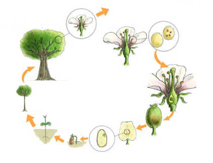
21. Label the diagram of alternation of generation. Include the sporophyte and gametophyte generations, the chromosome number (2n or 1n), and where mitosis and meiosis occur.

Plant Divisions

22. Plants are divided into __________ groups based on the presence or absence of an ___________ _____________ ___________ for carrying water and dissolved _____________.

23. What is the transport system for water and minerals called?

24. ______________ plants lack vascular tissue and are called _______________.

25. In what type of environment must nonvascular plants live?

26. Give an example of a bryophyte.

27. Nonvascular plants can’t grow as tall as vascular plants. Explain why.


28. The cells of nonvascular plants must be in _________ contact with water because water moves by _______________ from cell to cell.

29. How does the sperm get to the egg in nonvascular plants?

30. Name 3 divisions of nonvascular plants and give and organism found in each division.

     a.

     b.

     c.

31.Vascular plants are also called _______________.

32. What are the 2 subdivisions of vascular plants?


33. Name 4 divisions of seedless vascular plants and give an example of a plant in each group.

     a.

     b.

     c.

     d.

34. Name the 2 groups of seed-bearing vascular plants.


35. Gymnosperms have ____________ seeds found inside cones.

36. Angiosperms have ___________ to attract ____________ so seeds can be produced.

37. Name the division known as conifers and tell several plants in this group/


38. Name 2 other divisions of gymnosperms and tell a plant in each group.

     a.

     b.

39. Name the oldest living plant.

40. Name the tallest living plant.

41. What group are these 2 plants in?

42. Angiosperms are called ____________ plants.

43. How are seeds formed in angiosperms?


44. Where is the ovary found?

45. Name the male and female parts of a flower.

46. How are fruits formed?

47. Angiosperms are the division ______________.

48. What are the 2 subgroups of Anthophyta.

49. Describe the characteristics of monocots.


50. Describe the characteristics of dicots.


Homeostasis Worksheet Ch5 BI


Homeostasis & Transport


Section 5-1 Passive Transport

1. What is the purpose of the cell membrane?

2. Explain passive transport.

3. What is the simplest type of passive transport?

4. In which direction does diffusion occur?

5. What is a concentration gradient?

6. Sugar dissolving in water is an example of _______________________.

7. What supplies the energy for diffusion?

8. Molecules are constantly _____________________.

9. What is meant by equilibrium?

10. Do molecules stop moving when equilibrium is reached? Explain.

11. List three things that determine if a molecule will be able to diffuse across a membrane.

12. Name the 2 parts of a solution.

13. Define osmosis. Is it passive or active transport?

14. The direction water moves across a cell membrane depends on the concentration of what on either side of the cell membrane?

15. Explain what is true about solutes if the outside of the cell is hypotonic to the cytosol? Which way does water move?

16. Explain the solute conditions if the outside is hypertonic to the cytosol. Which way does water move?

17. What occurs if the solute concentration on each side of the cell membrane is isotonic?

18. If the inside & outside of a cell are both isotonic, does water still move across the cell membrane? Explain.

19. If the inside of the cell is hypotonic, the outside will be _________________________.

20. Water tends to diffuse from ____________________ to ___________________ solutions.

21. How does a unicellular paramecium get rid of its excess water? Is energy used?

22. Many cells in multicellular organisms have _________________ pumps to prevent them from taking in too much water in hypotonic solutions.

23. What structure around the outside of plant cells keeps hem from rupturing from too much water?

24. What is turgor pressure & how does it help plant cells?

25. What happens to plant cells placed in a hypertonic solution? Name this process.

26. What is cytolysis & what causes it?

27. Another type of passive transport is __________________________ diffusion.

28. Explain how carrier proteins help in facilitated.

29. Sketch the changes that take place in a carrier protein as it helps molecules move across the cell membrane.

30. What sugar moves across the cell membrane by facilitated diffusion?

31. What are ion channels & are they used in passive or active transport?

32. Name 4 ions that cross the cell membrane through ion channels.

33. Why can’t these ions diffuse across the lipid bilayer of the cell membrane?

34. Ion channels may be always ________________ or have ___________________.

35. Name 3 stimuli that open & close gated channels.

Section 5-2 Active Transport

36. Define active transport.

37. Why are carrier proteins in the cell membrane that are used for active transport called “pumps”?

38. What is the best-known carrier protein pump in animal cells?

39. What 2 ions move up their concentration gradient in this pump?

40. ___________________ ions are pumped out, while ______________ ions are pumped into the cell.

41. Is energy required for active transport? Explain.

42. Sodium ions are exchanged for potassium ions at a ____________ to ____________ ratio.

43. Name 2 processes used to move macromolecules & food particles across the cell membrane. Is energy required?

44. Explain how cells move large particles into the cell by endocytosis.

45. Name & describe the 2 types of endocytosis.

46. How do phagocytes protect cells?

47. What process moves large materials such as wastes & proteins out of the cell?

BACK


How Surface Area to Volume Ratio Limits Cell Size


How Surface Area to Volume Ratio Limits Cell Size

  1. A cell is a metabolic compartment where a multitude of chemical reactions occur.
  2. The number of reactions increase as the volume of metabolic volume within a cell increases. (The larger the volume the larger the number of reactions)
  3. 3.All raw materials necessary for metabolism can enter the cell only through its cell membrane.
  4. The greater the surface area the larger the amount of raw materials that can enter at only one time.
  5. Each unit of volume requires a specific amount of surface area to supply its metabolism with raw materials. The amount of surface area available to each unit of volume varies with the size of a cell.
  6. As a cell grows its SA/V decreases.
  7. At some point in its growth its SA/V becomes so small that its surface area is too small to supply its raw materials to its volume. At this point the cell cannot get larger.