Codon Bingo


Codon Bingo

Introduction:

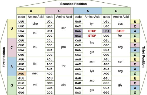
DNA is simply a storage form of information, like a recipe book.  In order to make useful proteins from this recipe, we must first transcribe the selected recipe from the DNA into messenger RNA (m-RNA) which then leaves the nucleus & goes to the ribosomes where it is “read” to link amino acids (building blocks of proteins). The code is “read” three bases at a time called a codon. The triplet code allows for a total of 4x4x4 or 64 different codons (groups of three RNA bases) –far more than needed to code for 20 amino acids. It was discovered that each amino acid is coded for by more than one codon. Codon Bingo is a simple exercise to learn how to use a codon table to translate mRNA into its associated amino acids.

Materials:  Bingo cards, pencil, codon table, beans or pennies

Procedure:

1. Pass out blank bingo cards.

2. Students should fill out each of the blanks with an amino acid from the codon chart.

3. Teacher will call out 3 bases (A, T, G, C)

4. Students find the amino acid that is associated with the codon and mark the square (use bingo chips, pennies, beans, or other miscellaneous items)


BIOLOGY BINGO


Chlorophyll Fluorescence


Chlorophyll Fluorescence

INTRODUCTION

When a pigment absorbs light, electrons of certain atoms in the pigment molecules are boosted to a higher energy level. The energy of an absorbed photon is converted to the potential energy of the electron that has been raised to an excited state. In most pigments, the excited electron drops back to its ground-state, or normal orbit, and releases the excess energy as heat. Some pigments, including chlorophyll, emit light as well as heat after absorbing photons.
In the chloroplast, these excited electrons jump from the chlorophyll molecule to a protein molecule in the thylakoid membrane, and are replaced by electrons from the splitting of water. The energy thus transferred, is used in carbohydrate production.
This release of light is called fluorescence. Chlorophyll will fluoresce in the red part of the spectrum, and also give off heat. In this lab, you will observe this fluorescence by separating the chlorophyll from the thylakoid membrane.

MATERIALS


Spinach leaves Flashlight or small lab light
Mortar and pestle Test tube
Acetone Filter paper
25-mL graduated cylinder Funnel
Ring stand or funnel rack Safety goggles

PROCEDURE

1. Grind the spinach leaves using a mortar and pestle.

2. Add acetone to the ground leaves, using enough acetone and spinach leaves to get between 10 and 15 mL of extract.

3. Set up your filtering apparatus, and using proper filtering technique, filter the extract to a test tube. NOTE: Use a small amount of acetone to wet the filter paper, to hold it into place, instead of water.

4. Shine a flashlight, or other similar light source, through the test tube and extract.

5. Observe the fluorescence of the chlorophyll at a 90 degree angle to the flashlight.


Ecosystem Quiz

Name: 


Ecosystem Interactions


True/False
Indicate whether the sentence or statement is true or false.
1.
Freshwater habitats are independent of terrestrial habitats.
2.
An ecosystem consists of biotic and abiotic factors.
3.
Clearing a forest would reduce the amount of energy available to the consumers.
4.
While an understanding of the interactions between organisms and their environment was very important to early hunter and gatherer humans, it is even more important today because humans are having significant effects on the environment.
5.
Cutting down trees in a forest alters the habitat of the organisms living in the forest.
6.
An organism’s niche includes its habitat.
 

Multiple Choice
Identify the letter of the choice that best completes the statement or answers the question.
7.
Abiotic factors affect an ecosystem by all of these except the
a.
quantity and quality of water
c.
nitrogen-fixing bacteria
b.
amount of light available
d.
quantity of minerals
8.
The most important single factor affecting the biosphere is
a.
solar radiation
c.
precipitation
b.
the biotic community
d.
wind
9.
Which of the following is the smallest ecological unit?
a.
a community
c.
a population
b.
a biome
d.
an ecosystem
10.
Collectively, physical factors such as light, temperature, and moisture that affect an organism’s life and survival are called the
a.
biotic environment
c.
ecosystem
b.
abiotic environment
d.
niche
11.
The rate of photosynthesis carried on by plants living in a body of water depends upon the
a.
oxygen content of the water
c.
amount of light that penetrates the water
b.
nitrogen content of the water
d.
elevation of the body of water
12.
Which of the following would not be included in a description of an organism’s niche?
a.
its trophic level
c.
its color
b.
the humidity it prefers
d.
when it reproduces
13.
Ecology is the study of the interaction of living organisms
a.
with each other and their habitat.
b.
and their communities.
c.
with each other and their physical environment.
d.
and the food they eat.
14.
The destruction of the ozone layer may be responsible for an increase in
a.
cataracts.
c.
cancer of the retina.
b.
melanoma.
d.
All of the above
15.
Ozone in the atmosphere
a.
leads to formation of acid precipitation.
b.
combines readily with water vapor.
c.
absorbs harmful radiation from the sun.
d.
All of the above
16.
Ecological models are useful for
a.
making predictions about future ecological changes.
b.
testing predictions about future ecological changes.
c.
evaluating proposed solutions to environmental problems.
d.
All of the above
17.
The physical location of an ecosystem in which a given species lives is called a
a.
habitat.
c.
community.
b.
tropical level.
d.
food zone.
18.
Which of the following is not an adaptation for avoiding unfavorable conditions?
a.
acclimation
c.
dormancy
b.
body temperature regulation
d.
migration
19.
Which of the following would not be included in a description of an organism’s niche?
a.
its trophic level
b.
the humidity and temperature it prefers
c.
its number of chromosomes
d.
when it reproduces


Check Your Work     Reset

Chromatography Plant Pigments


Chromatography of Plant Pigments

INTRODUCTION:

Chlorophyll often hides the other pigments present in leaves. In Autumn, chlorophyll breaks down, allowing xanthophyll and carotene, and newly made anthocyanin, to show their colors.
The mix of pigments in a leaf may be separated into bands of color by the technique of paper chromatography. Chromatography involves the separation of mixtures into individual components. Chromatography means “color writing.” With this technique the components of a mixture in a liquid medium are separated. The separation takes place by absorption and capillarity. The paper holds the substances by absorption; capillarity pulls the substances up the paper at different rates. Pigments are separated on the paper and show up as colored streaks. The pattern of separated components on the paper is called a chromatogram.

PRELAB PREPARATION:

Gather leaves from several different plants. CAUTION: Avoid poisonous plants. Autumn leaves from deciduous trees are especially interesting. Sort the leaves by kind (maple, etc.) and color. Review a diagram of a plant cell . Find the grana and the chloroplasts of the cell.
MATERIALS:

Safety goggles
Chromatography solvent (92 parts Petroleum ether to 8 parts acetone)
Chromatography paper (or filter paper) about 1 cm x 15 cm
Ethyl alcohol
Fresh spinach
Test tube
Test tube rack
Scissors and Ruler
Fresh leaves of plants
Glass stirring rod
Paper clip
Cork (to fit test tube)
Mortar and pestle
Sand (optional)
10-ml Graduated cylinder

PROCEDURE:

Leaves should be grouped by kind (maple, etc.) and color. Work with a spinach leaf and with one or more other types. CAUTION: Chromatography solvents are flammable and toxic. Have no open flames; maintain good ventilation; avoid inhaling fumes.

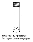
1. Cut a strip of filter paper or chromatography paper so that it just fits inside a 15-cm (or larger) test tube. Cut a point at one end. Draw a faint pencil line as shown in figure 1. Bend a paper clip and attach it to a cork stopper. Attach the paper strip so that it hangs inside the tube, as shown. The sides of the strip should not touch the glass.

2. Tear a spinach leaf into pieces about the size of a postage stamp. Put them into a mortar along with a pinch or two of sand to help with grinding. Add about 5 ml ethyl alcohol to the leaf pieces. Crush leaves with the pestle, using a circular motion, until the mixture is finely ground. The liquid in which the leaf pigments are now for paper chromatography dissolved is called the pigment extract.

3. Use a glass rod to touch a drop of the pigment extract to the center of the pencil line on the paper strip. Let it dry. Repeat as many as 20 times, to build up the pigment spot. NOTE: You must let the dot dry after each drop is added. The drying keeps the pigment dot from spreading out too much.

4. Pour 5 ml chromatography solvent into the test tube. Fit the paper and cork assembly inside. Adjust it so that the paper point just touches the solvent (but not the sides of the tube). The pigment dot must be above the level of the solvent. Watch the solvent rise up the paper, carrying and separating the pigments as it goes. At the instant the solvent reaches the top, remove the paper and let it dry. Observe the bands of pigment. The order, from the top, should be carotenes (orange), xanthophylls (yellow), chlorophyll a (yellow-green), chlorophyll b (blue-green), and anthocyanin (red). Identify and label the pigment bands on the dry strip. Write the species of leaf on the strip as well.
Record the species, external color, and chromatogram pigments in the DATA TABLE of your report sheet.

5. Each pigment has an Rf value, the speed at which it moves over the paper compared with the speed of the solvent.

Rf = Distance moved by the pigment / Distance moved by the solvent

Measure the distance in cm from the starting point (pencil line) to the center of each pigment band. Then measure the entire distance traveled by the solvent. Remember, the starting point for the solvent is also the pencil line and the ending point for the solvent is the top edge of the paper. Do the required divisions and record your Rf values in the DATA TABLE of your report sheet.

6. Wash the mortar and pestle thoroughly, using a little alcohol to remove any remaining pigment.

7. Repeat steps 1 through 6 for each species.

DATA TABLE:

Chromatography Data

Leaf Type (species) External color Chromatogram Pigments
Colors from the Top Pigment Names Rf Values