DNA Replication Lab

Modeling DNA Replication


Introduction

Within the nucleus of every cell are long strings of DNA, the code that holds all the information needed to make and control every cell within a living organism. DNA, which stands for deoxyribonucleic acid, resembles a long, spiraling ladder. It consists of just a few kinds of atoms: carbon, hydrogen, oxygen, nitrogen, and phosphorus. Combinations of these atoms form the sugar-phosphate backbone of the DNA — the sides of the ladder, in other words.

Other combinations of the atoms form the four bases: thymine (T), adenine (A), cytosine (C), and guanine (G). These bases are the rungs of the DNA ladder. (It takes two bases to form a rung — one for each side of the ladder.) A sugar molecule, a base, and a phosphate molecule group together to make up a nucleotide. Nucleotides are abundant in the cell’s nucleus. Nucleotides are the units which, when linked sugar to phosphate, make up one side of a DNA ladder.

During DNA replication, special enzymes move up along the DNA ladder, unzipping the molecule as it moves along. New nucleotides move in to each side of the unzipped ladder. The bases on these nucleotides are very particular about what they connect to. When the enzyme has passed the end of the DNA, two identical molecules of DNA are left behind. Cytosine (C) will “pair” to guanine (G), and adenine (A) will “pair” to thymine (T). How the bases are arranged in the DNA is what determines the genetic code.


When the enzyme has passed the end of the DNA, two identical molecules of DNA are left behind. Each contains one side of the original DNA and one side made of “new” nucleotides. It is possible that mistakes were made along the way — in other words, that a base pair in one DNA molecule doesn’t match the corresponding pair in the other molecule. On average, one mistake may exist in every billion base pairs. That’s the same as typing out the entire Encyclopedia Britannica five times and typing in a wrong letter only once!

Objectives

The replication of DNA before cell division can be shown using paper templates for the components of DNA nucleotides.

Materials

  • Cut Outs of basic subunits of DNA
  • Colors or markers
  • Scissors
  • Tape or glue
  • Paper & pencil

Procedure:

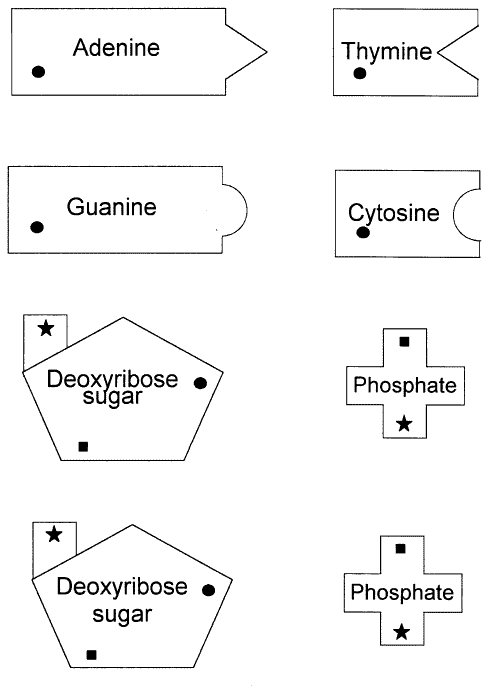
  1. Cut out all of the units needed to make the nucleotides from the handout provided.
  2. Color code the Nitrogenous bases, phosphorus, and deoxyribose sugar as follows —
    Adenine = red, Guanine = green, Thymine = yellow, Cytosine = blue, Phosphate = brown, and Deoxyribose = purple.
  3. Using the small squares and stars as guides, line up the bases, phosphates and sugars.
  4. Now glue the appropriate parts together forming nucleotides.
  5. Construct DNA model using the following sequence to form a row from top to bottom – cytosine (topmost), thymine, guanine, and adenine (bottommost).
  6. Let this arrangement represent the left half of your DNA molecule.
  7. Complete the right side of the ladder by adding the complementary bases. You will have to turn them upside down in order to make them fit.
  8. Your finished model should look like a ladder.
  9. To show replication, separate the left side from the right side, leaving a space of about 6-8 inches.
  10. Use the remaining nucleotides to complete the molecule using the left side as the base.
  11. Build a second DNA model by adding new nucleotides to the right half of the original piece of the molecule.
  12. Tape the nucleotides together to form 2 complete DNA ladders.

Questions

1. Of the 4 bases, which other base does adenine most closely resemble?

2. List the 4 different nucleotides.

3. Which 2 molecules of a nucleotide form the sides of a DNA ladder?

4. If 30% of a DNA molecule is Adenine, what percent is Cytosine?

5. What does the term replication mean?

6. What is another name for adenine and three phosphate molecules attached to it?


Effect of Solutions on Cells

 Effect of  Solutions on Cells

What happens when cells are place in different kinds of solutions

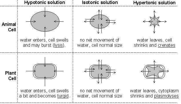
Plant cells placed in a hypertonic solution will undergo plasmolysis, a condition where the plasma membrane pulls away from the cell wall as the cell shrinks. The cell wall is rigid and does not shrink. 

   The Elodea cells  have been placed in a 10% NaCl solution. The contents of the cells have been reduced to the spherical structures shown.  


   Normal Elodea cells


Animal cells placed in a hypertonic solution will undergo crenation, a condition where the cell shrivels up as it loses water. Red blood cells in a hypotonic solution will swell and burst or lyse.


BACK

Egg Osmosis Sample 1 Lab

Osmosis through the Cell Membrane of an Egg

Introduction:
The cell or plasma membrane is made up of phospholipids and different types of proteins that move laterally. These include peripheral proteins, which are attached to the interior and exterior surface of the cell membrane. Integral proteins are embedded in the lipid bilayer. Attached to these integral proteins are carbohydrate chains. These carbohydrates may hold adjoining cells together, or act as sites where viruses or chemical messengers such as hormones can attach. Cell membranes are selectively permeable. They allow some substances to pass through, but not others. Small molecules that are usually nonpolar, such as oxygen, water, and carbon dioxide, easily move through the lipid bilayer. Larger molecules, such as glucose, the food for all living things, must seek aid from the carrier proteins in a process called facilitated diffusion. Facilitated diffusion is a process used for molecules that cannot diffuse rapidly through cell membranes. Integral proteins are used by calcium, potassium, and sodium ions to move through the cell membrane. The muscles and nerves use these ions.
Diffusion is the movement of molecules from an area of higher concentration to an area of lower concentration. This difference in the concentration of molecules across a space is called a concentration gradient. Diffusion is a type of passive transport, meaning it does not require energy input by the cell. This type of transport and osmosis are the two processes used in this lab. Osmosis is the process by which water molecules diffuse across a cell membrane from an area of higher concentration to an area of lower concentration. When the concentration of the solute is higher outside of the cell, it is known as a hypertonic solution. When the concentration of the solute is lower outside of the cell, it is known as a hypotonic solution.

Hypothesis:
The substance, syrup, which has a higher solute concentration than the interior of the eggs, will cause water to leave the eggs’ membrane; the other substance, distilled water, which has a lower solute concentration than the eggs’ interior, will cause liquid to enter the eggs’ membrane.

Materials:
The materials necessary for this lab are: two fresh eggs in their shells, a felt tip marker, 200mL graduated cylinder, five jars, clear Saran wrap, white vinegar, clear sugar syrup (Karo), distilled water, tap water, pencil, paper, eraser, computer, electronic scale, and a plastic tray.

Methods:
Day One: On day one, label the five jars, with the felt tip marker: one labeled vinegar, two labeled syrup, and two labeled distilled water. Also put the group number on each jar. Find the mass of each egg and record this information in the data table. Place the two eggs in the jar labeled vinegar. Add vinegar until both eggs are submerged by it. Cover the jar with the clear Saran wrap. Place the jar on the plastic tray and allow to set for 24 hours.

Day Two: On day two, observe what has happened to your eggs. Record this in a data table. Now that the eggs’ shells are dissolved, gently remove the eggs from the vinegar. Rinse each egg with tap water. Pat the eggs dry with paper towels and mass them separately on the electronic balance. Record this in the data table. Place the eggs in the jars labeled syrup. Add syrup to each jar (labeled egg 1 or egg 2) until the eggs are submerged in syrup. Loosely cover each jar with Saran wrap. Place the jars on the tray and allow them to soak for 24 hours.

Day Three: On day three, observe what has happened to the eggs and record this information in the data table. Carefully remove the eggs from the syrup and rinse them with tap water. Pat dry with paper towels. Using the electronic balance, find the mass of each egg separately and record these masses in the data table. Place the eggs in the jars labeled distilled water (labeled egg 1 and egg 2). Add distilled water to each jar until the eggs are covered. Cover the jars with the Saran wrap and allow them to sit on the tray for 24 hours.

Day Four: On day four, remove the eggs from the jars and record the eggs’ appearance. Mass each egg on the electronic balance. Record this in the data table. Dispose of the eggs in the container provided by the teacher.

Results:

Egg 1 Data Table


Substance egg submerged inEgg’s mass before placed in substanceEgg’s mass after removed from substanceObservations of egg before placed in solutionObservations of egg after removed from substance
Vinegar59.2 g86.0 gThe egg’s shell is intact and is included in the first mass.The egg’s shell dissolved and wasn’t included in the 2nd mass.
Syrup86.0 g53.2 gThe egg is swollen and soft, yet firm to touch.The liquid inside the egg diffused into the syrup.
Distilled Water53.2 g86.5 gThe egg has lost some of its firmness.The water diffused into the egg, increasing the egg’s mass.


Egg 2 Data Table


Substance egg submerged inEgg’s mass before place in substanceEgg’s mass after removed from substanceObservations of egg before placed in solutionObservations of egg after removed from substance
Vinegar58.8 g85.6 gThe egg’s shell is intact and is included in the first mass.The egg’s shell is mostly dissolved and so wasn’t included in 2nd mass.
Syrup85.6 g52.2 gThe egg is rough to touch and feels rather sturdy.The liquid inside the egg diffused into the syrup.
Distilled Water52.2 g88.9 gThe egg feels more fragile and lighter in weight.The water diffused into the egg increasing the egg’s mass.


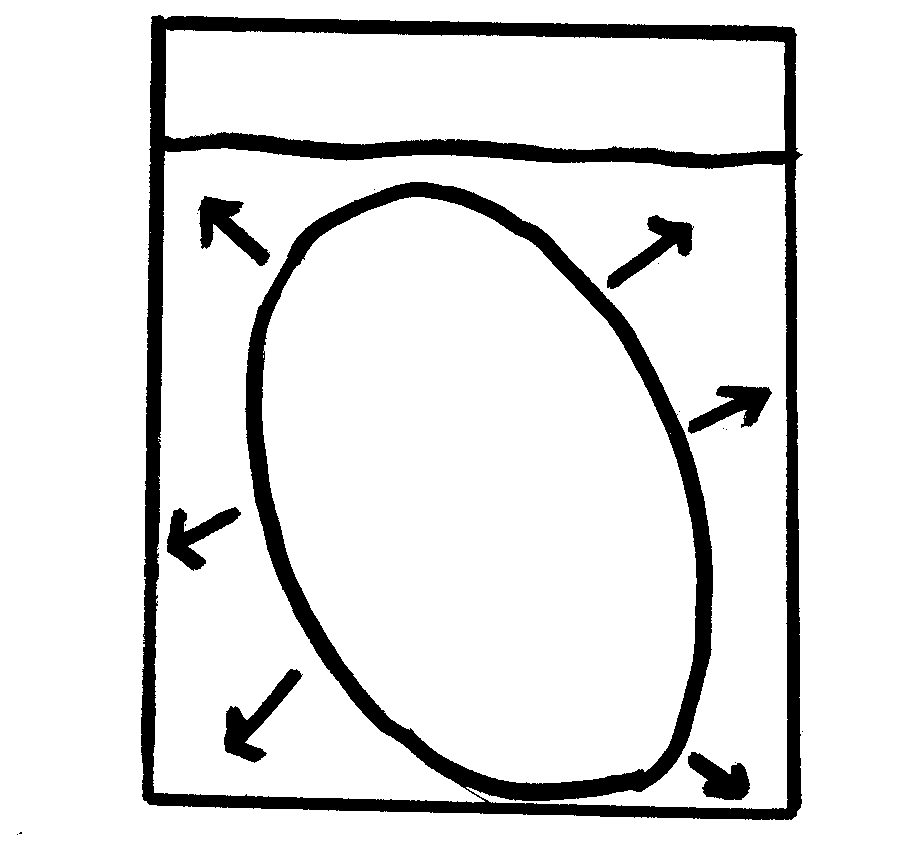
Egg in Hypotonic Solution of Vinegar & Plasmolyzed Egg in Distilled WaterEgg in Hypertonic Solution of Syrup


1. When the egg was place in the water, in which direction did the water molecules move? The water moved into the eggs from the surrounding environment.

2. On what evidence do you base this? The eggs’ masses had increased from the time they were placed in the water to when the eggs were removed.

3. How do you explain the volume of liquid remaining when the egg was removed from the syrup? The volume of the liquid remaining when the egg was removed from the syrup must have increased because the eggs’ masses had decreased. The liquid within the eggs left the eggs and diffused into the surrounding syrup.

4. When the egg was place in the water after being removed from the syrup, in which direction did the water move? The water moved into the eggs.

Error Analysis:
Several errors may have occurred during this lab. When finding the eggs’ masses, on each occasion, an error may have occurred. Mistakes may have been made when recording these masses on the data table. Some of the eggs’ shell may have been left on the eggs’ membranes and changed the outcome of this lab. When the eggs were rinsed, after being placed in the vinegar and syrup, a small amount of water could have entered through the membranes of the eggs, effecting their masses. These are just a few of the errors that may have taken place throughout the lab.

Discussion and Conclusion:
The hypothesis was correct. When the eggs were placed in the syrup, their masses decreased greatly. This shows that the interior of the eggs must have had a lower solute concentration than their surrounding environment of syrup. The water within the eggs left through the membrane and diffused into the syrup, decreasing its solute concentration. When the eggs were placed in the distilled water, their masses greatly increased. This shows that the interior of the eggs must have had a higher solute concentration than their surrounding environment of distilled water. The distilled water diffused into the eggs’ membrane, decreasing the interior of the eggs’ solute concentration.

Back

Egg Osmosis Sample 2 lab


Osmosis through the Cell Membrane of an Egg


Introduction:
Transport can be either passive or active. Passive transport is the movement of substances across the membrane without any input of energy by the cell. Active transport is the movement of materials where a cell is required to expend energy. In the case of this lab the discussion will be centered on passive transport.
The simplest type of passive transport is diffusion. Diffusion is the movement of molecules from an area of higher to an area of lower concentration without any energy input. Diffusion is driven by the kinetic energy found in the molecules. Diffusion will eventually cause the concentration of molecules to be the same throughout the space the molecules occupy, causing a state of equilibrium to exist.
Another type of passive transport is that of osmosis. Osmosis is the movement of water across a semi-permeable membrane. The process by which osmosis occurs is when water molecules diffuse across a cell membrane from an area of higher concentration to an area of lower concentration. The direction of osmosis depends on the relative concentration of the solutes on the two sides. In osmosis, water can travel in three different ways.
If the molecules outside the cell are lower than the concentration in the cytosol, the solution is said to be hypotonic to the cytosol, in this process, water diffuses into the cell until equilibrium is established. If the molecules outside the cell are higher than the concentration in the cytosol, the solution is said to be hypertonic to the cytosol, in this process, water diffuses out of the cell until equilibrium exists. If the molecules outside and inside the cell are equal, the solution is said to be isotonic to the cytosol, in this process, water diffuses into and out of the cell at equal rates, causing no net movement of water.
In osmosis the cell is selectively permeable, meaning that it only allows certain substances to be transferred into and out of the cell. In osmosis, the proteins only on the surface are called peripheral proteins, which form carbohydrate chains whose purpose is used like antennae for communication. Embedded in the peripheral proteins are integral proteins that can either be solid or have a pore called channel proteins. Channel proteins allow glucose, or food that all living things need to live, pass through.


Hypothesis:
In the syrup solution, there will be a net movement of molecules out of the egg, and in the water solution, the molecules will diffuse in and out of the cell at equal rates.


Materials:
The materials used in this lab were 2 fresh eggs in the shell, an overhead marker, 400 ml of water, graduated cylinder, 1 large beaker, 2 medium beakers, 1 small beaker, white vinegar, Karo syrup, distilled water, pencil, paper, lab apron, lab goggles, saran wrap, masking tape, plastic tray, tongs, electronic balance, osmosis lab sheet, and computer.


Methods:
On day 1, measure the masses of both the eggs with the shell. Label 1 beaker vinegar, and then use the graduated cylinder to measure 400 mL of vinegar to put in the labeled beaker. Place both eggs in the solution (place a small beaker on top of the eggs, if necessary) then cover. Let the eggs stand for 24 hours or more to remove the shell.


On day 2, record the observations of what happened to the eggs in the vinegar solution. Carefully, remove the eggs from the vinegar, gently rinsing the eggs off in water. Clean the beakers used for the vinegar solution preparing them for the syrup solution, and then label the 2 medium beakers syrup. Before the eggs are placed in the syrup solution record the mass of both eggs then put it on the datasheet. After that has been done, place the eggs in the beaker, pouring enough syrup to cover the eggs, cover them loosely and let them stand for 24 hours.

On day 3, record the observations of the egg from the syrup solution. Carefully, remove the eggs from the beakers, gently rinsing the syrup off of the eggs. Pour the remaining syrup in the container provided by the teacher. Clean the two beakers used in the syrup solution, preparing them for the water solution. Before the eggs are placed in the water solution record the mass of both eggs then put it on the datasheet. After that has been done, using a graduated cylinder, measure out 200 mL of water for each beaker. Place the eggs in the water solution, cover and let stand 24 hours.

On day 4, record the observations of the egg from the water solution. Carefully remove the eggs from the beakers, gently rinsing them off. Mass both of the eggs. After the teacher has came and looked at the eggs, discard in the proper place.


Results:


Isotonic SolutionHypotonic (Vinegar is acid in Water)
Hypertonic


Table 1- Egg 1 Data


Egg mass before added into the solution (g)


Egg mass after added into the solution (g)


Observations


Vinegar

70.8 g (with shell)98.0 g (without shell)Before the egg was added into the vinegar, it was large, but the after effect was that the egg increased in size and had become hard. After two days, the shell was completely removed.
 

Syrup

98.0 g65.0 gWhen the egg was removed from the syrup, it had shrunk and it was softer than before it was added into the solution
 

Water

65.0 g105.3 gWhen the egg was removed out of the water, the color looked of a pale yellow. The water had diffused into the egg, because the egg was larger in size before it was added into the water.


Table 2- Egg 2 Data


Egg mass before added into the solution (g)


Egg mass after added into the solution (g)


Observations


Vinegar

71.6 g (with shell)99.1 g (without shell)Egg 2s’ mass was greater than egg 1s’ mass before and after it was added into the vinegar solution. The mass had increased some 20 grams with the shell off.
 

Syrup

99.1 g64.0 gThe mass of the egg had decreased some 30 grams after it the egg was removed from the syrup solution. The mass of the egg 2 was smaller than the mass of egg1.
 

Water

64.0 g105.2 gThe mass of egg 2 had increased some 50 grams after being added into the water solution. The mass of egg 1, though, was larger than the mass of 2 by 1 gram. If the egg would have remained in the water a little while longer, the egg would have probably went through cytolysis.


1. When the egg was placed in the water in which direction did the water molecules move?     The water molecules moved in the egg.

2. On what evidence do you base this? The molecules moved in, because the size of the egg increased

3. How do you explain the volume of liquid remaining when the egg was removed from the syrup? Since, the cell is selectively permeable, it only allowed a certain amount of the syrup to be present in the cell, just enough to shrink it and also equilibrium was reached..

4. When the egg was placed in the water after being removed from the syrup in which direction did the water move? The water moved in.

5. Why did the water molecules travel better inside the cell than the syrup molecules? The water molecules traveled better into the cell because smaller molecules travel better than other larger molecules.

6. What was the purpose of placing the egg in vinegar? The  vinegar solution was only used to remove the shell off the egg.

Error Analysis:
A possible error in this lab occurred by having to leave the egg in vinegar for two days instead of one to remove the shell. This caused the egg to initially take in more water.


Discussion and Conclusion:
Based on the data collected and the results of the experiment, the hypothesis was  correct. The egg appeared shriveled after removing it from the syrup because of the movement of water out of the egg. The syrup solution was hypertonic so water moved out of the egg from an area where water was more concentrated to the outside of the egg where water was less concentrated due to the high amount of sugar or solute. The acetic acid in vinegar did remove the shell from the egg, because the egg required two days to completely remove the shell, some water did move into the egg causing its initial mass without the shell to be higher than the egg’s mass with its shell. Whenever the egg was transferred from the syrup to the distilled water, the concentration of water outside the shriveled egg was greater than the water concentration inside the egg; therefore, water moved into the egg until equilibrium was reached. At that point, movement into and out of the egg continued with no net movement of water molecules.
Additional research  to see if the egg would have went through cytolysis in another 24 or more hours in the water solution would have been interesting.
BACK

Energy in food


The Heat is On – The Energy Stored in Food
Introduction:

Plants utilize sunlight during photosynthesis to convert carbon dioxide and water into glucose (sugar) and oxygen. This glucose has energy stored in its chemical bonds that can be used by other organisms. This stored energy is released whenever these chemical bonds are broken in metabolic processes such as cellular respiration.

Cellular respiration is the process by which the chemical energy of “food” molecules is released and partially captured in the form of ATP. Cellular respiration is the general term which describes all metabolic reactions involved in the formation of usable energy from the breakdown of nutrients. In living organisms, the “universal” source of energy is adenosine triphosphate (ATP). Carbohydrates, fats, and proteins can all be used as fuels in cellular respiration, but glucose is most commonly used as an example to examine the reactions and pathways involved.

Marathon runners eat a large plate of pasta the night before a competition because pasta is a good source of energy, or fuel for the body. All foods contain energy, but the amount of potential energy stored will vary greatly depending on the type of food. Moreover, not all of the stored energy is available to do work. When we eat food, our bodies convert the stored energy, known as Calories, to chemical energy, thereby allowing us to do work. A calorie is the amount of heat (energy) required to raise the temperature of 1 gram (g) of water 1 degree Celsius (°C). The density of water is 1 gram per milliliter (1g/ml) therefore 1 g of water is equal to 1 ml of water. When we talk about caloric values of food, we refer to them as Calories (notice the capital “C”), which are actually kilocalories. There are 1000 calories in a kilocalorie. So in reality, a food item that is listed as having 38 Calories has 38,000 calories. Calories are a way to measure the energy you get from the food you eat.

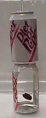
Just as pasta can provide a runner energy to run a marathon, a tiny peanut contains stored energy that can be used to heat a container of water. For this lab exercise, you will indirectly measure the amount of Calories in couple of food items using a calorimeter. A calorimeter (calor = Latin for heat) is a device that measures the heat generated by a chemical reaction, change of state, or formation of a solution. There are several types of calorimeters but the main emphasis of all calorimeters is to insulate the reaction to prevent heat loss. We will be using a homemade calorimeter modeled after a constant-volume calorimeter. A particular food item will be ignited, the homemade calorimeter will trap the heat of the burning food, and the water above will absorb the heat, thereby causing the temperature (T) of the water to increase. By measuring the change in temperature (∆T) of a known volume of water, you will be able to calculate the amount of energy in the food tested


Objective:


In this experiment, you will measure the amount of energy available for use from three types of nuts, a plant product. This process of measuring the energy stored in food is known as calorimetry.

Materials:
large paper clip, oC thermometer, soft drink can, soft drink can with openings cut into the side, mixed nuts, matches, water, electronic balance, pencil & paper, 100 ml graduated cylinder, calculator

Procedure:

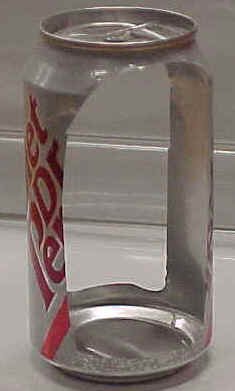
  1. Carefully, cut out two openings along the side of a soft drink can. This will serve as your support for the second drink can that will contain water & sit on top.

  1. Bend a large size paper clip so that a nut can be attached on one end and the other end will sit flat inside the cut-out soft drink can.


  1. Use the graduated cylinder to accurately measure 100g (100ml) of water. Pour this water into the uncut soft drink can.
  2. Place the thermometer in the uncut can and measure the water temperature after 3 minutes.  Record this temperature on  data table 1.

  1. Mass the nut (g) that you will burn and record this mass on  data table 1.
  2. Attach the nut to the bent end of your paper clip and carefully set the clip & nut into the cut-out soft drink can on bottom. Make sure the cans are sitting on a flat, nonflammable surface!

  1. Carefully light the nut from the bottom using a match and record the change in water temperature as the nut burns (thermometer in the can during burning). Immediately after the nut finishes burning, record the final (highest) water temperature on data table 1.
  2. Measure the mass (g) of the remaining nut & record this in the data table 1. (Mass the burned nut and paper clip together and then subtract the mass of the nut to get the mass of the nut alone.)
  3. Complete the data table1 by calculating the change in mass of the nut.
  4. Repeat this experiment with the other two types of nuts .
  5. When all three nuts have been burned, complete the analysis on data table 2.

Results:


Table 1 – Results of Burning

PECANWALNUTALMOND
oC  H2O temperature Before burning
oC
 
oC  H2O temperature After burning
oC
Difference in oC H2O temperature
oC
Mass of Paper Clip
g
Mass of Nut Before Burning
Mass of Paper Clip and Nut After Burning
g
Mass of Nut ALONE After Burning
(Subtract paper clip mass from mass of nut & paper clip after burning)
g
(Subtract paper clip mass from mass of nut & paper clip after burning)
g


Table 2 – Data Analysis from Nut Calorimetry

PECAN WALNUT ALMOND
Mass Difference of Nut Before & After Burning

(Subtract mass of nut after burning from Mass of nut before burning)
g

Temperature Difference of H2O Before & After Burning
(Subtract original water temp. from final water temp.)
oC
Calories Required to Change the Temperature of 100 g of H2O
(Multiply temperature change by 100)Cal
Average Calories per gram in the Nut
(Divide the total calories by the mass difference of the nut before & after burning)Cal/g
Average kilocalories or food calories per gram
(Divide the calories per gram by 1000)kcal/g


Questions & Conclusion:

  1. Where did the energy stored in the nut originally come from?
  2. During what process was this energy stored in the nut, & where specifically was it stored?
  3. What simple sugar made by plants is a common source for stored energy?
  4. Which group of macromolecules would a nut contain — carbohydrates, lipids, or protein?
  5. What is the name for stored energy?
  6. Give some examples of how organisms would use this stored energy.
  7. In this experiment, discuss what happened to the energy stored in the nut.
  8. Why was the final mass of the nut less than the original mass of the nut? (Remember that matter can’t be destroyed in a chemical reaction.)


BACK