Biochemistry Notes BI Chapter 3


Biochemistry   All Materials © Cmassengale

I. Cells Contain Organic Molecules

A. Most Common Elements

1. Most common elements in living things are carbon, hydrogen, nitrogen, and oxygen.

2. These four elements constitute about 95% of your body weight.

3. Chemistry of carbon allows the formation of an enormous variety of organic molecules.

4. Organic molecules have carbon and hydrogen; determine structure and function of living things.

5. Inorganic molecules do not contain carbon and hydrogen together; inorganic molecules (e.g., NaCl) can play important roles in living things.

B. Small Molecules Have Functional Groups

1. Carbon has four electrons in outer shell; bonds with up to four other atoms (usually H, O, N, or another C).

2. Ability of carbon to bond to itself makes possible carbon chains and rings; these structures serve as the backbones of organic molecules.

3. Functional groups are clusters of atoms with characteristic structure and functions.

a. Polar molecules (with +/- charges) are attracted to water molecules and are hydrophilic.  

b. Nonpolar molecules are repelled by water and do not dissolve in water; are hydrophobic.

c. Hydrocarbon is hydrophobic except when it has an attached ionized functional group such as carboxyl (acid) ( COOH); then molecule is hydrophilic.  


d. Cells are 70-90% water; degree organic molecules interact with water affects their function.

4. Isomers are molecules with identical molecular formulas but differ in arrangement of their atoms


C. Large Organic Molecules Have Monomers

1. Each small organic molecule can be a unit of a large organic molecule called a macromolecule.

2. Small organic molecules (e.g., monosaccharides, glycerol and fatty acid, amino acids, and nucleotides) that can serve as monomers, the subunits of polymers.

3. Polymers are the large macromolecules composed of three to millions of monomer subunits.

4. Four classes of macromolecules (polysaccharides or carbohydrates, triglycerides or lipids, polypeptides or proteins, & nucleic acids such as DNA & RNA) provide great diversity.

D. Condensation Is the Reverse of Hydration

1. Macromolecules build by different bonding of different monomers; mechanism of joining and breaking these bonds is condensation and hydrolysis.

2. Cellular enzymes carry out condensation and hydrolysis of polymers.

3. Condensation involves a dehydration synthesis because a water is removed (dehydration) and a bond is made (synthesis).

a. When two monomers join, a hydroxyl ( OH) group is removed from one monomer and a hydrogen is removed from the other.

b. This produces the water given off during a condensation reaction.

4. Hydrolysis (hydration) reactions break down polymers in reverse of condensation; a hydroxyl
( OH) group from water attaches to one monomer and hydrogen ( H) attaches to the other.

II. Carbohydrates

A. Monosaccharides, Disaccharides, and Polysaccharides

1. Monosaccharides are simple sugars with a carbon backbone of three to seven carbon atoms.

a. Best known sugars have six carbons (hexoses).

[Glucose Straight Structure]

1) Glucose and fructose isomers have same formula (C6H12O6) but differ in structure.

2) Glucose is commonly found in blood of animals; is immediate energy source to cells.

3) Fructose is commonly found in fruit.

4) Shape of molecules is very important in determining how they interact with one another.

2. Ribose and deoxyribose are five-carbon sugars (pentoses); contribute to the backbones of RNA and DNA, respectively.

3. Disaccharides contain two monosaccharides joined by condensation.

a. Sucrose is composed of glucose and fructose and is transported within plants.

sucrose molecule - Sucrose

b. Lactose is composed of galactose and glucose and is found in milk.

c. Maltose is two glucose molecules; forms in digestive tract of humans during starch digestion.


Sugar Sweetness
fructose 173%
sucrose 100%
glucose 74%
maltose 33%
galactose 33%
lactose 16%


B. Polysaccharides Are Varied in Structure and Function

1. Polysaccharides are chains of glucose molecules or modified glucose molecules

a. Starch is straight chain of glucose molecules with few side branches.

b. Glycogen is highly branched polymer of glucose with many side branches; called “animal starch,” it is storage carbohydrate in the liver of animals.

c. Cellulose is glucose bonded to form microfibrils; primary constituent of plant cell walls.

d. Chitin is polymer of glucose with amino acid attached to each; it is primary constituent of crabs and related animals like lobsters and insects.

III. Lipids

A. Lipids

1. Lipids are varied in structure.

2. Many are insoluble in water because they lack polar groups.

B. Fats and Oils Are Similar

1. Each fatty acid is a long hydrocarbon chain with a carboxyl (acid) group at one end.

a. Because the carboxyl group is a polar group, fatty acids are soluble in water.

b. Most fatty acids in cells contain 16 to 18 carbon atoms per molecule.

c. Saturated fatty acids have no double bonds between their carbon atoms. (C-C-C-)

d. Unsaturated fatty acids have double bonds in the carbon chain.(C-C-C-C=C-C-)

e. Saturated animal fats are associated with circulatory disorders; plant oils can be substituted for animal fats in the diet.

2. Glycerol is a water-soluble compound with three hydroxyl groups.

3. Triglycerides are glycerol joined to three fatty acids by condensation

4. Fats are triglycerides containing saturated fatty acids (e.g., butter is solid at room temperature).

5. Oils are triglycerides with unsaturated fatty acids (e.g., corn oil is liquid at room temperature).

6. Fats function in long-term energy storage in organisms; store six times the energy as glycogen.

C. Waxes Are Nonpolar Also

1. Waxes are a long-chain fatty acid bonded to a long-chain alcohol.

a. Solid at room temperature; have a high melting point; are waterproof and resist degradation.

b. Form protective covering that retards water loss in plants; maintain animal skin and fur.

D. Phospholipids Have a Polar Group

1. Phospholipids are like neutral fats except one fatty acid is replaced by phosphate group or a group with both phosphate and nitrogen

[Lecithin]

2.Phosphate group is the polar head: hydrocarbon chain becomes nonpolar tails

3. Phospholipids arrange themselves in a double layer in water, so the polar heads face outward toward water molecules and nonpolar tails face toward each other away from water molecules.

[Phospholipid Bilayer]

4. This property enables them to form an interface or separation between two solutions (e.g., the interior and exterior of a cell); the plasma membrane is a phospholipid bilayer.  

E. Steroids Have Carbon Rings

1. Steroids differ from neutral fats; steroids have a backbone of four fused carbon rings; vary according to attached functional groups.

2. Cholesterol is a precursor of other steroids, including aldosterone and sex hormones.

3. Testosterone is the male sex hormone.

4. Functions vary due primarily to different attached functional groups.

IV. Proteins

A. Amino Acids  

1. Amino acids are the monomers that condense to form proteins, which are very large molecules with structural and metabolic functions.

2. Structural proteins include keratin, which makes up hair and nails, and collagen fibers, which support many organs.

3. Myosin and actin proteins make up the bulk of muscle.

4. Enzymes are proteins that act as organic catalysts to speed chemical reactions within cells.

5. Insulin protein is a hormone that regulates glucose content of blood.

6. Hemoglobin transports oxygen in blood.

7. Proteins embedded in the plasma membrane have varied enzymatic and transport functions.

B. Peptide Bonds Join Amino Acids

1. All amino acids contain a carboxyl (acid) group ( COOH) and an amino group ( NH2).

2. Both ionize at normal body pH to produce COO- and NH+; thus, amino acids are hydrophilic.

3. Peptide bond is a covalent bond between amino acids in a peptide; results from condensation reaction.

a. Atoms of a peptide bond share electrons unevenly (oxygen is more electronegative than nitrogen).

b. Polarity of the peptide bond permits hydrogen bonding between parts of a polypeptide.

Diagram representing the above description

4. Amino acids differ in nature of R group, ranging from single hydrogen to complicated ring compounds.

a. R group of amino acid cysteine ends with a sulfhydryl ( SH) that serves to connect one chain of amino acids to another by a disulfide bond ( S S).

b. There are 20 different amino acids commonly found in cells.

5. A peptide is two or more amino acids joined together.

a. Polypeptides are chains of many amino acids joined by peptide bonds.

b. Protein may contain more than one polypeptide chain; it can have large numbers of amino acids.

C. Proteins Can Be Denatured

1. Both temperature and pH can change polypeptide shape.

a. Examples: heating egg white causes albumin to congeal; adding acid to milk causes curdling. When such proteins lose their normal configuration, the protein is denatured.

b. Once a protein loses its normal shape, it cannot perform its usual function.

2. The sequence of amino acids, therefore, forecasts the protein’s final shape.

D. Proteins Have Levels of Structure

1. Final 3-D shape of a protein determines function of the protein in the organism.

a. Primary structure is sequence of amino acids joined by peptide bonds.

1) Frederick Sanger determined first protein sequence, with hormone insulin, in 1953.

a) First broke insulin into fragments and determined amino acid sequence of fragments.

b) Then determined sequence of the fragments themselves.

c) Required ten years research; modern automated sequencers analyze sequences in hours.

2) Since amino acids differ by R group, proteins differ by a particular sequence of the R groups.

b. Secondary structure results when a polypeptide takes a particular shape.

1) The (alpha) helix was the first pattern discovered by Linus Pauling and Robert Corey.

a) In peptide bonds, oxygen is partially negative, hydrogen is partially positive.

b) Allows hydrogen bonding between the C O of one amino acid and the N H of another.

c) Hydrogen bonding between every fourth amino acid holds spiral shape of a helix.

d) helices covalently bonded by disulfide (S S) linkages between two cysteine amino acids.

2) The sheet was the second pattern discovered.

a) Pleated sheet polypeptides turn back upon themselves; hydrogen bonding occurs between extended lengths.

b) keratin includes keratin of feathers, hooves, claws, beaks, scales, and horns; silk also is protein with sheet secondary structure.

3. Tertiary structure results when proteins of secondary structure are folded, due to various interactions between the R groups of their constituent amino acids

4. Quaternary structure results when two or more polypeptides combine.

1) Hemoglobin is globular protein with a quaternary structure of four polypeptides.

2) Most enzymes have a quaternary structure.

V. Nucleic Acids

A. Nucleotides

1. Nucleotides are a molecular complex of three types of molecules: a phosphate (phosphoric acid), a pentose sugar, and a nitrogen-containing base.  

2. Nucleotides have metabolic functions in cells.

a. Coenzymes are molecules, which facilitate enzymatic reactions.

b. ATP (adenosine triphosphate) is a nucleotide used to supply energy.

c. Nucleotides also serve as nucleic acid monomers.

B. Nucleic Acids

1. Nucleic acids are huge polymers of nucleotides with very specific functions in cells.

2. DNA (deoxyribonucleic acid) is the nucleic acid whose nucleotide sequence stores the genetic code for its own replication and for the sequence of amino acids in proteins.  

3. RNA (ribonucleic acid) is a single-stranded nucleic acid that translates the genetic code of DNA into the amino acid sequence of proteins.

4. DNA and RNA differ in the following ways:

a. Nucleotides of DNA contain deoxyribose sugar; nucleotides of RNA contain ribose.  

b. In RNA, the base uracil occurs instead of the base thymine, as in DNA.

c. DNA is double-stranded with complementary base pairing; RNA is single-stranded.

1) Complementary base pairing occurs where two strands of DNA are held together by hydrogen bonds between purine and pyrimidine bases

2) The number of purine bases always equals the number of pyrimidine bases; called Chargaff’s rule

3) Adenine pairs with Thymine & guanine pairs with cytoseine on DNA

4) Guanine & adenine are purines; Cytosine & thymine are pyrimidines

d. Two strands of DNA twist to form a double; RNA generally does not form helices.

C. ATP (Adenosine Triphosphate)

1. ATP (adenosine triphosphate) is a nucleotide of adenosine composed of ribose and adenine.

2. Derives its name from three phosphates attached to the five-carbon portion of the molecule.

3. ATP is a high-energy molecule because the last two unstable phosphate bonds are easily broken.

4. Usually in cells, a terminal phosphate bond is hydrolyzed, leaving ADP (adenosine diphosphate).

5. ATP is used in cells to supply energy for energy-requiring processes (e.g., synthetic reactions); whenever a cell carries out an activity or builds molecules, it “spends” ATP.


Summary of Biological Macromolecules:


 Macromolecule  Building Blocks  Functions
 Polysaccharides Sugars (monosaccharides)
  • Energy storage (4 Cal/gm)
  • Structure (cell walls, exoskeletons)
 Lipids (Triglycerides)
Fatty acids, glycerol
  • Energy storage (9 Cal/gm)
 Lipids (Phospholipids) Fatty acids, glycerol, phosphate group
  • Cell membranes
 Proteins Amino acids (20 types)
  • Cell structure
  • Enzymes
  • Molecular motors (muscle, etc)
  • Membrane pumps & channels
  • Hormones & receptors
  • Immune system: antibodies
 Nucleic Acids: DNA
(forms a double helix)
  • 4 Bases: A, C, G, T
  • Deoxyribose sugar
  • Phosphate
  • Subunits called nucleotides
  • Storage of hereditary information (genetic code)
Nucleic Acids (RNA)


3 types:


  • m-RNA
  • t-RNA
  • r-RNA

(usually a single strand)

  • 4 Bases: A, C, G, U
  • Ribose sugar
  • Phosphate
  • Subunits called nucleotides


Protein synthesis:

  • m-RNA: working copy of genetic code for a gene (transcription)
  • t-RNA & r-RNA: translation of the code


BACK

Bioenergetics Powerpoint Worksheet

Bioenergetics
ppt Questions

Energy

1. What is bioenergetics?


2. All organisms require ____________ to perform their functions.

3. Name the 2 main kinds of energy.

4. What is kinetic energy?


5. Give 2 examples of kinetic energy.


6. What is potential energy?


7. Potential energy is stored in ____________ __________.

Two Types of Energy Reactions

8. What is an endergonic reaction?


9. Give an example of an endergonic reaction.


10. What serves as the energy for photosynthesis?

11. During photosynthesis, the light energy is stored in the chemical bonds of what sugar?

12. What are the two raw materials (reactants) for photosynthesis?

13. What is an exergonic reaction?


14. Give an example of an exergonic reaction.

15. Where does the energy for cellular respiration come from?


16. Energy released during cellular respiration that can be used by cells is called _________.

Metabolic Reactions of Cells

17. Define metabolism of cells.


18. From what compound do animals get their energy (ATP) to do cellular work?

19. Name the 2 types of metabolism.


20. Explain anabolic pathways and give an example.


21. Explain catabolic pathways and give an example.


22. The energy that drives catabolic pathways in organisms comes from breaking _____________ ___________ and producing the energy molecule ___________.

Cellular Energy – ATP

23. Name the 3 components that make up ATP.

     a.

     b.

     c.

24. How many phosphate groups are in ATP?

25. The last two phosphate groups on ATP are bonded with ___________ ___________ bonds.

26. Which phosphate bond contains the MOST energy?

27. Give the formula for a phosphate group.

28. Name the process that breaks the bonds of ATP to release energy.

29. How often does phosphorylation occur in cells?


30. What enzyme weakens the last phosphate bond so it can be broken?

31. Organisms use ___________ to break down energy-rich __________ to release the potential  energy stored in its bonds.

32. Energy released from the chemical bonds of glucose are trapped & stored in ________ until a cell needs energy.

33. What does ATP stand for?

34. How much ATP do cells use?


35. What coupled reactions make ATP and then release its energy.


36. Is hydrolysis exergonic or endergonic?

37. Is energy stored or released during hydrolysis?

38. Is dehydration exergonic or endergonic?

39. Is energy stored or released in dehydration?

40. When ATP is broken down to release energy, what two things form?


41. During hydrolysis of ATP, a molecule of ___________ is added to split the ________ phosphate bond.

42. What happens to the energy released from the hydrolysis of ATP?


43. During dehydration of ATP, a molecule of ___________ is removed to join a free phosphate and __________ making more ATP again.

44. Where is the energy stored in the dehydration process to form ATP?


Review

45. How many high-energy phosphate bonds does ATP have?

46. Is photosynthesis anabolic or catabolic?

47. Is photosynthesis exergonic or endergonic?

48. The breakdown of ATP is due to hydrolysis or dehydration?

49. Water is added or removed in the breakdown of ATP?

50. Which of the following are coupled reactions in organisms:

     a. hydrolysis – dehydration?

     b. Anabolism – Catabolism?

     c. Endergonic – Exergonic?


Ap Lab 1 Sample 5


Osmosis & Diffusion – Lab 1 

Introduction:

All molecules have kinetic energy and are constantly in motion.  This motion causes the molecules to bump into each other and move in different directions.  The result is diffusion.  Diffusion is the random movement of molecules from an area of high concentration to an area of low concentration. This will continue until dynamic equilibrium is reached; no net movement will occur.  Osmosis is a special kind of diffusion.  It is the diffusion of water through a selectively permeable membrane. A selectively permeable membrane means that the membrane will only allow certain molecules through such as water, small solutes, oxygen, carbon dioxide, and glucose, because no additional ATP is required. The membrane will not let ions, nonpolar molecules, or large molecules through because extra ATP is needed for them to travel across the membrane.  Active transport is how molecules (such as ions) move against the concentration gradient.  Additional ATP is required to perform this process.

Water will travel from an area of high water potential to an area of low water potential.  Water potential is the measure of free energy of water in a certain solution.  It is measured by using the Greek letter psi (ψ).  The formula for figuring water potential is:

ψ          =             ψp             +           ψs

Water Potential   =   Pressure Potential   +  Solute Potential

Water potential is affected by 2 different factors.  They are the addition of a solute and the pressure potential.  If a solute is added to the water, then the water potential is lowered.  If more pressure is placed on the water, then the potential is raised. The addition of a solute and water potential are inversely proportional.  Pressure being placed onto the water and the potential of the water are directly proportional.

Solutions can have three relationships with each other; isotonic, hypertonic, or hypotonic.  When the solutions have the same concentration of solutes, they are isotonic.  There is no net change in the amount of water on each side of the membrane.  If the solutions differ in their solute concentrations, the solution that has the most solute is hypertonic to the other solution.  The solution with the smaller amount of solute is hypotonic to the other solution. The net movement of water will be from the hypertonic solution to the hypotonic solution. Net movement will occur until dynamic equilibrium is reached, then there will be no net movement of water.

Hypothesis:

In this lab, osmosis and diffusion will occur between the solutions of different concentration until dynamic equilibrium is reached and there is no net movement of water.

Materials:

Exercise 1A:

The materials used include a 30cm piece of 2.5cm dialysis tubing, string, scissors, 15mL of 15% glucose/1% starch solution, 250mL beaker, distilled water, and 4mL of Lugol’s solution (Iodine Potassium-Iodine or IKI).

Exercise 1B:

This exercise required six 30cm strips of presoaked dialysis tuning, six 250mL cups or beakers, string, scissors, a balance, and 25mL of  these solutions: distilled water, 0.2M sucrose, 0.4M sucrose, 0.6M sucrose, 0.8M sucrose, and 1.0M sucrose.

Exercise 1C:

The materials that were required include 100mL of these solutions: distilled water, 0.2M sucrose, 0.4M sucrose, 0.6M sucrose, 0.8M sucrose, and 1.0M sucrose, six 250mL beakers or cups, a potato, a cork borer, a balance, paper towel, and plastic wrap.

Exercise 1D:

The materials used include a calculator, and a pencil.

Procedure:

Exercise 1A:

Soak the dialysis tubing in water.  Tie off one end of the tubing to form a bag.  Open the bag and place the glucose/starch solution in it.  Tie off the other end of the bag, leaving enough room for expansion of the contents in the bag.  Record the color of the solution in Table 1.1.  Next, test the glucose/starch solution for the presence of glucose.  Record the results in Table 1.1.  Fill a 250mL beaker or cup with 2/3 full with distilled water.  Add 4mL of Lugol’s solution to the distilled water and record the color of the solution in Table 1.1.  Test the solution for glucose and record the results in Table 1.1.  Immerse the bag in the beaker of solution.  Allow the beaker and bag to stand for approximately 30 minutes or until you see a distinct color change in the bag and the beaker.  Record the final color of the solution in the bag, and the solution in the beaker, in Table 1.1.  Test the liquid in the beaker and in the bag for the presence of glucose.  Record the results in Table 1.1.

Exercise 1B:

Obtain the six strips of presoaked dialysis tubing and create a bag out of each one by tying off one end.  Pour 25mL of the 6 solutions into separate bags. Tie off the other end of the 6 bags.  Rinse each bag gently with distilled water and blot dry.  Determine the mass of each bag and record it in Table 1.2.  Immerse each bag in one beaker filled will distilled water and label the beaker to indicate the molarity of the solution in the bag.  Let the setups stand for 30 minutes.  Remove the bags from the water.  Carefully blot them dry and determine their masses.  Record them in Table 1.2.  Obtain the other lab groups data to complete Table 1.3.

Exercise 1C:

Pour 100mL of the solutions into a labeled 250mL beaker.  Use a cork borer to cut potato cylinders.  You need 4 cylinders for each cup.  Determine the mass of the 4 cylinders together and record the amount in Table 1.4.  Place the cylinders into the beaker of sucrose solution.  Cover the beaker with plastic wrap to prevent evaporation.  Let it stand overnight.  Remove the cores from the beaker and blot them gently on a paper towel and determine their total mass.  Record the results in Table 1.4.  Calculate the percentage change.  Do this for the individual and class data.  Graph the class average percentage change in mass.

Exercise 1D:

Determine the solute, pressure, and water potential of the sucrose solution.  Then, graph the information that is given about the zucchini cores.

Results:

Exercise 1A:

 Table 1.1


Initial Contents Initial Color Final Color Initial Presence of Glucose Final Presence of Glucose
Bag 15% glucose & 1% starch Cloudy White Purple Yes Yes
Beaker Water & IKI Brown Orange No Yes


  1. Which substances are entering the bag and which are leaving the bag? What evidence supports the answer?  Distilled water and IKI are  leaving and entering.  Glucose is able to leave the bag.
  2. Explain the results that were obtained.  Include the concentration differences and membrane pore size in the discussion.  Glucose and small molecules were able to move through the pores.  Water and IKI moved from high to low concentration.
  3. How could this experiment be modified so that quantitative data could be collected to show that water diffused into the dialysis bag?  You could mass the bag before and after it was placed into the solution.
  4. Based on your observations, rank the following by relative size, beginning with the smallest: glucose molecules, water molecules, IKI molecules, membrane pores, and starch molecules.  Water molecules, IKI molecules, Glucose molecules, Membrane pores, and Starch molecules
  5. What results would you expect if the experiment started with a glucose and IKI solution inside the bag and only starch and water outside?  The glucose and IKI would move out of the bag and turn the starch and water solution purple/blue.  The starch couldn’t move inside the bag because its molecules are too big to pass through the membrane of the tubing.

Exercise 1B:


Table 1.2: Dialysis Bag Results: Individual Data


Contents in dialysis bag Initial mass (g) Final mass (g) Mass difference (g) % Change in mass
Distilled Water 24.7 23.7 1 4.1
0.2M 26.7 27.4 .7 2.62
0.4M 27.4 29 1.6 5.84
0.6M 25.9 29 3.1 12
0.8M 29 32.6 3.6 12.41
1.0M 28 33.7 5.7 20.4


Table 1.3: Dialysis Bag Results: Class Data


Group 1

Group 2

Group 3

Total Class Average
Distilled Water 4.1% .7% 1.6% 6.4% 2.13%
0.2M 2.62% 6.4% 4.1% 13.12% 4.37%
0.4M 5.84% 9.9% 9.5% 25.24% 8.41%
0.6M 12% 13.4% 9.3% 34.37% 11.57%
0.8M 12.41% 14.6% 15.2% 42.21% 14.07%
1.0M 20.4% 19.7% 15.9% 56% 18.67%


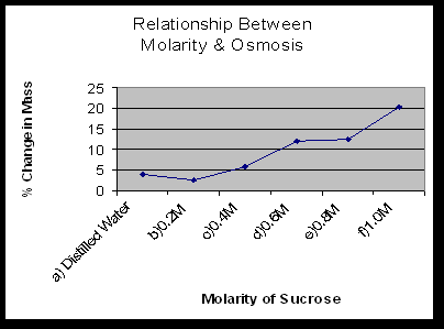
  1. Explain the relationship between the change in mass and the molarity of sucrose within the dialysis bags.  The solute is hypertonic and water will move into the bag.  As the molarity increases the water moves into the bag.
  2. Predict what would happen to the mass of each bag in this experiment if all the bags were placed in a 0.4M sucrose solution instead of distilled water.  Explain.  With the 0.2M bag, the water would move out.  With the 0.4M bag, there will be no net movement of water because the solutions reach dynamic equilibrium.  With the 0.6M-1M bags, the water would move into the bag.
  3. Why did you calculate the percent change in mass rather than simply using the change in mass?  This was calculated because each group began with different initial masses and we would have different data.  All the groups needed consistent data.
  4. A dialysis bag is filled with distilled water and then places in a sucrose solution.  The bag’s initial mass is 20g and its final mass is 18g.  Calculate the percent change of mass, showing your calculations.  ((18-20)/20) x 100 = 10%
  5. The sucrose solution in the beaker would have been hypotonic to the distilled water in the bag.

Exercise 1C


Table 1.4: Potato Core: Individual Data


Contents of Beaker Initial Mass (g) Final Mass (g) Difference in Mass % Change in Mass
Distilled Water 2.8 3.7 .9 32.14
0.2M 2.9 3.1 .2 7
0.4M 2.5 2.2 .3 12
0.6M 2.3 1.9 .4 17.39
0.8M 2.5 1.9 .6 24
1.0M 2.3 1.8 .5 21.74


Table 1.5: Potato Core: Class Data


Group 1 Group 2 Total Class Average
Distilled Water 32.14% 21.1% 53.24% 26.62%
0.2M 7% 6.7% 13.7% 6.85%
0.4M -12% -6.5% -18.5% -9.25%
0.6M -17.39% -15.2% -32.59% -16.30%
0.8M -24% -20% -44% -22%
1.0M -21.74% -19% -40.74% -20.37%


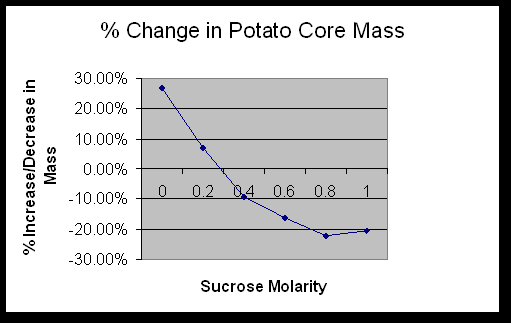
Determine the molar concentration of the potato core.  0.3M

Exercise 1D


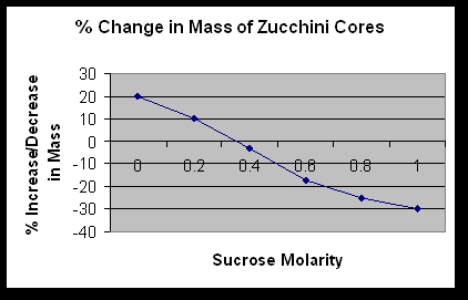
What is the molar concentration of the zucchini cores? .35M


  1. If a potato core is allowed to dehydrate by sitting in the open air, would the water potential of the potato cells decrease or increase? Why?  It would decrease because the water would leave the cells and cause the water potential to go down.
  2. If a plant cell has a lower water potential than its surrounding environment and if pressure is equal to zero, is the cell hypertonic or hypotonic to its environment? Will the cell gain water or lose water?  It is hypotonic and it will gain water.
  3. The beaker is open to the atmosphere.  What is the pressure potential of the system?  The pressure potential is zero.
  4. Where is the greatest water potential?  In the dialysis bag.
  5. Water will diffuse out of the bag. Why? It is because the water moves from and area of high water potential to an area of lower water potential.
  6. What effect does adding solute have on the solute potential component of that solution? Why?  It makes is more negative.
  7. Consider what would happen to a red blood cell placed in distilled water: a) Which would have the higher concentration of water molecules?  Distilled Water  b) Which would have the higher water potential?  Distilled Water  c)  What would happen to the red blood cell? Why?  It would lyce, because it would take on too much water.

Error Analysis:

Possible errors that could have affected the results of the lab include incorrectly mixing the solutions, ineffectively tying the dialysis tubing, inaccurately measuring , and inaccurately calculating.

Conclusion:

            During Exercise 1A the data that was collected help determine which molecules can and can not move across a cell membrane. Obviously, because of the color change in the bag, the IKI was able to move across the membrane.  It is small enough to fit through the pores in the selectively permeable membrane, along with water.  Starch was too large to move across the membrane. Glucose, as the Benedict’s test proves, was able to move freely along with the water and IKI solution.

In Exercise 1B, it was proven that water moves faster across the cell membrane than sucrose.  The water moved to help reach dynamic equilibrium between the 2 solutions.  The sucrose molecules are too big to move across the membrane as fast as water can.

The data in Exercise 1C showed that the potatoes contained sucrose.  The sucrose in the potato raised the solute potential, which lowered the water potential.  The beaker of distilled water had a high water potential.  Water moves down the concentration gradient, causing the potato cores to take on water.

Exercise 1D helped better understand the lab with simple algebra equations.  It proved that the data that was collected was correct through mathematics.


Ap Unit 6 Heredity Study Guide


Unit 5    Molecular Genetics  Study Guide 


ü     Be able to describe & explain the experiments of the following scientists:
Frederick Griffith
Erwin Chargraff
Alfred Hershey
Martha Chase
Watson & Crick
Meselson & Stahl
Beadle & Tatum
“Dolly” experiment

ü     Know how DNA replicates including steps & the enzymes involved, energy required, nucleotides, leading & lagging strands, proofreading

ü     Be able to describe the ultrastructure of each component of the DNA & RNA molecules

ü     Know the steps of transcription, enzymes involved, etc

ü     Know the steps of translation, enzymes involved, etc.

ü     Be able to describe the structure & function of free and bound ribosomes

ü     Know the processing steps of newly made mRNA

ü     Know the types of mutations and their effects

ü     Know viral structure, reproduction, host recognition, viral genome, etc

ü     Be able to describe the prokaryotic genome, mechanisms for genetic recombination, & defenses against phages

ü     Differentiate between hetero- & euchromatin

ü     Know the function and use of restriction enzymes

ü     Be able to describe genomic differentiation

BACK