Cell Death

Nobel Prize in Medicine 2002

Genetic Regulation of Organ Development and Programmed Cell Death

Sydney Brenner H. Robert Horvitz John E. Sulston

Introduction

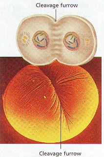
Sydney Brenner, Robert Horvitz and John Sulston’s discoveries concerning the genetic regulation of organ development and programmed cell death have truly opened new avenues for biological and medical research. We have all begun our lives in a seemingly modest way – as the fertilized egg cell, a tenth of a millimeter in size. From this small cell, the adult human being develops, with its hundred thousand billion cells, through cell division, cell differentiation and by formation of the various organs. To only make new cells is however not sufficient, certain cells must also die at specific time points as a natural part of the growth process. Think for example about how we for a short period during fetal life have web between our fingers and toes, and how this is removed by cell death.

The importance of cell differentiation and organ development was understood by many, but progress was slow. This was largely an effect of our complexity, with the large number of cells and many cell types – the forest could not be seen because of all the trees. Could the task to find the genetic principles be made simpler? Were there a species simpler than humans, but still sufficiently complex to allow for general principles to be deduced?

Sydney Brenner in Cambridge, UK, took on the challenge, and his choice was the nematode Caenorhabditis elegans. This may at first seem odd, a spool-shaped approximately 1 millimeter long worm with 959 cells that eats bacteria, but Brenner realized in the early 1960s that it was, what we today would call, “loaded with features”. It was genetically amenable and it was transparent, so that every cell division and differentiation could be directly followed in the worm under the microscope. Brenner demonstrated in 1974 that mutations could be introduced into many genes and visualized as distinct changes in organ formation. Through his visionary work, Brenner created an important research tool. The nematode had made into the inner circle of research.

John Sulston came to Brenner’s laboratory in 1969. He took advantage of that cell divisions could be followed under the microscope and assembled the cell lineage in the worm, showing which cells are siblings, first and second cousins. He found that cell divisions occurred with a very high degree of precision, the cell lineage was identical between different individuals. He also realized that certain cells in the lineage always died at a certain time point. This meant that programmed cell death was not a stochastic process, but rather occurred with a very high degree of precision. During the course of this work Sulston identified the first gene important for the cell death process: nuc-1.

Robert Horvitz came to work with Brenner and Sulston in 1974. Horvitz started a systematic search for genes controlling programmed cell death. He identified the key genes for the cell death process proper. The discovery of these central death genes, ced-3, ced-4 and ced-9, changed the view on programmed cell death from something rather obscure to a process with a strict genetic program. Horvitz also showed that there are human homologues to the death genes in the worm and that those have corresponding functions – the cell death machinery had deep evolutionary roots.

This year’s Nobel Prize celebrates the Joy of Worms. Brenner’s almost prophetic visions from the early 1960s of the advantages of this model organism have been fulfilled. It has given us new insights into the development of organs and tissues and why specific cells are destined to die. This knowledge has proven valuable, for instance, in understanding how certain viruses and bacteria attack our cells, and how cells die in heart attack and stroke.

Cell lineage – from egg to adult

All cells in our body are descendents from the fertilized egg cell. Their relationship can be referred to as a cellular pedigree or cell lineage. Cells differentiate and specialize to form various tissues and organs, for example muscle, blood, heart and the nervous system. The human body consists of several hundreds of cell types, and the cooperation between specialized cells makes the body function as an integrated unit. To maintain the appropriate number of cells in the tissues, a fine-tuned balance between cell division and cell death is required. Cells have to differentiate in a correct manner and at the right time during development in order to generate the correct cell type.

It is of considerable biological and medical importance to understand how these complicated processes are controlled. In unicellular model organisms, e.g. bacteria and yeast, organ development and the interplay between different cells cannot be studied. Mammals, on the other hand, are too complex for these basic studies, as they are composed of an enormous number of cells. The nematode C. elegans, being multi-cellular, yet relatively simple, was therefore chosen as the most appropriate model system, which has then led to characterization of these processes also in humans.

Programmed cell death

Normal life requires cell division to generate new cells but also the presence of cell death, so that a balance is maintained in our organs. In an adult human being, more than a thousand billion cells are created every day. At the same time, an equal number of cells die through a controlled “suicide process”, referred to as programmed cell death.

Developmental biologists first described programmed cell death. They noted that cell death was necessary for embryonic development, for example when tadpoles undergo metamorphosis to become adult frogs. In the human foetus, the interdigital mesoderm initially formed between fingers and toes is removed by programmed cell death. The vast excess of neuronal cells present during the early stages of brain development is also eliminated by the same mechanism.

The seminal breakthrough in our understanding of programmed cell death was made by this year’s Nobel Laureates. They discovered that specific genes control the cellular death program in the nematode C. elegans. Detailed studies in this simple model organism demonstrated that 131 of totally 1090 cells die reproducibly during development, and that this natural cell death is controlled by a unique set of genes.

The model organism C. elegans

Sydney Brenner realized, in the early 1960s, that fundamental questions regarding cell differentiation and organ development were hard to tackle in higher animals. Therefore, a genetically amenable and multicellular model organism simpler than mammals, was required. The ideal solution proved to be the nematode Caenorhabditis elegans. This worm, approximately 1 mm long, has a short generation time and is transparent, which made it possible to follow cell division directly under the microscope.

Brenner provided the basis in a publication from 1974, in which he broke new ground by demonstrating that specific gene mutations could be induced in the genome of C. elegans by the chemical compound EMS (ethyl methane sulphonate). Different mutations could be linked to specific genes and to specific effects on organ development. This combination of genetic analysis and visualization of cell divisions observed under the microscope initiated the discoveries that are awarded by this year’s Nobel Prize.

Mapping the cell lineage

John Sulston extended Brenner’s work with C. elegans and developed techniques to study all cell divisions in the nematode, from the fertilized egg to the 959 cells in the adult organism. In a publication from 1976, Sulston described the cell lineage for a part of the developing nervous system. He showed that the cell lineage is invariant, i.e. every nematode underwent exactly the same program of cell division and differentiation.

As a result of these findings Sulston made the seminal discovery that specific cells in the cell lineage always die through programmed cell death and that this could be monitored in the living organism. He described the visible steps in the cellular death process and demonstrated the first mutations of genes participating in programmed cell death, including the nuc-1 gene. Sulston also showed that the protein encoded by the nuc-1 gene is required for degradation of the DNA of the dead cell.

Identification of “death genes”

Robert Horvitz continued Brenner’s and Sulston’s work on the genetics and cell lineage of C. elegans. In a series of elegant experiments that started during the 1970s, Horvitz used C. elegans to investigate whether there was a genetic program controlling cell death. In a pioneering publication from 1986, he identified the first two bona fide “death genes”, ced-3 and ced-4. He showed that functional ced-3 and ced-4 genes were a prerequisite for cell death to be executed.

Later, Horvitz showed that another gene, ced-9, protects against cell death by interacting with ced-4 and ced-3. He also identified a number of genes that direct how the dead cell is eliminated. Horvitz showed that the human genome contains a ced-3-like gene. We now know that most genes that are involved in controlling cell death in C. elegans, have counterparts in humans.

Of importance for many research disciplines

The development of C. elegans as a novel experimental model system, the characterization of its invariant cell lineage, and the possibility to link this to genetic analysis have proven valuable for many research disciplines. For example, this is true for developmental biology and for analysis of the functions of various signaling pathways in a multicellular organism. The characterization of genes controlling programmed cell death in C. elegans soon made it possible to identify related genes with similar functions in humans. It is now clear that one of the signaling pathways in humans leading to cell death is evolutionarily well conserved. In this pathway ced-3-, ced-4- and ced-9-like molecules participate. Understanding perturbations in this and other signaling pathways controlling cell death are of prime importance for medicine.

Disease and programmed cell death

Knowledge of programmed cell death has helped us to understand the mechanisms by which some viruses and bacteria invade our cells. We also know that in AIDS, neurodegenerative diseases, stroke and myocardial infarction, cells are lost as a result of excessive cell death. Other diseases, like autoimmune conditions and cancer, are characterized by a reduction in cell death, leading to the survival of cells normally destined to die.

Research on programmed cell death is intense, including in the field of cancer. Many treatment strategies are based on stimulation of the cellular “suicide program”. This is, for the future, a most interesting and challenging task to further explore in order to reach a more refined manner to induce cell death in cancer cells.

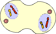
Using the nematode C. elegans this year’s Nobel Laureates have demonstrated how organ development and programmed cell death are genetically regulated. They have identified key genes regulating programmed cell death and demonstrated that corresponding genes exist also in higher animals, including man. The figure schematically illustrates the cell lineage (top left) and the programmed cell death (below) in C. elegans. The fertilized egg cell undergoes a series of cell divisions leading to cell differentiation and cell specialization, eventually producing the adult organism (top right). In C. elegans, all cell divisions and differentiations are invariant, i.e. identical from individual to individual, which made it possible to construct a cell lineage for all cell divisions. During development, 1090 cells are generated, but precisely 131 of these cells are eliminated by programmed cell death. This results in an adult nematode (the hermaphrodite), composed of 959 somatic cells.

Source: http://nobelprize.org/nobel_prizes/medicine/laureates/2002/press.html


Campbell Problem 10

Molecular Genetics Problem 10
10. An aneuploid person is obviously female, but her cells have two Barr bodies. What is the probable complement of sex chromosomes in this individual?

This individual probably is XXX.

The individual is a female. Nondisjunction of sex chromosomes produces a variety of aneuploid conditions in humans. Most of these conditions appear to upset genetic balance less than aneuploid conditions involving autosomes. Extra copies of the X chromosome are deactivated as Barr bodies in the somatic cells. Females with trisomy of the X chromosome (XXX), which occurs about once in approximately 1000 live births, are healthy and cannot be distinguished from XX females except by karyotype.

An Example of nondisjunction:

Klinefelter’s syndrome

49 ,XXXXY

This karyotype shows a variant of Klinefelter’s syndrome.

Individuals with this syndrome are male, typically with the karyotype 47,XXY.

Individuals with Klinefelter’s syndrome exhibit a characteristic phenotype including tall stature, infertility, gynecomastia and hypogonadism.

Aneuploidy above one extra chromosome is usually fatal but because of X-inactivation, which “turns off” all but one X chromosome per cell, the effects of 3 extra chromosomes are reduced.

BACK 

Cell Cycle & Division


Cell Cycle & Division
All Materials © Cmassengale


Cell Division:

  • All cells are derived from preexisting cells (Cell Theory)
  • Cell division is the process by which cells produce new cells
  • Cell division differs in prokaryotes (bacteria) and eukaryotes (protists, fungi, plants, & animals)
  • Some tissues must be repaired often such as the lining of gut, white blood cells, skin cells with a short lifespan 
  • Other cells do not divide at all after birth such as muscle & nerve 

Reasons for Cell Division:

  • Cell growth
  • Repair & replacement of damaged cell parts
  • Reproduction of the species

Copying DNA: 

  • Since the instructions for making cell parts are encoded in the DNA, each new cell must get a complete set of the DNA molecules
  • This requires that the DNA be copied (replicated, duplicated) before cell division

Replication process

Chromosomes & Their Structure:

  • The plans for making cells are coded in DNA
  • DNA, deoxyribose nucleic acid, is a long thin molecule that stores genetic information
  • DNA in a human cell is estimated to consist of six billion pairs of nucleotides
  • DNA is organized into giant molecules called chromosomes
  • Chromosomes are made of protein & a long, single, tightly-coiled DNA molecule visible only when the cell divides
  • When a cell is not dividing the DNA is less visible & is called chromatin
  • DNA in eukaryotic cells wraps tightly around proteins called histones to help pack the DNA during cell division
  • Nonhistone proteins help control the activity of specific DNA genes
  • Kinetochore proteins bind to centromere and attach chromosome to the spindle in mitosis
  • Centromeres hold duplicated chromosomes together before they are separated in mitosis
  • Telomeres are the ends of chromosomes which are important in cell aging
  • When DNA makes copies of itself before cell division, each half of the chromosome is called a sister chromatid

  • DNA of prokaryotes (bacteria) is one, circular chromosome attached to the inside of the cell membrane

Chromosome Numbers:

  • Humans somatic or body cells have 23 pairs of chromosomes or 46 chromosomes (diploid or 2n number)
  • The 2 chromatids of a chromosome pair are called homologues (have genes for the same trait at the same location)


Homologs

  • Human reproductive cells or gametes (sperms & eggs) have one set or 23 chromosomes (haploid or n number)
  • Every organism has a specific chromosome number


Organism Chromosome Number (2n)
Human 46
Fruit fly 8
Lettuce 14
Goldfish 94


  • Fertilization, joining of the egg & sperm, restores the diploid chromosome number in the zygote (fertilized egg cell)
  • Sex chromosomes, either X or Y, determine the sex of the organism
  • Two X chromosomes, XX, will be female and XY will be male
  • All other chromosomes, except X & Y, are called autosomes
  • Chromosomes from a cell may be arranged in pairs by size starting with the longest pair and ending with the sex chromosomes to make a karyotype
  • A human karyotype has 22 pairs of autosomes and 1 pair of sex chromosomes (23 total)


Human Male Karyotype

Genes:

  • A section of DNA which codes for a protein is called a gene
  • Each gene codes for one protein
  • Humans have approximately 50,000 genes or 2000 per chromosome
  • About 95% of the DNA in chromosome is “junk” that does not code for any proteins

Cell Cycle:

  • Cells go through phases or a cell cycle during their life before they divide to form new cells 
  • The cell cycle includes 2 main parts — interphase, and cell division
  • Cell division includes mitosis (nuclear division) and cytokinesis (division of the cytoplasm)
  • Interphase is the longest part of a cell’s life cycle and is called the “resting stage” because the cell isn’t dividing
  • Cells grow, develop, & carry on all their normal metabolic functions during interphase
  • Interphase consists of 3 parts — G1, S, & G2phases

Interphase:

  • G1 or 1st Growth Phase occurs after a cell has undergone cell division
  • Cells mature & increase in size by making more cytoplasm & organelles while carrying normal metabolic activities in G1 
  • S or Synthesis Phase follows  G1  and the genetic material of the cell (DNA) is copied or replicated 

  • G2 or 2nd Growth Phase occurs after S Phase and the cell makes all the structures needed to divide

Cell division in Prokaryotes:

  • Prokaryotes such as bacteria do not have a nucleus
  • Prokaryotes divide into two identical new cells by the process of binary fission
  • Binary fission is an asexual method of reproduction
  • In binary fission,  the chromosome, attached to cell membrane, makes a copy of itself and the cell grows to about twice its normal size
  • Next, a cell wall forms between the chromosomes & the parent cell splits into 2 new identical daughter cells (clones)


Cell Division in Eukaryotes:

  • Eukaryotes have a nucleus & membrane-bound organelles which must be copied exactly so the 2 new cells formed from division will be exactly alike
  • The original parent cell & 2 new daughter cells must have identical chromosomes
  • DNA is copied in the S phase of the cell cycle & organelles, found in the cytoplasm,  are copied in the Growth phases
  • Both the nucleus (mitosis) and the cytoplasm (cytokinesis) must be divided during cell division in eukaryotes

Stages of Mitosis:

  • Division of the nucleus or mitosis occurs first
  • Mitosis is an asexual method of reproduction
  • Mitosis consists of 4 stages — Prophase, Metaphase, anaphase, & Telophase

  • Prophase:
    • Chromosomes become visible when they condense into sister chromatids
    • Sister chromatids attach to each other by the centromere
    • Centrioles in animal cells move to opposite ends of cell
    • Spindle forms from centriole (animals) or microtubules (plants)
    • Kinetochore fibers of spindle attach to centromere
    • Polar fibers of spindle extend across cell from pole to pole
    • Nuclear membrane dissolves
    • Nucleolus disintegrates
  • Metaphase:
    • Chromosomes line up in center or equator of the cell attached to kinetochore fibers of the spindle
  • Anaphase:
    • Kinetochore fibers attached to the centromere pull the sister chromatids apart
    • Chromosomes move toward opposite ends of cell
  • Telophase:
    • Nuclear membrane forms at each end of the cell around the chromosomes
    • Nucleolus reform
    • Chromosomes become less tightly coiled & appear as chromatin again
    • Cytokinesis begins

Cytokinesis:

  • Cytoplasm of the cell and its organelles separate into 2 new daughter cells
  • In animals, a groove called the cleavage furrow forms pinching the parent cell in two

  • In plants, a cell plate forms down the middle of the cell where the new cell wall will be

Summary of Mitosis:


 Interphase


  1. Cell matures & carries on normal activities
  2. DNA copied & appears as chromatin
  3. Nucleolus visible
 Early Prophase  


  1. Chromosomes condense & become visible
  2. Centrioles separate & spindle starts forming
 
 Late Prophase

  1. Spindle forms with aster at each pole
  2. Nuclear membrane & nucleolus disintegrate
  3. Centromere of chromosomes attaches to spindle fibers
 Metaphase

  1. Chromosomes line up at the equator of the cell attached to kinetochore fibers of spindle
 Anaphase

  1. Centromeres split apart
  2. Homologs move to opposite poles of the cell
 Telophase/Cytokinesis  

  1. Nuclear membrane & nucleolus reform
  2. Cell pinches into 2 cells in animals
  3. In plants, a cell plate separates the 2 new cells


Cancer is Uncontrolled Mitosis:

  • Mitosis must be controlled, otherwise growth will occur without limit (cancer)
  • Control is by special proteins produced by oncogenes
  • Mutations in control proteins can cause cancer

Meiosis & Sexual Reproduction

  • Reduces the number of chromosomes in new cells to half the number in the original cell
  • New cells have a single copy of chromosomes (23 total) but are not identical to each other or the original parent cell
  • Used for making gametes ( sperm and eggs) with the haploid or n number
  • In meiosis, cells divide twice after a single DNA duplication
  • Meiosis I separates homologs & the Meiosis II separates sister chromatids
  • Meiosis I stages are Prophase I, Metaphase I, Anaphase I, & Telophase I
  • Meiosis II stages are Prophase II, Metaphase II, Anaphase II, & Telophase II
  • Produces 4 haploid cells or gametes
  • When a sperm fertilizes an egg to form a zygote, the diploid number of chromosomes is restored (23 + 23 = 46)
  • Egg cells or ova (ovum, singular) are larger , nonmotile cells
  • Gametoogenesis is meiosis producing eggs & occurs in the female’s ovaries


Oogenesis

  • Sperms contain less cytoplasm so they’re smaller & have a flagellum to swim to the egg
  • Spermatogenesis is meiosis producing sperm cells & occurs in the testes


Spermatogenesis

Meiosis I:

  • The cell that undergoes Meiosis I is a primary spermatocyte or oocyte
  • Prophase I:
    • Chromosomes coil tightly & are visible
    • Nuclear membrane & nucleolus disintegrate
    • Spindle forms
    • Synapsis (joining) of homologous chromosomes occurs making tetrads
    • Kinetochore fiber forms on each chromosome
    • Chromosomes in tetrad exchange fragments by a process called crossing over

  • Metaphase I:
    • Tetrads become aligned in the center of the cell attached to spindle fibers
  • Anaphase I:
    • Homologous chromosomes separate
  • Telophase I:
    • May not occur in all species
    • Cytokinesis occurs producing 2 cells
    • In females,  2nd cell in females is called the 1st Polar Body
    • 1st Polar Body dies due to uneven splitting of the cytoplasm

  • Prophase II:
    • Cells called Secondary Spermatocytes or oocytes
    • DNA is not copied before cell divides
    • Chromatids attach to spindle fiber
  • Metaphase II:
    • Chromosomes become aligned in the center of the cell attached to spindle fibers
  • Anaphase II:
    • Sister chromatids separate randomly
    • Called independent assortment
  • Telophase I:
    • Cytokinesis occurs producing 4 cells in males called spermatids
    • Spermatids mature & form flagellum to become sperm
    • Cytokinesis in females produces a 2nd Polar Body that dies and an ootid 
    • Ootids mature to become ovum or egg

Asexual & Sexual reproduction:

  • Evolution is the slow process of change in living populations over time
  • Variations are differences that occur due to crossing-over among members of a sexually reproducing population
  • Variations are important to the survival of individuals in a population (some must survive to reproduce)
  • Asexually reproducing organisms rarely show variations because the organisms have identical genes

Campbell Problem 12

Molecular Genetics Problem 12
12. About 5% of individuals with Downs syndrome are the result of chromosomal translocation. In most of these cases, one copy of chromosome 21 becomes attached to chromosome 14. How does this translocation lead to children with Down syndrome?


The case of having a chromosomal fragment joining to a nonhomologous chromosome is called translocation.

Children who have Down’s Syndrome will either have an extra chromosome 21, thus having a total of 47 chromosomes, or about 5% receive a combined 14-21 chromosome combination. (Chromosome 21 links to #14).

In meiosis, the combined 14-21 chromosome will actually behave as a single chromosome. Should this mutated gamete join a normal one during fertilization there will be three chromosome 21’s in the resulting zygote.


BACK 

Cell Division Study Guide BI

Cell Division Study Guide

What molecule contains the information needed to direct all the activities of a cell?
Where in a cell are prokaryotic chromosomes found? eukaryotic chromosomes?
A human somatic cell contains how many homologous chromosomes?
How many chromosomes are in an human egg cell? sperm cell?
What is a karyotype?
Are gametes diploid or haploid?
Zygotes will have what chromosome number?
Does cell division in bacteria take place in the same way as it does in eukaryotes? Explain.
In what stage do cells spend most of their life cycle?
Is mitosis asexual or sexual reproduction?
A new nuclear envelope develops during cell division in what stage?
In what stage do chromatids separate from each other?
How does the number of chromosomes in newly divided cells compare with the number of chromosomes in the original cell?
During what type of cell division do haploid cells develop from diploid cells?
In order for DNA to fit into a cell, what must be done to compact it?
What is a centromere?
How many chromosomes are in a human skin cell? a human ovum?
Bacteria reproduce by a method known as _____________  ______________.
What is the shape of a bacterial chromosome?
Chromosomes are arranged along the equator of a cell during which stage of cell division?
Spindle fibers are made of ________________.
Be able to recognize sketches of the stages of mitosis.
What happens during cytokinesis in a plant cell?
Homologs separate during ________________.
BACK