Animal Symmetry and Phyla

Annelids
Segmented Worms
All Materials © Cmassengale
Phylum Annelida
Characteristics


PARAPODIA
![]()
![]()
![]()
![]()
![]()
Class Oligochaeta
Characteristics

EARTHWORM

![]()
![]()
![]()
![]()
![]()
Class Hirudenia
Characteristics

LEECH
![]()
![]()
![]()
![]()
![]()
Class Polychaeta
Characteristics

SANDWORM
Antibiotic resistance of bacteria
Procedure Using a sterile loop, pick an isolated colony from you bacterial plate. Try to find one that grew well but is all by itself. Move the colony (don’t scoop up the agar) to a new plate.  Using a moist, sterile, cotton swab, spread the bacteria around on the plate.
The goal is to get an complete, even, coverage of bacterial growth on the plate (called a “lawn”). Remember to open the plate only minimally, using the lid as an “umbrella” to prevent contamination (see image below). Label each plate on the bottom (agar contaning side) and store it for examination during next week’s lab.Each new prepared plate will receive four paper discs containing antibiotics. We will be using several different types of Antibiotics and/or antimicrobials.
(please fill in which antibiotics you used below)
The antibiotic discs come in a little tube-like dispenser. To remove the discs take a sterile toothpick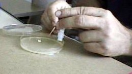 and push out a disc into your plate. Use the toothpick to gently press the disc onto the agar. Once you have added the five antibiotic discs to your plates, make sure the plates are labelled and store them in the back of the lab until next week.
If the bacteria are susceptible to the antibiotic a zone of inhibited growth will be evident next week. Measuring the size of this zone is a relative indication of the effect of the antibiotic on the particular bacteria.
Bacteria possess several characteristics that enable them to become resistant to antimicrobial drugs:
Some Information on Antibiotics
Questions –
1. Name two ways (1. and 2. ) that common human practices towards antimicrobials aids bacteria in becoming resistant.
2. Name two reasons your Physician will perform cultures such as the ones you have done in this lab.
4. How are materials are collected for cultures?
5. Why is neccesary to use sterile technique when obtaining cultures?
AP Calendar 2006

| week | topics | text chapters/activities | supplemental readings/URLs |
| see you in late August!!
week 1 Aug 24-26 |

|
ch 1, 50-55-due Aug. 24-your first day back!!**check study guide (SG) and question set (QS)
powers of ten see the size of everything! ch 2
handouts/AP website |
scientific method -The Biology Project size and biology -The Biology Project
dissociation of water acid-base explained water movement click on animations water movement ions, simulations of pH solutions
|

| week | topics | text chapters/activities | supplemental readings/URLs |
| week 2
Aug 29-Sept 2 |
|
ch 3
BBC guru condensation and structure of molecules
|
central dogma animation
molecules -animated (rotate) dehydration synthesis -animation the biological basis of life for condensation |
| week 3
Sept 6-9 Sept 5 off-Labor Day |
|
ch 6 | interactives catalysis, enzyme inhibition
BBC guru this is the enzyme site we used in class
|
| week 4
CAMP KERN (but you are responsible for everything!!) Sept 12-16 |
|
population clock population estimation
climate graphs/biomes dissolved O2 lab
|
Nova-The World in Balance Human Numbers/Time The Earth in Peril Be a Demographer Population Trend quiz
nitrogen cycle -animation
|
| week 5
Sept 19-23
|
|
|
interesting ecology simulations, including: biomass succession
|

| week | topics | text chapters/activities | supplemental readings/URLs |
| week 6
Sept 26- 30
|
|
ch 20 |
burgess shale -resources
early life -Burgess Shale |
| week 7
Oct 3-7 |
|
ch 4
ch 7
|
microscopy -CellsAlive
eukaryotic vs pro, quizzes on plant and animal cell show big is? (cell biology) -CellsAlive binary fission movie lysogenic cycle -animation |
| week 8
Oct 10-14 Oct 11 off -teacher inservice |
|
ch 4
these are the links that we used in class- virtual cell -virtual cell textbook cell comparison -check yourself the cell -ThinkQuest
|
inside the cell -NIH/NIGMS
cell parts -cells.de link of the month drop/drag organelles plant and animal cells -structure and function problem sets and tutuorials -The Biology Project, includes: studying cells tutorial and cytoskeleton among others-best site cell similarity -GeoCities-fun stuff click on animations -HHMI |
| week 9
Oct 17-21 Oct 22-end of Q1 |
|
ch 5
membrane structure -simple pathways in and out of the cell antibiotic resistance |
drop/drag membrane ZeroBio
membrane structure power point tutorial-advanced cell signaling Access Excellence osmosis tutorial Cornell BioG101-104 osmosis simulation Colorado osmosis animation Oklahoma State osmotic pressure sanger reverse osmosis gearfiltration ion channels (cell biology, myocyte) CellsAlive membrane animation HHMI cell transport animation Na/K pump Brookscole membrane tutorial The Biology Project |
| week 10
Oct 24-28
|
|
ch 7
not active…. cell respiration -tutorial overview -respiration electron transport chain and ATP synthesis -movies glycolysis and fermentation -excellent molecular animations |
glucose catabolism U. Alberta-overview
intro to metabolism animation metabolic process location animation citric acid cycle animation oxydative phosphorylation electron transport chain animation cellular respiration -Kimball’s pages cellular respiration -showing molecular models glycolysis explained in animations oxydative phosphorylation explained inanimations |


| week | topics | text chapters/ activities | supplemental readings/URLs |
| week 11
Oct 31-Nov 4
|
|
ch 8
|
photosynthesis light rxn -The Biology Project
photosynthesis carbo formation -The Biology Project photosynthesis sites -Arizona State University photosynthesis animation oxygenic photosynthesis animation U. Alberta photosynthesis -Maricopa bio181
|
| week 12
Nov 7-11
|
|
ch 9 Online Onion Tips -The biology Project more animations -BSC Courseware |
cell cycle -The Biology Project
cell cycle animation, myocyctes, cytoskeleton, apoptosis (cell biology, cell models, cell gallery) -CellsAlive mitosis review -Nebraska Wesleyan interactive mitosis (cell biology)-CellsAlive mitosis -The Biology Project modeling mitosis -Cornell BioG101-104 click either random or assignment…… mitosis/meiosis animations -About: homework help mitosis and meiosis tutorial -Cornell BioG101-104 meiosis vs mitosis -WGBH (PBS) cancer growth -WGBH (PBS) metastasis -animation |
| week 13
Nov 14-18
|
|
ch 10, 15 |
mendelian genetics -The Biology Project
human karyotyping -The Biology Project genetics -University of Utah harlequin chromosomes -J. Kimball drop/drag genetic cross ZeroBio
|
| week 14
Nov 21-22
|
Nov 23-25 off Thanksgiving!
|
ch 11
edu/genobc/animations/
|
Central Dogma animation nucleic acids -The Biology Project Hershey/Chase -AccessExcellence DNA basics -The Biology Project DNA replication animation Cracking the code (PBS) DNA structure -subunit animation
|

| week | topics | text chapters/ activities | supplemental readings/URLs |
| week 15
Nov 28-Dec 2
|
|
transformation movie
translation animation ZeroBio protein synthesis animation Beginner’s Guide to Molecular Biology -Molecular Biology Notebook DNA from the Beginning -DNA from the Beginning |
|
|
week 16 Dec 5-9
|
|
ch 13, 16
|
gene regulation -Indiana State University
RNA splicing -movie cloning animation developement -The Biology Project gene expression -The Biology Project gene expression in prokaryotes -The Biology Project signal transduction animation |
| week 17
Dec 12-16
|
|
ch 14
|
Blackett family -The Biology Project
DNA profiling -The Biology Project Western Blot -The Biology Project recombinant DNA -The Biology Project PCR animation human genome project -National Human Genome Research Institute bacterial genetics/recombinant DNA tutorial -Cornell BioG101-104 forensics -The Biology Project
|
|
Dec 21 Winter Vacation starts at end of day
|
ch 17 are yours for the break–Happy Holidays!! (you would be bored without this work…. |
End of First semester

| week | topics | text chapters/activities | supplemental readings/URLs |
| week 18
Jan 3-7
|
|
ch 17–18
|
dating techniques -Geology Labs on-line
Darwin’s life and works -Cal. State University, Dr. C. Urbanowicz population drift -simulation genetic drift -simulation |
| week 19
Jan 10-14
Jan 14-end of Sem I |
semester I final exam week
|
AP sample exam |
| Semester II calendar 2005
home | AP Bio | Honors Bio | CP Bio | calendars | Visit the Lab
© Mariemont City Schools 2002, Halsall (05/23/06 )
|

 |
Viruses Worksheet | |
Structure of Viruses
1. Are viruses living or nonliving?
2. How can viruses be useful?
3. What odes a virologist do for a living?
4. Construct a Venn diagram comparing viruses and cells.
5. Explain how viruses were discovered and by whom.
6. Compare the size of viruses, bacteria, and eukaryotic cells.
7. What must be true for viruses to be able to replicate?
8. Name the two main parts of all viruses.
9. Discuss the hereditary material of viruses.
10. Compare & contrast capsids and envelopes of viruses.
11. Name 2 enveloped viruses that cause sexually transmitted disease.
12. What type of virus causes flu?
13. Where are glycoproteins found & what is there purpose?
14. What characteristics are used to group viruses?
15. How are these viruses grouped — retrovirus, adenovirus, and herpes virus?
16. Compare & contrast helical & icosahedral viral shapes & diseases.
17. Explain how RNA viruses replicate.
18. Do viruses contain enzymes? Explain.
19. Compare 7 contrast viroids & prions by constructing a Venn diagram.
Viral Replication
20. Why are viruses considered to be obligate intracellular parasites?
21. What is the best known bacteriophage, and what virus does it attack?
22. Sketch & label a bacteriophage and tell the function of each labeled part.
23. Name the steps of the lytic cycle & tell what happens to the host cell & virus at each stage.
24. What are temperate phages and how do they affect a cell?
25. Name the steps of the lysogenic cycle & tell what happens to the host cell & virus at each stage.
26. How does a prophage form?
27.Name a sexually transmitted virus that uses the lysogenic cycle to attack host cells.
28. Why is the influenza virus so hard to combat?
Viruses & Human Disease
29. Name some of the most common viral disease that attack humans.
30. How are shingles & chickenpox alike? How are they different?
31.What two methods are used to control viral diseases?
32. What is the CDC and what is its job?
33.What eradication program did the World Health Organization undertake in 1967, and what were the results?
34. What virus do we vaccinate our pets against each year?
35. How does AZT work?
36. What drugs prevent viruses from making capsids?
37. Why is rain forest clearing dangerous to humans?
38. Some lysogenic viruses can trigger certain types of _________________.


