Plant Pigments and Photosynthesis
Introduction:
Photosynthesis has two main parts, which are the light dependent and the light –independent. In the light-dependent reactions pigments trap energy from light, and this energy is used to split water molecules (photolysis). The light-independent reactions or dark phase of photosynthesis involve the fixing of carbon dioxide. It makes glucose and fructose chains and also releases oxygen , which passes through the stomata of the plant.
Organisms that carry out photosynthesis making their own organic molecules are called autotrophic. Some autotrophic organisms include plants, algae, and blue-green bacteria. Plants have many varieties of pigments, all of which absorb different colors of light. Chlorophyll a is the primary plant pigment and makes up about three-fourths of all the plant pigments. It absorbs red and blue light and is not found in photosynthetic bacteria.
Chlorophyll b is another plant pigment. It absorbs blue-green and orange-red light. Carotenoids are a type of accessory pigment that absorb blue and blue-green light. These pigments are fat soluble and usually masked by chlorophyll a. Anthocyanin is another accessory pigment that absorbs bright red colors. There is also chlorophyll c and d that sometimes take the place of chlorophyll b.
Chromatography is a process used to separate mixtures that can separate plant pigments. This lab uses paper chromatography where a piece of paper is used to wick solvent up to the pigments and separate them according to solubilities. The rate of migration on a chromatogram is the Rf value.
Hypothesis
Plants contain several different pigments, and the rate of photosynthesis in plant cells is directly related to light and temperature.
Materials
Exercise 4A: Plant Pigment Chromatography
This exercise required 1 50-mL graduated cylinder, a small amount of a solvent, a stopper, filter paper, scissors, a pencil, spinach leaves, and a quarter.
Exercise 4B: Photosynthesis/The Light Reaction
The materials needed for this part of the lab were a spectrophotometer, a light, a water flask, a test tube rack, ice, 5 labeled cuvettes, lens tissue, foil, and parafilm. The substances put in the cuvettes were 5 mL of phosphate buffer, approximately 16 mL of distilled water, 9 drops of unboiled chloroplasts, and 3 drops of boiled chloroplasts.
Methods
Exercise 4A: Plant Pigment Chromatography
A 50-mL graduated cylinder was filled with about 1 cm of solvent and then tightly stoppered. The filter paper was then cut to a point on one end, and a line was drawn 1.5 cm above the point. Using the ribbed edge of a quarter, spinach cells were extracted onto the pencil line. This procedure was repeated 8-10 times using a new portion of the leaf each time. The filter paper was then placed in the cylinder with the tip barely touching the solvent and none of the edges touching the sides. When the solvent reached 1 cm below the top of the paper, it was removed from the cylinder. The solvent location was immediately marked, and then the bottom of each pigment band was also marked.
Exercise 4B: Photosynthesis/The Light Reaction
The spectrophotometer was set to 605 nm and allowed to warm up. The chloroplast suspensions were prepared the previous day, part of which were boiled, and stored on ice until they were ready for use. An incubation area was prepared with a flood light, water flask, and test tube rack, by using the flask as a heat sink between the light and the rack. Five cuvettes were numbered respectively and then wiped with lens tissue. The walls and bottom of cuvette 2 were covered with foil and a foil cap was made for the top.
To each cuvette 1 mL of phosphate buffer was added. Then, to cuvette 1 4 mL of distilled water was added, but to cuvettes 2, 3, and 4 3 mL of distilled water was added. Next, 1 mL of DPIP was added to cuvettes 2, 3,and 4. To cuvette 5, 3 mL plus 3 drops of distilled water were added and 1 mL of DPIP. To cuvette 1, 3 drops of unboiled chloroplasts were added.
The spectrophotometer was brought back to zero and the contents of cuvette 1 were mixed by inverting and placed in the sample holder. Cuvette 1 was used periodically through this experiment to recalibrate the spectrophotometer. Three drops of unboiled chloroplasts were added to cuvette 2. After removing the foil sleeve, it was placed in the sample holder and the transmittance was recorded. Additional readings were also taken at 5, 10, and 15 minutes. Next, three drops of unboiled chloroplasts were transferred to cuvette 3. The percent transmittance was recorded at 0, 5, 10, and 15 minutes. Three drops of boiled chloroplasts were added to cuvette 4, and the transmittances were recorded at the same times. Finally, cuvette 5 was mixed and placed in the sample holder. The transmittance readings were recorded.
Results
Table 4.1 Distance Moved by Pigment Band
|
Band Number |
Distance (mm) |
Band Color |
| 1. | 0 mm | Yellow-brown |
| 2. | 5 mm | Light green |
| 3. | 30 mm | Green |
| 4. | 48 mm | Yellow |
Distance Solvent Front Moved 60 mm.
Table 4.2 Rf Values
| 0.8 = Rf for xanthophyll (yellow)
0.5 = Rf for chlorophyll a (bright green to blue green) 0 = Rf for chlorophyll b (yellow green to olive green) |
Table 4.4 Transmittance (%)
|
Cuvette |
0 |
5 |
10 |
15 |
|
2 Unboiled/Dark |
41% | 43% | 44% | 43% |
|
3 Unboiled/Light |
35% | 38% | 39% | 37% |
|
4 Boiled/Light |
51% | 52% | 53% | 54% |
|
5 No Chloroplasts |
57% | 57% | 56% | 55% |
Lab 4B Color Chart
|
Cuvette |
Initial Color |
Final Color |
|
1 |
Clear | Clear |
|
2 |
Light clear blue | Blue/green |
|
3 |
Light clear blue | Dark clear blue |
|
4 |
Light clear blue | Light clear blue |
|
5 |
Light clear blue | Dark clear blue |
Questions:
Exercise 4A: Plant Pigment Chromatography
What factors are involved in the separation of the pigments?
The solubility, size of particles, and their attractiveness to the paper are all involved in the separation.
Would you expect the Rf value of the pigment to be the same if a different solvent were used? Explain.
No, the different solubilities of the pigments would change the Rf values. For example chlorophyll b is only soluble to fat solutions.
What type of chlorophyll does the reaction center contain? What are the roles of the other pigments?
The reaction center contains chlorophyll a. The other pigments collect different light waves and transfer the energy to chlorophyll a.
Exercise 4B: Photosynthesis/The Light Reaction
What is the purpose of DPIP in this experiment?
DPIP is the electron acceptor in this experiment.
What molecule found in chloroplasts does DPIP “replace” in this experiment?
DPIP substitutes for the NADP molecules.
What is the source of the electrons that will reduce DPIP?
The electrons come from the photolysis of water.
What was measured with the spectrophotometer in this experiment?
The spectrophotometer measures the percentage of light transmittance through the cuvette due to DPIP reduction.
What is the effect of darkness on the reduction of DPIP? Explain.
The effect of darkness is that no reaction will occur.
What is the effect of boiling the chloroplasts on the subsequent reduction of DPIP? Explain.
Boiling denatures the protein molecules and stops the reduction.
What reasons can you give for the difference in the percent transmittance between the live chloroplasts that were incubated in the light and those that were kept in the dark?
In the dark cuvette, there was no light energy available, so there was no flow of electrons and no photolysis of water, while in the lighted cuvette these processes were allowed to continue.
Error Analysis
In Lab 4A, several mistakes could have been made with the chromatography paper. Too much handling, bending, or allowing the paper to touch the sides of the cylinder could all have affected the outcome of this experiment. The different pigment bands were also difficult to distinguish.
In Lab 4B, the procedure was very complicated and required a lot of pre-lab planning and reading. Incorrect usage of the spectrophotometer or neglecting to recalibrate often enough could have caused errors in this portion of the lab.
Discussion and Conclusion
Lab 4A demonstrated the different plant pigments by chromatography and showed how to calculate Rf values and explained their importance. There are 4-5 main pigments present in plants ranging from green to yellow in color. Lab 4B proves that light and chloroplasts are required for the light reactions of photosynthesis to occur. It showed the effects of boiling, denaturing, which did not allow photosynthesis to occur.
| BACK |


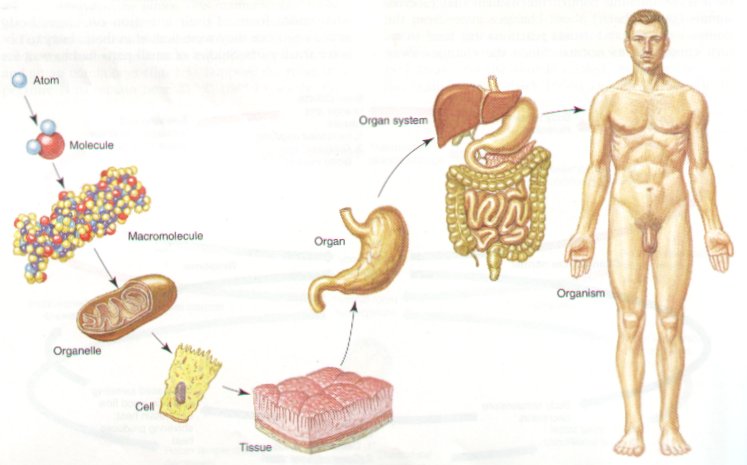

 4. ORGAN LEVEL
 1. All of the above systems function together to help the Human Body to Maintain HOMEOSTASIS.
 1. Many organs and organ systems in the human body are housed in compartments called BODY CAVITIES. (Figure 46-2)