| Cell Cycle & Division All Materials © Cmassengale |
 |
Cell Division:
- All cells are derived from preexisting cells (Cell Theory)
- Cell division is the process by which cells produce new cells
- Cell division differs in prokaryotes (bacteria) and eukaryotes (protists, fungi, plants, & animals)
- Some tissues must be repaired often such as the lining of gut, white blood cells, skin cells with a short lifespan
- Other cells do not divide at all after birth such as muscle & nerve
Reasons for Cell Division:
- Cell growth
- Repair & replacement of damaged cell parts
- Reproduction of the species
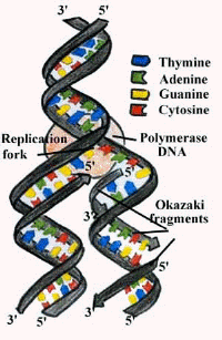
Copying DNA:
- Since the instructions for making cell parts are encoded in the DNA, each new cell must get a complete set of the DNA molecules
- This requires that the DNA be copied (replicated, duplicated) before cell division
Chromosomes & Their Structure:
- The plans for making cells are coded in DNA
- DNA, deoxyribose nucleic acid, is a long thin molecule that stores genetic information
- DNA in a human cell is estimated to consist of six billion pairs of nucleotides
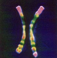
- DNA is organized into giant molecules called chromosomes
- Chromosomes are made of protein & a long, single, tightly-coiled DNA molecule visible only when the cell divides
- When a cell is not dividing the DNA is less visible & is called chromatin
- DNA in eukaryotic cells wraps tightly around proteins called histones to help pack the DNA during cell division
- Nonhistone proteins help control the activity of specific DNA genes
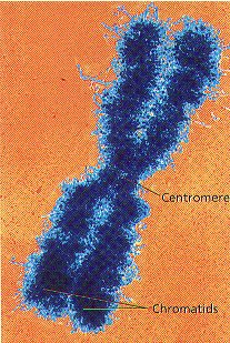
- Kinetochore proteins bind to centromere and attach chromosome to the spindle in mitosis
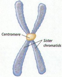
- Centromeres hold duplicated chromosomes together before they are separated in mitosis
- Telomeres are the ends of chromosomes which are important in cell aging
- When DNA makes copies of itself before cell division, each half of the chromosome is called a sister chromatid
- DNA of prokaryotes (bacteria) is one, circular chromosome attached to the inside of the cell membrane
Chromosome Numbers:
- Humans somatic or body cells have 23 pairs of chromosomes or 46 chromosomes (diploid or 2n number)
- The 2 chromatids of a chromosome pair are called homologues (have genes for the same trait at the same location)
Homologs
- Human reproductive cells or gametes (sperms & eggs) have one set or 23 chromosomes (haploid or n number)
- Every organism has a specific chromosome number
| Organism | Chromosome Number (2n) |
| Human | 46 |
| Fruit fly | 8 |
| Lettuce | 14 |
| Goldfish | 94 |
- Fertilization, joining of the egg & sperm, restores the diploid chromosome number in the zygote (fertilized egg cell)
- Sex chromosomes, either X or Y, determine the sex of the organism
- Two X chromosomes, XX, will be female and XY will be male
- All other chromosomes, except X & Y, are called autosomes
- Chromosomes from a cell may be arranged in pairs by size starting with the longest pair and ending with the sex chromosomes to make a karyotype
- A human karyotype has 22 pairs of autosomes and 1 pair of sex chromosomes (23 total)

Human Male Karyotype
Genes:
- A section of DNA which codes for a protein is called a gene
- Each gene codes for one protein
- Humans have approximately 50,000 genes or 2000 per chromosome
- About 95% of the DNA in chromosome is “junk” that does not code for any proteins
Cell Cycle:
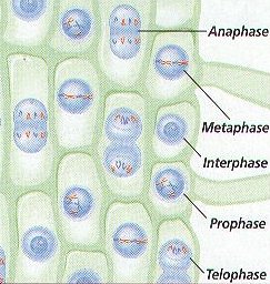
- Cells go through phases or a cell cycle during their life before they divide to form new cells
- The cell cycle includes 2 main parts — interphase, and cell division

- Cell division includes mitosis (nuclear division) and cytokinesis (division of the cytoplasm)
- Interphase is the longest part of a cell’s life cycle and is called the “resting stage” because the cell isn’t dividing
- Cells grow, develop, & carry on all their normal metabolic functions during interphase
- Interphase consists of 3 parts — G1, S, & G2phases
Interphase:
- G1 or 1st Growth Phase occurs after a cell has undergone cell division
- Cells mature & increase in size by making more cytoplasm & organelles while carrying normal metabolic activities in G1
- S or Synthesis Phase follows G1 and the genetic material of the cell (DNA) is copied or replicated
- G2 or 2nd Growth Phase occurs after S Phase and the cell makes all the structures needed to divide
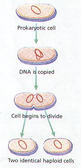
Cell division in Prokaryotes:
- Prokaryotes such as bacteria do not have a nucleus
- Prokaryotes divide into two identical new cells by the process of binary fission
- Binary fission is an asexual method of reproduction
- In binary fission, the chromosome, attached to cell membrane, makes a copy of itself and the cell grows to about twice its normal size
- Next, a cell wall forms between the chromosomes & the parent cell splits into 2 new identical daughter cells (clones)
Cell Division in Eukaryotes:
- Eukaryotes have a nucleus & membrane-bound organelles which must be copied exactly so the 2 new cells formed from division will be exactly alike
- The original parent cell & 2 new daughter cells must have identical chromosomes
- DNA is copied in the S phase of the cell cycle & organelles, found in the cytoplasm, are copied in the Growth phases
- Both the nucleus (mitosis) and the cytoplasm (cytokinesis) must be divided during cell division in eukaryotes
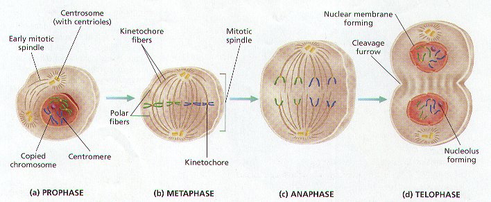
Stages of Mitosis:
- Division of the nucleus or mitosis occurs first
- Mitosis is an asexual method of reproduction
- Mitosis consists of 4 stages — Prophase, Metaphase, anaphase, & Telophase
- Prophase:
- Chromosomes become visible when they condense into sister chromatids
- Sister chromatids attach to each other by the centromere
- Centrioles in animal cells move to opposite ends of cell
- Spindle forms from centriole (animals) or microtubules (plants)
- Kinetochore fibers of spindle attach to centromere
- Polar fibers of spindle extend across cell from pole to pole
- Nuclear membrane dissolves
- Nucleolus disintegrates
- Metaphase:
- Chromosomes line up in center or equator of the cell attached to kinetochore fibers of the spindle
- Anaphase:
- Kinetochore fibers attached to the centromere pull the sister chromatids apart
- Chromosomes move toward opposite ends of cell
- Telophase:
- Nuclear membrane forms at each end of the cell around the chromosomes
- Nucleolus reform
- Chromosomes become less tightly coiled & appear as chromatin again
- Cytokinesis begins
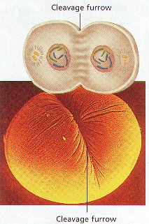
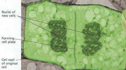
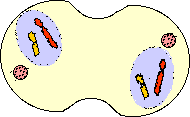
Cytokinesis:
- Cytoplasm of the cell and its organelles separate into 2 new daughter cells
- In animals, a groove called the cleavage furrow forms pinching the parent cell in two
- In plants, a cell plate forms down the middle of the cell where the new cell wall will be
Summary of Mitosis:
 |
 |
| Interphase
|
Early Prophase
|
 |
 |
Late Prophase
|
Metaphase
|
 |
 |
Anaphase
|
Telophase/Cytokinesis
|
Cancer is Uncontrolled Mitosis:
- Mitosis must be controlled, otherwise growth will occur without limit (cancer)
- Control is by special proteins produced by oncogenes
- Mutations in control proteins can cause cancer
Meiosis & Sexual Reproduction
- Reduces the number of chromosomes in new cells to half the number in the original cell
- New cells have a single copy of chromosomes (23 total) but are not identical to each other or the original parent cell
- Used for making gametes ( sperm and eggs) with the haploid or n number
- In meiosis, cells divide twice after a single DNA duplication
- Meiosis I separates homologs & the Meiosis II separates sister chromatids
- Meiosis I stages are Prophase I, Metaphase I, Anaphase I, & Telophase I
- Meiosis II stages are Prophase II, Metaphase II, Anaphase II, & Telophase II
- Produces 4 haploid cells or gametes
- When a sperm fertilizes an egg to form a zygote, the diploid number of chromosomes is restored (23 + 23 = 46)
- Egg cells or ova (ovum, singular) are larger , nonmotile cells
- Gametoogenesis is meiosis producing eggs & occurs in the female’s ovaries
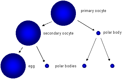
Oogenesis
- Sperms contain less cytoplasm so they’re smaller & have a flagellum to swim to the egg
- Spermatogenesis is meiosis producing sperm cells & occurs in the testes
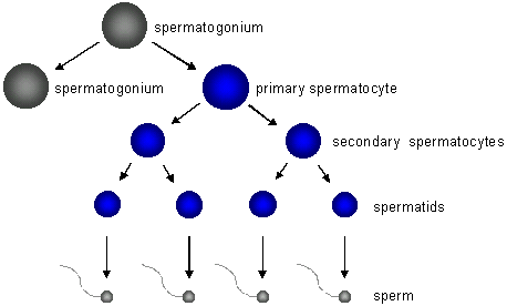
Spermatogenesis
Meiosis I:
- The cell that undergoes Meiosis I is a primary spermatocyte or oocyte
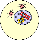
- Prophase I:
- Chromosomes coil tightly & are visible
- Nuclear membrane & nucleolus disintegrate
- Spindle forms
- Synapsis (joining) of homologous chromosomes occurs making tetrads
- Kinetochore fiber forms on each chromosome
- Chromosomes in tetrad exchange fragments by a process called crossing over

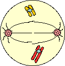
- Metaphase I:
- Tetrads become aligned in the center of the cell attached to spindle fibers
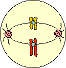
- Anaphase I:
- Homologous chromosomes separate
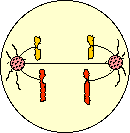
- Telophase I:
- May not occur in all species
- Cytokinesis occurs producing 2 cells
- In females, 2nd cell in females is called the 1st Polar Body
- 1st Polar Body dies due to uneven splitting of the cytoplasm

- Prophase II:
- Cells called Secondary Spermatocytes or oocytes
- DNA is not copied before cell divides
- Chromatids attach to spindle fiber
- Metaphase II:
- Chromosomes become aligned in the center of the cell attached to spindle fibers
- Anaphase II:
- Sister chromatids separate randomly
- Called independent assortment
- Telophase I:
- Cytokinesis occurs producing 4 cells in males called spermatids
- Spermatids mature & form flagellum to become sperm
- Cytokinesis in females produces a 2nd Polar Body that dies and an ootid
- Ootids mature to become ovum or egg

Asexual & Sexual reproduction:
- Evolution is the slow process of change in living populations over time
- Variations are differences that occur due to crossing-over among members of a sexually reproducing population
- Variations are important to the survival of individuals in a population (some must survive to reproduce)
- Asexually reproducing organisms rarely show variations because the organisms have identical genes


