Human Genetics Notes BI


Home      General Biology 1      General Biology 2      Human Biology      Anatomy and Physiology
Chapter 16 – Genetics, Part 3: Human Genetics Introduction

This chapter is a review of patterns of inheritance in humans including a review of genetic diseases.

The genetic diseases are divided into two categories: chromosomal abnormalities and gene abnormalities.  Chromosomal abnormalities are caused by cells that have extra or missing chromosomes or parts of chromosomes.  Gene abnormalities (gene mutations) occur when the genetic instructions stored in the DNA are altered so that the protein product coded for by the gene is less functional or nonfunctional.

Prenatal Diagnosis

The techniques listed below enable physicians to diagnose many kinds of genetic abnormalities by examining some of the cells from the developing fetus.


Amniocentesis


The fetus is surrounded by a layer of liquid called amniotic fluid. Amniocentesis is a technique in which a sample of amniotic fluid is removed and cells that it contains are grown on a culture dish. Because these cells are of fetal origin, any chromosomal abnormalities present in the fetus will also be present in the cells.

In addition to chromosomal analysis, a number of biochemical tests can be done on the fluid to determine if any problems exist.

Amniocentesis cannot be done until the 14th to 16th week of pregnancy. Cells must then be cultured on a laboratory culture dish for 2 weeks to obtain sufficient numbers of cells.

The risk of inducing a spontaneous abortion by this procedure is 0.5 to 1% above the background rate of spontaneous abortion.


Chorionic Villi Sampling


Chorionic villi sampling is a procedure in which a small amount of the placenta is removed.

It is normally done during the 10th to 12th week but it can be done as early as the 5th week of pregnancy. Karyotype analysis can be performed on these cells immediately after sampling.

Although Chorionic villi sampling can be performed earlier in the pregnancy than amniocentesis, the risk of inducing a spontaneous abortion is 1 to 2% higher than the background rate.

Karyotypes

Karyotypes are prepared using cells from amniocentesis, chorionic villi sampling, or white blood cells.

Cells are photographed while dividing. cells are normally stained so that banding patterns appear on the chromosomes. The bands make it easier to identify the chromosomes. Banding patterns are not visible in the photograph below due to the staining technique.

Pictures of the chromosomes are cut out and arranged in pairs according to size and banding patterns.

Karyotypes can be used to determine if there is an abnormality in chromosome number or structure.

Nondisjunction

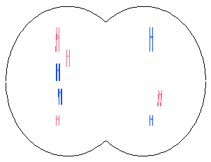
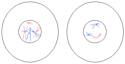
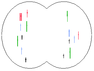
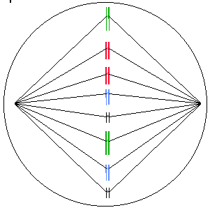
Nondisjunction occurs when chromosomes fail to “disjoin” during meiosis or mitosis.

Meiosis

Metaphase I
img017.gif (3781 bytes)

Anaphase I

img018.gif (3680 bytes)

Telophase I

img019.gif (3168 bytes)

Prophase II

img020.gif (4544 bytes)

Meiosis II and Mitosis

The diagrams below show nondisjunction during mitosis in a hypothetical species with 2N=8 chromosomes.

Metaphase
img013.gif (5064 bytes)

Anaphase

img014.gif (5337 bytes)

Telophase

img015.gif (3798 bytes)

G1 Interphase

img016.gif (4787 bytes)

The probability of nondisjunction increases with age. It increases rapidly after age 35 years in women and after 55 years in men.

Aneuploidy

Cells that have extra chromosomes or chromosomes missing are aneuploid. Two types of aneuploidy are discussed below.

Monosomy refers to a condition in which there is one chromosome is missing. It is abbreviated 2N – 1. For example, monosomy X is a condition in which cells have only one X chromosome.

A trisomy has one extra chromosome and is abbreviated 2N + 1. Trisomy 21 is an example of a trisomy in which cells have an extra chromosome 21.

Monosomies and trisomies usually result from nondisjunction during meiosis but can also occur in mitosis. They are more common in meiosis 1 than meiosis 2.

They are generally lethal except monosomy X (female with one X chromosome) and trisomy 21 (Down’s Syndrome).

Affected indivisuals have a distinctive set of physical and mental characteristics called a syndrome. For example, trisomy 21 is Down syndrome.

Incidence of Genetic Abnormalities

Maternal Age

At 25 years, 17% of secondary oocytes may have chromosomal abnormalities. At 40 years, up to 74% may contain abnormalities.

Spontaneous Abortion (Miscarriage)

Two-thirds of all pregnancies are lost. These miscarriages are called spontaneous abortions.

Genetic mutation causes an estimated 60% of these spontaneous abortions.

Autosomal Abnormalities

Nine percent of spontaneous abortions are trisomy 13, 18, or 21; but 0.1% of newborns have these trisomies.


Down Syndrome


Down syndrome is trisomy 21. It is characterized by mental retardation, an abnormal pattern of palm creases, a flat face, sparse, straight hair, and short stature. People with Down syndrome have a high risk of having cardiac anomalies, leukemia, cataracts, and digestive blockages.

Life expectancy of Down syndrome individuals is in the middle teens but some live much longer.

The gene responsible for Alzheimer’s is on chromosome 21. Down’s are at increased risk for developing Alzheimer’s.

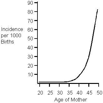
Down Syndrome is associated with maternal age. Older women, particularly those older than 40, are more likely to have a Down Syndrome child.
 

Translocation Down Syndrome


A translocation is the movement of a chromosomal segment from one chromosome to another nonhomologous chromosome.

Five percent of Down Syndrome cases involve a translocation.

The translocation often involves chromosome 14.

In the translocation diagrammed below, chromosome #21 has become fused with chromosome #14.
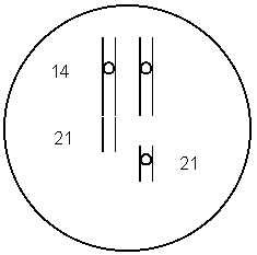
During meiosis, the two chromosomes might align so that each daughter cell receives one chromosome 21 as shown below. This will produce a normal egg.
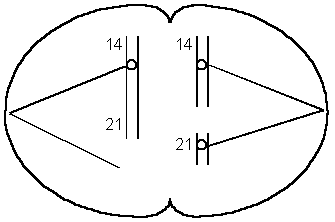
If the chromosomes align as illustrated below, one daughter cell will receive two chromosome 21s and the other will not receive any.  When a gamete with two 21s fuses with a normal gamete, the result is a zygote with three chromosome 21s.
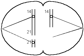
This form runs in families and is not age-related.


Mosaic Down Syndrome


Some of the cells of mosaic Down’s sydrome are trisomy 21 but others are normal.

This is due to nondisjunction that occurs during mitosis (after fertilization).

Mosaic Down Syndrome is likely to be less severe because some of the cells are normal.


Trisomy 18 (Edward Syndrome)


Trisomy 18 is associated with mental and physical retardation, skull and facial abnormalities, defects in all organ systems, and poor muscle tone.

Mean survival is 2 to 4 months.


Trisomy 13 (Patau Syndrome)


Trisomy 13 produces mental and physical retardation, skull and facial abnormalities, and defects in all organ systems. It is also associated with a left lip, a large, triangular nose, and extra digits.

One half die in first month; the mean survival time is 6 months.

Polyploidy

Polyploidy is a condition in which there is more than 2 sets of chromosomes.

Triploids (3N), tetraploids (4N), pentaploids (5N) etc. are polyploids.


Polyploidy in Plants


Polyploidy is a major evolutionary mechanism in plants. Approximately 47% of all flowering plants are polyploid.

Some examples of polyploid plant species are corn, wheat, cotton, sugarcane, apples, bananas, watermelons, and many flowers.

Polyploid plants are often more vigorous than the diploid parent species.

Polyploid plants are fertile.


Polyploidy in Humans


Polyploids have defects in nearly all organs.

Most die as embryos or fetuses. Occasionally an infant survives for a few days.

Abnormalities of the Sex Chromosomes

Turner Syndrome – XO

Characteristics of Turner syndrome include the following:

Sexually underdeveloped

Short stature

Folds of skin on the back of the neck

Wide-spaced nipples

Narrow aorta

Pigmented moles

97% die before birth

Malformed elbows

Infertile

Normal Intelligence

The incidence of Turner syndrome is 1 in 2000 female births.

Turner syndrome individuals that are treated with hormones lead fairly normal lives.

XXX – Triple-X Syndrome (also XXXX and XXXXX)

Triple-X individuals are tall and thin and have menstrual irregularities. Their IQ is in the normal range but it is slightly reduced.

The incidence of Triple-X Syndrome is 1 in 1,500 female births.

Additional X chromosomes are associated with an increased mental handicap.


XXY – Klinefelter Syndrome (also XXXY)


Males with two or more X chromosomes have Klinefelter Syndrome.

The incidence of Klinefelter Syndrome is 1 in 1000 male births.

Symptoms include reduced sexual maturity and secondary sexual characteristics, breast swelling, and no sperm. Klinefelter males are slow to learn and individuals with additional X’s (XXXY) may be mentally retarded.


XYY – Jacob Syndrome


XYY males are tall, have acne, speech, and reading problems.

Although there are a disproportionate number in penal institutions, 96% of Jacob’s Syndrome men are normal.

In the early 1970’s screening began in hospitals in England, Canada, Denmark and US. Families with XYY boys were offered “anticipatory guidance”. These types of programs were stopped because they were self-fulfilling prophesies.

Other Chromosomal Abnormalities

Deletions


Deletions are fragments of chromosomes that are missing. They are usually lethal when homozygous and cause abnormalities when heterozygous.

Radiation, viruses, chemicals, and unequal crossing-over may cause them.


Cri du Chat Syndrome


Cri du chat syndrome is due to a deletion of a portion of chromosome 5.

Cri du chat individuals are mentally retarded.

“Cri du chat” is French for “cry of the cat”. The infants cry sounds like a cat.


Duplication


A chromosome segment that is repeated is called a duplication.

It can be due to unequal crossing over which produces a deletion on one chromosome and a duplication on the other.

Often, multiple copies of genes from duplication can mutate without harming the individual because they still have one good copy of the gene. This type of mutation may be a source of variation for species. For example, the gene for human globin has given rise to several different genes that produce similar types of proteins. The different globins produced by these genes have very similar amino acid sequences.

An example of a family of genes that have been produced by duplication is the beta globin family. This family contains five functioning genes and a pseudogene.

Epsilon globin

G-gamma globin

A-gamma globin

delta globin

beta globin

a pseudogene

All of these genes have similar amino acid sequences due to their evolution from the same ancestral gene.

Some families of genes contain hundreds of genes.


Repeated Sequences


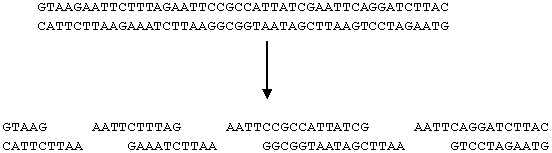
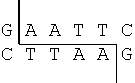
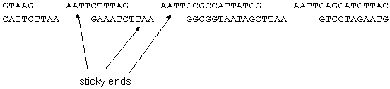
Repeated sequences are short segments of DNA that are repeated hundreds or thousands of times. For example: In the segment of DNA illustrated below, CCG is repeated several times.



The cause is unknown.

Fragile X Syndrome

This is the second most common cause of mental retardation (Down Syndrome is first).

The characteristic long, narrow face becomes more pronounced with age.

The symptoms of fragile-X syndrome appear to be caused by an abnormal number of repeats (CCG) on the X chromosome. Normal DNA has 6 – 50 copies of “CCG” at the locus in question. Carrier males have 50 – 230 copies. This is referred to as a premutation (pre-fragile-X). The full mutation involves more than 230 repeats of CCG.

The chance of being affected increases in successive generations because extra copies of CCG are added during the gamete-formation process.

Females are more likely to add repeats than males. At most, males pass on 230 repeats to their children but females pass on more than 230 repeats.

Mental problems are more common if the fragile X is inherited from the mother. This is an example of genomic imprinting discussed in the previous chapter. Fragile-X is more common in males because males inherit their X chromosome from their mother.

The repeats cause the X to have a thread-like portion. It is called a fragile site because it breaks if cultured under certain conditions in the laboratory.


Translocation


Chromosomes that break usually rejoin at the same place but sometimes the broken ends rejoin in different places.

Translocation is the movement of a chromosome or part of a chromosome to another (nonhomologous) chromosome.


Inversion


A segment of a chromosome may become turned around forming an inversion.

This can cause altered gene activity, a loss of crossing-over, or a duplication/deletion if crossing-over does occur.

Pedigrees

It is often easy to visualize relationships within an extended family by using symbols to represent people and relationships. A family tree which uses these symbols is called a pedigree. A sample pedigree is below.

In a pedigree, squares represent males and circles represent females. Horizontal lines connecting a male and female represent mating. Vertical lines extending downward from a couple represent their children. Subsequent generations are therefore written underneath the parental generations and the oldest individuals are found at the top of the pedigree.

If the purpose of a pedigree is to analyze the pattern of inheritance of a particular trait, it is customary to shade in the symbol of all individuals that possess this trait.

In the pedigree above, the grandparents had two children, a son and a daughter. The son had the trait in question. One of his four children also had the trait.

Autosomal Recessive

Characteristics of autosomal recessive inheritance


It often skips generations; children that have the trait can have parents that do not.

Heterozygotes (carriers) do not have the trait. People with the trait have two copies of the genes.

If both parents are have the trait, all offspring will.

Males and females are affected equally.

Inbreeding results in a greater-than-expected number of rare autosomal recessive phenotypes.


Cystic Fibrosis


Thick mucous forms in the digestive tract and lungs of people with CF. As a result, they have difficult breathing and are susceptible to lung infections.

People with cystic fibrosis have a life expectancy of approximately 30 years.

The gene that causes the disease is on chromosome 7. One particular mutation of this allele causes 70-75% of the cases.

It is somewhat difficult to detect prenatally.

Gene therapy may be a possibility in the future. The normal gene was inserted into cells in laboratory cultures.

Viruses have been engineered to deliver the gene. An aerosol spray is used to deliver the virus to the lungs.

There has been some success reported in treating human patients in 1994.

Cystic fibrosis is the most common lethal genetic disease among Caucasians in the US.

One in 25 is a carrier; one in 2500 is affected.


Tay Sachs


A fatty substance builds up in the neurons (nerve cells) of people with Tay Sachs. This causes a gradual paralysis and loss of nervous function that leads to death by age 4 or 5.

It is due to a single defective enzyme which normally digests the fatty material.

Heterozygotes (Aa) are not affected and are resistant to tuberculosis.

Prenatal diagnosis is available.

It is a common genetic disease among the Jewish population in the US (central and eastern European descent). Up to 11% are carriers. It is also common in people of French-Canadian or Cajun descent.


PKU – Phenylketonuria


PKU is a recessive genetic disease in which the person does not have the ability to break down the amino acid phenylalanine. The level of phenylalanine in the persons blood builds up and interferes with the development of the nervous system.

Children that are raised on a phenylalanine-restricted diet may develop normally but children that are not raised on a special diet will become severely mentally retarded. The diet should be followed for life because high phenylalanine levels affect cognitive functioning.

Genetic screening is the routine testing of individuals for specific genotypes. Newborns in U.S. hospitals are screened for PKU.

PKU women must resume the diet several months before conception

The incidence of PKU in the United States is 1 in 13,500 to 1 in 19,000.


Sickle-Cell Anemia


Sickle-cell anemia is an abnormality of hemoglobin, the molecule that carries oxygen in our blood. Hemoglobin is contained within red blood cells. When the oxygen concentration in the hemoglobin molecules becomes low, the molecules stick together forming long rods that distort the cell (picture below). The cells break down or clog blood vessels causing pain, poor circulation, jaundice, anemia, internal hemorrhaging, low resistance, and damage to internal organs. Death usually occurs before age 50.
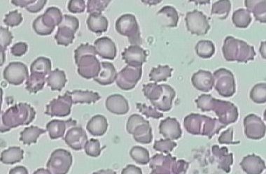
Heterozygotes (carriers) are not affected with anemia and are resistant to malaria.

Eight to ten percent of African Americans carry the allele (have sickle-cell trait).

Hemochromatosis

Hemochromatosis is a disease that causes the body to absorb more iron from food than normal. High iron levels can lead to organ damage if it is left untreated for many years.

Symptoms include joint pain, fatigue, and abdominal pain.

There are two different mutations of the gene that causes hemochromatosis (the HFE gene) and the severity of symptoms depends on the mutations that are inherited.

One in 200 people in the United States carry the gene and it is the most common genetic disease in people of northern European descent.

There is also a form of this disease that is not due to genetic factors, it is acquired.

Autosomal Dominant

Severe dominant diseases are rare because carriers die before they get a chance to reproduce and pass on the disease to their offspring.

Heterozygotes (Aa) have the trait.

Children with the trait have at least one parent that has the trait.

Two parents with the trait can produce a child that does not have the trait.

Both males and females are affected equally.


Neurofibromatosis (NF)


Neurofibromatosis is sometimes called elephant man disease.

People with this gene have 6 or more large tan spots on the skin which may increase in size, number and darkness. The nerve cells form benign tumors which may vary in size. There may be learning disabilities and hyperactivity.

The disease is usually mild but may be severe causing deformities and even death.

The incidence is 1 in 3000 newborns.

The gene is on chromosome 17.


Huntington’s Disease


The brain cells of Huntington’s victims slowly degenerate, producing jerking muscles, slurred speech, swallowing difficulty, loss of balance, mood swings, reasoning and memory loss, incapacitation, and eventually death (usually from pneumonia or heart failure).

The onset of Huntington’s disease is typically 35 to 45 years.

It is caused by a repeated DNA sequence (AGC).  The normal allele has 11-34 copies; affected people have 42 – 120 copies.

The severity and time of onset depends on the number of repeats.

People who are most at risk inherit the gene from their father.  This is an example of genomic imprinting.

The gene is on chromosome 4.  A diagnostic test is available.

X-Linked Recessive

More males than females have x-linked recessive traits.

A son with the trait can have parents that do not have the trait.

There is no father to son transmission of the gene.

The trait can skip generations; grandfather to grandson transmission can occur.

If a female has the trait, her father has it, her mother is a carrier (or has it), and all her sons will have it.


Color Blindness


3 different kinds

2 X-linked forms: 1 for green insensitivity (6% of all males), one for red insensitivity (2% of all males); 1 in 12 males have some form of colorblindness.


Hemophilia


People with hemophilia lack a clotting factor in their blood and as a result, their blood does not form clots normally. This results in excessive bleeding from even minor cuts. Internal hemorrhaging from bruises is common and leads to painful complications.

The incidence is in 1,500 newborn males. Most (75%) have hemophilia A, a lack of clotting factor VIII. Hemophilia B- “Christmas Disease” is a defect in clotting factor IX.

Transfusions of fresh whole blood or plasma or factor concentrates control bleeding but have previously caused AIDS infections.

The human gene has been isolated and cloned using recombinant DNA techniques. This is leading to improved treatment.


Royal Families of Europe


Victoria (granddaughter of George III) was a carrier and spread the gene to the royal families of Europe. Her granddaughter Alix- married Czar Nicholas II of Russia. The Czar’s son Alexis, heir to the throne, had hemophilia.

The Czar’s preoccupation with Alexis’ health contributed to the revolution that overthrew the throne and eventually led to the communist government.


Duchenne Muscular Dystrophy


There are four different kinds of X-linked muscular dystrophy. They are multiple alleles at a single locus.

Duchenne’s is the most common and most severe form of muscular dystrophy.

1 in 5,000 live male births (Duchenne’s)

One in 4000 newborn males have some form of muscular dystrophy. One third of these are new mutations.

Muscular deterioration begins between ages 3 to 5. Affected individuals are confined to a wheelchair by age12 and rarely survive past age 20. Death is usually due to breathing or heart problems.

It is transmitted primarily by female carriers (males rarely reproduce)

Sex-Influenced Inheritance

Sex-influenced traits are those that are dominant in one sex but recessive in the other

This difference is due to the different hormonal environments between the sexes.

Sex-influenced genes are not necessarily located on the X chromosomes. Don’t confuse this with X-linked inheritance.


Examples

Pattern baldness is male dominant.

A gene that causes the index finger to be longer than the third finger is female dominant.


Home      General Biology 1      General Biology 2      Human Biology      Anatomy and Physiology

Insect

Insects   All Materials © Cmassengale  

Phylum Arthropoda        Subphylum Uniramia          Class Insecta

Characteristics

  • Largest arthropod group
  • Found in freshwater & terrestrial habitats, especially tropical areas
  • Legs, mouthparts, & antenna jointed
  • Body segmented into three sections — head, thorax, & abdomen
  • Six legs & up to two pairs of wings located on thorax
  • Have compound & simple eyes
  • One pair of antennae on head
  • Abdomen has 11 segments
  • Exoskeleton, covering & protecting body, is made of chitin & must be molted to grow
  • Elaborate mouthparts include:
         *  Mandibles – jaws
    *
       Maxillae – paired sensory structures that move food to mouth
      Labium – lower lip
      Labrum – upper lip
      Palpi – used for tasting
  • Known as mandibulates
  • Spiracles on abdomen open into tracheal tubes for oxygen & carbon dioxide exchange
  • Tympanic membranes on 1st abdominal segment aid in hearing
  • Thorax divided into 3 sections — prothorax, mesothorax, & metathorax
  • One pair of legs on each thoracic segment
  • Wings located on mesothorax & metathorax
  • Ovipositor located on the end of the abdomen in female insects & used to dig hole & lay eggs

Common Insect Orders

  • Orthoptera – grasshoppers, crickets, & cockroaches 2 pairs of straight wings & chewing mouthparts)
  • Isoptera – termites (feed on wood)
  • Dermaptera – earwigs (pincers on end of abdomen)
  • Anoplura – sucking lice (wingless parasites)
  • Hemiptera – true bugs (have triangular-shaped scutellum & last 1/3 of wings membranous)
  • Homoptera – aphids & cicadas (membranous wings held roof-like over body
  • Ephemeroptera – mayflies (have 2 cerci on tail, membranous wings, & nonfunctional mouthparts in adults)
  • Odonata – dragonflies & damselflies (2 pairs of equal size, membranous wings, strong fliers, feed on other insects)
  • Neuroptera – Dobson flies &  lacewings (2 pairs of membranous wings)
  • Coleoptera – beetles (hard forewings or elytra, membranous hindwings)
  • Lepidoptera – butterflies & moths (powdery scales covered wings
  • Diptera – flies & mosquitoes (one pair of wings, 2nd pair modified into balancing structure called halteres)
  • Siphonaptera – fleas (parasites on birds & mammals, wingless as adults)
  • Hymenoptera – bees, ants, & wasps (stinger on abdomen for protection, may live together in groups, pollinators)

     Click Here for Pictures of Insect Orders


Success of Insects

  • Found everywhere except in deep part of ocean
  • Very short life span & rapidly adapt to new environments
  • Small size helps minimize competition in habitats
  • Flight helps escape predators & move into other environments

Environmental Impact

  • Pollinate almost 2/3’s of all plants
  • Serve as food for fish, birds, & mammals
  • Help recycle materials (termites recycle wood)
  • Make useful byproducts such as silk & honey
  • Some spread disease
  • Agricultural pests

Grasshoppers

External Structure

  • Head with antenna, compound eyes, & chewing mouthparts
  • Walking legs on prothorax & mesothorax; jumping legs on metathorax
  • Tarsus are lower leg segments with spines, hooks, & pads
  • Leathery, protective forewings on mesothorax & membranous hindwings for flight on metathorax
  • Covering over thorax called pronotum

Internal Structure
Digestive & Excretory Systems

  • Cutting & chewing mouthparts (labium, labrum, mandibles, & maxillae)
  • Saliva added to food in mouth
  • Esophagus carries food to crop for temporary storage
  • Gizzard has chitinous plates to grind food
  • Midgut (insect’s stomach) has gastric caeca (pouches) to secrete digestive enzymes to break down food
  • Food is absorbed into the body cavity or coelom in the hindgut (composed of the colon & rectum)
  • Malpighian tubules filter chemical wastes from the blood & deposit them in the rectum where they leave through the anus

Circulatory System

  • Open circulation of blood
  • Aorta is the largest blood vessel carrying blood to the body cells
  • Hearts are muscular regions of the aorta in the posterior end of the abdomen that pump blood toward head
  • Blood flows back toward abdomen carrying digested food & re-enters the aorta through openings called ostia

Respiratory System

  • Air enters through openings called spiracles along the sides of the abdomen & enters into tracheal tubes that branch into smaller tracheoles where gas exchange with body cells occurs 
  • Tracheal tubes carry oxygen to body cells & return carbon dioxide to leave the body though spiracles

Nervous System

  • Simple brain, nerve cords, & ganglia 
  • Three simple eyes or ocelli (detect light) & a pair of compound eyes (can detect movement but not images)
  • Tympanic membrane on 1st abdominal segment
  • Pair of antenna contains sense organs for touch, taste, & smell detects sound
  • Sensory hairs found on parts of the body
  • Palpi for taste

Reproductive System

  • Reproductive organs (ovaries & testes) located  in abdomen
  • Male deposits sperm into female’s seminal receptacle
  • Stored sperm fertilizes eggs as they  are released by female
  • Ovipositor on tip of female’s abdomen is used to lay eggs
  • Separate sexes
  • Lay large number of eggs to ensure survival

Development

  • Most insects go through changes in form & size called metamorphosis
  • Some insects such as silverfish don’t go through metamorphosis
  • Incomplete metamorphosis goes from egg to nymph (immature form that looks like adult but without fully developed wings) to adult (3 stages)
  • Instars are growth periods between molts of nymphs & larva
  • Grasshoppers, termites, & true bugs go through incomplete metamorphosis


HEMIPTERAN (TRUE BUG) NYMPH

  • Complete metamorphosis goes from egg to larva (segmented & wormlike) to pupa  to adult (4 stages)


BUTTERFLY LARVA (CATERPILLAR)

  • Butterflies, beetles, & flies go through complete metamorphosis
  • In pupal stage, larval tissues break down & cells called imaginal disk develops into tissues of the adult
  • Cocoon or chrysalis is a protective case formed around the pupa


BUTTERFLY COCOON

  • Metamorphosis controlled by hormones
    * Brain hormone stimulates the release of molting hormone (ecdysone)
    * When juvenile hormone level high, larva molts
    * When juvenile hormone level low, larva pupates
    * When juvenile hormone absent, adult emerges from pupal case
  • Different stages of metamorphosis eliminates competition between larva & adults for food & space
  • Multi-stage life cycle helps insects withstand harsh weather
  • Different stages have different functions (caterpillar/growth & adult/reproduction)

Defense Mechanisms

  • Bombardier beetle sprays noxious chemical


BOMBARDIER BEETLE

  • Wasps & bees can sting
  • Some insects use camouflage to blend into their environments
  • Some insects taste bad & have warning colorations 


PAPER WASP

  • Mullerian mimicry – poisonous or dangerous species have similar patterns of warning coloration so predators avoid all the species (black & yellow stripes on bees & wasps)
  • Batesian mimicry – species that are nonpoisonous or not bad tasting have colorations that mimic other poisonous or bad tasting species (Viceroy butterfly mimics bad tasting Monarch)

Insect Communication

  • Insects may communicate with each other using sound (cricket chirps), light (firefly), or “dances” (honeybee)
  • Pheromones are chemicals released by some insects to attract mates or mark trails

Insect Behavior

  • Insects may be solitary or social
  • Social insects (bees, ants, & some wasps) live together in groups & share work (division of labor)
  • Social insects have a caste system with different individuals doing different jobs
  • Honeybee caste system:
    * Workers
    – sterile females
    – care for queen & feed her honey and pollen
    – make beeswax for hive
    – fan wings to cool hive
    – eat honey
    – collect nectar, pollen, & royal jelly
    – live about 6 weeks
    – nurse bees care for larva
    – secrete royal jelly to feed new queen
    * Drones
    – males
    – mate with queen
    – feed by workers
    – driven out of hive to conserve food during winter
    * Queen
    – reproductive female
    – mate only once but store sperm for up to 5 years in seminal receptacles
    – feed by workers
    – secretes chemical called queen factor that prevents other females from sexually maturing
    – leaves hive with 1/2 the workers if there is overcrowding


HONEYBEE HIVE

BACK


Human Hand Adaptations


Human Hand Adaptation

Introduction:        Living things have bodies that are adapted for the places they live and the things they do. Fish have gills so that they can remove oxygen that is dissolved in water. Most plants have green leaves which contain chlorophyll so that they can make food. Jellyfish have stinging cells to capture prey. Birds have hollow spongy bones so that they will be light enough to fly. Arctic animals have layers of fat and thick coats of fur to keep warm in the frigid Arctic climate. There are hundreds of examples of ways that organisms are adapted for a successful lifestyle.       Humans, too, are adapted for the things they do. One of our adaptations is our hand. Humans, as well as monkeys, gorillas, and other primates, have a hand that can grasp objects. We are able to grasp objects because of our opposable thumb. When students first hear or read about the opposable thumb during discussions of human evolution, they may perceive it as an anatomical fact with little seeming importance. In this activity, students will discover which of their simplest daily activities are possible only because of their opposable thumbs, which activities take longer without the use of an opposable thumb, and what sort of human activities would not be likely in the absence of an opposable thumb.   In this lab exercise, you will perform several common actions. Then you will change your hand so that it resembles that of a non-primate animal. You will determine whether or not you can successfully perform the same actions. This will demonstrate how the human hand is adapted for the actions it performs. You will work with a partner to do this exercise.   Materials: (per group)

  • masking tape
  • scissors
  • paper clips
  • zip-lock storage bag
  • plastic fork and knife
  • small amounts of food items to be cut
  • pencil
  • jar with screw-on lid
  • paper
  • roll of tape
  • balloons
  • comb
  • book
  • lace-up shoe
  • clock with a second hand
  • Piece of yarn or string
  • balloon
  • clothes with zippers & buttons

Procedure: Using masking tape, have your partner tightly tape each of your thumbs to the palm of the hand. Then, try to complete the tasks that are listed below. Be careful not to use your thumbs. Have your partner record on your data table how long it takes to do each task with your thumb taped and then with your thumb free. If an activity takes longer than 2 minutes, record the event as unsuccessful . After completing each item, write out the answers to the following questions:

  • Is the task more difficult with or without an opposable thumb?
  • How did you have to change your usual technique in order to complete this task?
  • Do you think organisms without opposable thumbs would carry out this task on a regular basis? Why or why not?

Tasks:

  1. Pick up a single piece of paper. Put it down on your desk.
  2. Pick up a pen or pencil from the table top. Use it to write your name on the piece of paper.
  3. Open a book. Turn single pages in the book.
  4. Unscrew a bottle cap or jar cover.
  5. Use a fork and knife to cut a food item into small pieces.
  6. Tear off a small piece of tape.
  7. Turn on the water faucet. (Complete activity #8!) Turn it off.
  8. Moisten a paper towel and wash and dry the desktop.
  9. Sharpen a pencil.
  10. Cut a circle out of a piece of paper using scissors.
  11. Pick up all the scraps from activity #10 and throw them into the recycling box.
  12. Comb your hair.
  13. Open a door.
  14. Pick up one paper clip. Clip a pile of papers together.
  15. Tie your shoelaces.
  16. Button several buttons.
  17. Zip up your jacket.
  18. Blow up a balloon and tie it.
  19. Tie a knot in a piece of string.
  20. Close a zip-lock bag.

Data:

Table 1 – Time It Took To Perform Various Tasks


Task Time Taken for Event: Task Difficulty With Taped Thumb
(More/Less)
Modification Made to complete Task
Thumb Free Thumb Taped
Pick up paper
Write name
Turn book pages
Open jar
Use knife & fork
Tear off tape
Turn faucet on & off
Clean desk top
Sharpen a pencil
Cut out a circle
Pick up the scraps of paper
Comb hair
Open door
Clip papers together
Tie shoelaces
Button & unbutton garment
Use zipper
Blow up & tie balloon
Knot string
Close zip-lock bag

Conclusion:   1. Explain why dog and cat paws are not adapted for doing the six actions you tested.     2. What are cat and dog paws adapted for?     3. Describe how your hand is adapted for doing the actions you tested.       4. You have an opposable thumb. Explain what this means.     5. Why do you feel that human hand adaptations have helped to make humans such a successful species on earth?


Ink Chromatography

Chromatography of Inks

Introduction:

One of the main jobs of biochemists is to unravel the complexities of chemical compounds and reduce them to their individual components.  The term chromatography comes from two Greek words, “chromat” meaning color and the word “graphon” meaning to write.  Separation of the components of chemical compounds can be done by using several methods. Liquids can be separate by High Performance liquid Chromatography (HPLC), while the components of gases are separated by Gas Chromatography.  Chromatography is a method for analyzing complex mixtures (such as ink) by separating them into the chemicals from which they are made. Chromatography is used to separate and identify all sorts of substances in police work. Drugs from narcotics to aspirin can be identified in urine and blood samples, often with the aid of chromatography.

Chromatography was first used to separate pigments (colors) in leaves, berries, and natural dyes. Paper chromatography is a technique used to separate, isolate, and identify chemical components of a compound. In paper chromatography, the solid surface is the cellulose fibers in the chromatography paper.  A solvent or developer (water, alcohol, or acetone) is placed in the bottom of the chromatography chamber. The paper acts as a wick to pull the solvent up the paper. The solvent front will “wick” up the chromatography paper by capillary action.  A minute drop of the ink or chemical mixture to be separated is placed near the bottom of the strip of chromatography paper, but slightly above the level of the solvent in the chamber.  As the solvent passes over the drop of ink, the components of the ink dissolve in the solvent. Because the components of the ink do not all dissolve at the same rate, as the components of the mixture move upward, they show up as colored streaks.  The separated substances on the chromatography paper form a color pattern called a chromatogram.

To determine the rate of migration for each pigment or component of the ink, the Rf value for each pigment must be calculated. The Rf value represents the ratio of the distance a pigment moved on the chromatogram relative to the  distance the solvent front moved. Each pigment or compound will have a unique Rf value that scientists can use to identify the substance. The Rf value is calculated using the following formula:

Rf = distance traveled by the compound / distance traveled by the solvent

Objective:

Use the process of paper chromatography to separate the pigments in various markers and then determine the Rf value for each color on your chromatogram.

Materials:

Plastic vials, paper clips, markers in assorted colors, chromatography paper, scissors, pencil

Procedure:

  1. Obtain chromatography vials and chromatography strips, and different color markers so that each person in the group will have two chromatograms.
  2. Cut one end of the chromatography strip to a point. The bottom of the point will mark the starting point for movement of the solvent (H2O).
  3. About 2.0 centimeters from the bottom of the strip, draw a faint horizontal line with pencil. This will mark the starting point for measuring the migration distance of each color.
  4. Using a different color marker for each strip, drop a dot of ink on the center of the horizontal pencil line.  Let this dry a moment & then add more ink to the dot.
  5. Add a small amount of water to the bottom of the chromatography chamber. (The ink dot should be ABOVE the surface of the water.)
  6. Straighten a paper clip and poke a hole through the top of your chromatography strip
  7. Use the paper clip to hang the strip in your chamber. (The straighten paper clip will lay across the top of the chamber.)
  8. MAKE SURE THE TIP OF THE STRIP BUT NOT THE INK IS IMMERSED IN THE WATER!
  9. Notice the separation of the ink as both the solvent and ink travel up the chromatography strip.
  10. Once the solvent front has neared the top of the strip, remove the strip from the chamber and lay it on a piece of paper towel.
  11. Immediately mark the solvent front with a faint pencil line.
  12. Immediately mark the leading edge of each color with an “x”.
  13. Measure, in millimeters, the distance the solvent migrated from the tip of the strip to your solvent front pencil line.
  14. Measure, in millimeters, the distance each color migrated from the point of origin (pencil line where the ink dot was placed) to the leading edge of the color (marked with an “x”.
  15. Record all data in Data table 1.
  16. Calculate and record the Rf value for each color using the formula below.

Rf = distance traveled by the compound / distance traveled by the solvent

Data Table 1


Color pen/marker used:

Separated colors
(list top of strip to bottom)
Distance each color traveled

(mm)

Distance solvent (H2O)
(mm)
Rf Value for each color

(Distance color traveled / Distance solvent traveled)


Color pen/marker used:

Separated colors
(list top of strip to bottom)
Distance each color traveled

(mm)

Distance solvent (H2O)
(mm)
Rf Value for each color

(Distance color traveled / Distance solvent traveled)


Questions:

1. Which color of marker did you use?

2. which color separated out first from your ink dot?

3. Why did the inks separate?


4. What was your solvent?

5. If you had used markers that weren’t water-soluble, how would you have had to change this lab?


6. Why did some inks move a greater distance than others?


7. How do scientists use paper chromatography in their investigations?


Genetic Notes Bi


Mendelian Genetics 


Mendel 1862 Mendel 1868 Mendel 1880
1862 1868 1880


Genetic Terminology:

  • Trait – any characteristic that can be passed from parent to offspring
  • Heredity – passing of traits from parent to offspring
  • Genetics – study of heredity
  • Alleles – two forms of a gene (dominant & recessive)
  • Dominant – stronger of two genes expressed in the hybrid; represented by a capital letter (R)
  • Recessive – gene that shows up less often in a cross; represented by a lower case letter (r)
  • Genotype – gene combination for a trait (e.g. RR, Rr, rr)
  • Phenotype – the physical feature resulting from a genotype (e.g. tall, short)
  • Homozygous genotype – gene combination involving 2 dominant or 2 recessive genes (e.g. RR or Rr); also called pure 
  • Heterozygous genotype – gene combination of one dominant & one recessive allele    (e.g. Rr); also called hybrid
  • Monohybrid cross – cross involving a single trait
  • Dihybrid cross – cross involving two traits
  • Punnett Square – used to solve genetics problems

Blending Concept of Inheritance:

  • Accepted before Mendel’s experiments
  • Theory stated that offspring would have traits intermediate between those of its parents such as red & white flowers producing pink
  • The appearance of red or white flowers again was consider instability in genetic material
  • Blending theory was of no help to Charles Darwin’s theory of evolution 
  • Blending theory did not account for variation and could not explain species diversity
  • Particulate theory of Inheritance, proposed by Mendel, accounted for variation in a population generation after generation
  • Mendel’s work was unrecognized until 1900

Gregor Mendel:

  • Austrian monk
  • Studied science & math at the University of Vienna
  • Formulated the laws of heredity in the early 1860’s
  • Did a statistical study of  traits in garden peas over an eight year period


drawing of a flower cross-section showing both male and female sexual structures


Why peas, Pisum sativum?

  • Can be grown in a small area
  • Produce lots of offspring
  • Produce pure plants when allowed to self-pollinate several generations
  • Can be artificially cross-pollinate

Picture of Pisum sativum
GARDEN PEA

Mendel’s Experiments:

  • Mendel studied simple traits from 22 varieties of  pea plants (seed color & shape, pod color & shape, etc.)
  • Mendel traced the inheritance of individual traits & kept careful records of numbers of offspring
  • He used his math principles of probability to interpret results
  • Mendel studied pea traits, each of which had a dominant & a recessive form (alleles)
  • The dominant (shows up most often) gene or allele is represented with a capital letter, & the recessive gene with a lower case of that same letter (e.g. B, b)
  • Mendel’s traits included:

         a. Seed shape —  Round (R) or Wrinkled (r)
            b. Seed Color —- Yellow (Y) or  Green (y)
            c. Pod Shape — Smooth (S) or wrinkled (s)
            d. Pod Color —  Green (G) or Yellow (g)
            e. Seed Coat Color —  Gray (G) or White (g)
            f. Flower position — Axial (A) or Terminal (a)
            g. Plant Height — Tall (T) or Short (t)
            h. Flower color — Purple (P) or white (p)


  •  Mendel produced pure strains by allowing the plants to self-pollinate for several generations
  • These strains were called the Parental generation or P1 strain
  • Mendel cross-pollinated two strains and tracked each trait through two
    generations (e.g. TT  x  tt )


                  Trait – plant height

                  Alleles – T tall, t short

    P1 cross    TT  x  tt

    genotype      —    Tt
    t t phenotype    —    Tall
    T Tt Tt genotypic ratio –all alike
    T Tt Tt phenotypic ratio- all alike


  • The offspring of this cross were all hybrids showing only the dominant trait & were called the First Filial or F1 generation
  • Mendel then crossed two of his F1 plants and tracked their traits; known as an F1 cross


              Trait – plant height

              Alleles – T tall, t short

F1 cross    Tt  x  Tt

genotype      —    TT, Tt, tt
T t phenotype    —    Tall & short
T TT Tt genotypic ratio —1:2:1
t Tt tt phenotypic ratio- 3:1


  • When 2 hybrids were crossed, 75% (3/4) of the offspring showed the dominant trait & 25% (1/4) showed the recessive trait; always a 3:1 ratio
  • The offspring of this cross were called the F2 generation
  • Mendel then crossed a pure & a hybrid from his F2 generation; known as an F2 or test cross


Trait   –  Plant Height
Alleles – T  tall, t  short

F2 cross       TT  x Tt

F2 cross       tt  x Tt

T t T t
T TT Tt t Tt tt
T TT Tt t Tt tt
          genotype – TT, Tt           genotype – tt, Tt
          phenotype  –  Tall           phenotype  –  Tall & short
          genotypic ratio  – 1:1           genotypic ratio  – 1:1
          phenotypic ratio – all alike           phenotypic ratio – 1:1


  • 50% (1/2) of the offspring in a test cross showed the same genotype of one parent & the other 50% showed the genotype of the other parent; always a 1:1 ratio

Problems: Work the P1, F1, and both F2 crosses for all of the other pea plant traits & be sure to include genotypes, phenotypes, genotypic & phenotypic ratios.

  • Mendel also crossed plants that differed in two characteristics (Dihybrid Crosses)
    such as seed shape & seed color
  • In the P1 cross, RRYY  x  rryy, all of the F1 offspring showed only the dominant form for both traits; all hybrids, RrYy


Traits:      Seed Shape & Seed Color

Alleles:     R round                Y yellow
r wrinkled             y green

 P1 Cross:     RRYY          x     r r yy  


ry Genotype:      RrYy
RY RrYy
Phenotype:      Round yellow seed
Genotypic ratio:      All alike
Phenotypic ratio:      All Alike


  • When Mendel crossed 2 hybrid plants (F1 cross), he got the following results


Traits:       Seed Shape & Seed Color

Alleles:     R round                Y yellow
r wrinkled             y green

     F1 Cross:     RrYy           x     RrYy                   
RY Ry rY ry
RY
RRYY

RRYy

RrYY

RrYy
Ry
RRYy

RRyy

RrYy

Rryy
rY
RrYY

RrYy

r rYY

r rYy
ry
RrYy

Rryy

r rYy

r ryy


Genotypes Genotypic Ratios Phenotypes Phenotypic Ratios
RRYY 1 Round yellow seed
9
RRYy 2
RrYY 2
RrYy 4
RRyy 1 Round green seed
3
Rryy 2
r rYY 1 Wrinkled yellow seed
3
r rYy 2
r ryy 1 Wrinkled green seed
1


Problems: Choose two other pea plant traits and work the P1 and F1 dihybrid crosses. Be sure to show the trait, alleles, genotypes, phenotypes, and all ratios. 

Results of Mendel’s Experiments:

  • Inheritable factors or genes are responsible for all heritable characteristics
  • Phenotype is based on Genotype
  • Each trait is based on two genes, one from the mother and the other from the father
  • True-breeding individuals are homozygous ( both alleles) are the same
  • Law of Dominance states that when different alleles for a characteristic are inherited (heterozygous), the trait of only one (the dominant one) will be expressed. The recessive trait’s phenotype only appears in true-breeding (homozygous) individuals


Trait: Pod Color
Genotypes: Phenotype:
GG Green Pod
Gg Green Pod
gg Yellow Pod


  • Law of Segregation states that each genetic trait is produced by a pair of alleles which separate (segregate) during reproduction


Rr
R r


  • Law of Independent Assortment states that each factor (gene) is distributed (assorted) randomly and independently of one another in the formation of gametes


RrYy

RY Ry rY ry


Other Patterns of Inheritance:

  • Incomplete dominance occurs in the heterozygous or hybrid genotype where the 2 alleles blend to give a different phenotype
  • Flower color in snapdragons shows incomplete dominance whenever a red flower is crossed with a white flower to produce pink flowers

  • In some populations, multiple alleles (3 or more) may determine a trait such as in ABO Blood type
  • Alleles A & B are dominant, while O is recessive


Genotype Phenotype
IOIO Type O
IAIO Type A
IAIA Type A
IBIO Type B
IBIB Type B
IAIB Type AB


  • Polygenic inheritance occurs whenever many variations in the resulting phenotypes such as in hair, skin, & eye color
  • The expression of a gene is also influenced by environmental factors (example: seasonal change in fur color)