Cell Cycle & Division


Cell Cycle & Division
All Materials © Cmassengale


Cell Division:

  • All cells are derived from preexisting cells (Cell Theory)
  • Cell division is the process by which cells produce new cells
  • Cell division differs in prokaryotes (bacteria) and eukaryotes (protists, fungi, plants, & animals)
  • Some tissues must be repaired often such as the lining of gut, white blood cells, skin cells with a short lifespan 
  • Other cells do not divide at all after birth such as muscle & nerve 

Reasons for Cell Division:

  • Cell growth
  • Repair & replacement of damaged cell parts
  • Reproduction of the species

Copying DNA: 

  • Since the instructions for making cell parts are encoded in the DNA, each new cell must get a complete set of the DNA molecules
  • This requires that the DNA be copied (replicated, duplicated) before cell division
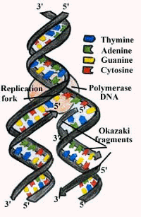
Replication process

Chromosomes & Their Structure:

  • The plans for making cells are coded in DNA
  • DNA, deoxyribose nucleic acid, is a long thin molecule that stores genetic information
  • DNA in a human cell is estimated to consist of six billion pairs of nucleotides
  • DNA is organized into giant molecules called chromosomes
  • Chromosomes are made of protein & a long, single, tightly-coiled DNA molecule visible only when the cell divides
  • When a cell is not dividing the DNA is less visible & is called chromatin
  • DNA in eukaryotic cells wraps tightly around proteins called histones to help pack the DNA during cell division
  • Nonhistone proteins help control the activity of specific DNA genes
  • Kinetochore proteins bind to centromere and attach chromosome to the spindle in mitosis
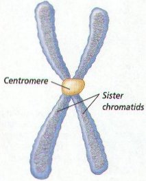
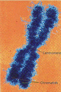
  • Centromeres hold duplicated chromosomes together before they are separated in mitosis
  • Telomeres are the ends of chromosomes which are important in cell aging
  • When DNA makes copies of itself before cell division, each half of the chromosome is called a sister chromatid
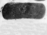
  • DNA of prokaryotes (bacteria) is one, circular chromosome attached to the inside of the cell membrane
Chromosome Numbers:

  • Humans somatic or body cells have 23 pairs of chromosomes or 46 chromosomes (diploid or 2n number)
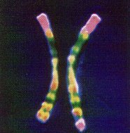
  • The 2 chromatids of a chromosome pair are called homologues (have genes for the same trait at the same location)

Homologs

  • Human reproductive cells or gametes (sperms & eggs) have one set or 23 chromosomes (haploid or n number)
  • Every organism has a specific chromosome number


Organism Chromosome Number (2n)
Human 46
Fruit fly 8
Lettuce 14
Goldfish 94


  • Fertilization, joining of the egg & sperm, restores the diploid chromosome number in the zygote (fertilized egg cell)
  • Sex chromosomes, either X or Y, determine the sex of the organism
  • Two X chromosomes, XX, will be female and XY will be male
  • All other chromosomes, except X & Y, are called autosomes
  • Chromosomes from a cell may be arranged in pairs by size starting with the longest pair and ending with the sex chromosomes to make a karyotype
  • A human karyotype has 22 pairs of autosomes and 1 pair of sex chromosomes (23 total)


Human Male Karyotype

Genes:

  • A section of DNA which codes for a protein is called a gene
  • Each gene codes for one protein
  • Humans have approximately 50,000 genes or 2000 per chromosome
  • About 95% of the DNA in chromosome is “junk” that does not code for any proteins

Cell Cycle:

  • Cells go through phases or a cell cycle during their life before they divide to form new cells 
  • The cell cycle includes 2 main parts — interphase, and cell division
  • Cell division includes mitosis (nuclear division) and cytokinesis (division of the cytoplasm)
  • Interphase is the longest part of a cell’s life cycle and is called the “resting stage” because the cell isn’t dividing
  • Cells grow, develop, & carry on all their normal metabolic functions during interphase
  • Interphase consists of 3 parts — G1, S, & G2phases

Interphase:

  • G1 or 1st Growth Phase occurs after a cell has undergone cell division
  • Cells mature & increase in size by making more cytoplasm & organelles while carrying normal metabolic activities in G1 
  • S or Synthesis Phase follows  G1  and the genetic material of the cell (DNA) is copied or replicated 
  • G2 or 2nd Growth Phase occurs after S Phase and the cell makes all the structures needed to divide

Cell division in Prokaryotes:

  • Prokaryotes such as bacteria do not have a nucleus
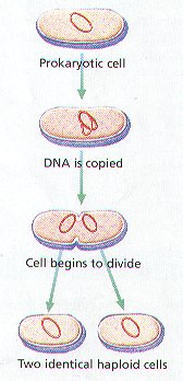
  • Prokaryotes divide into two identical new cells by the process of binary fission
  • Binary fission is an asexual method of reproduction
  • In binary fission,  the chromosome, attached to cell membrane, makes a copy of itself and the cell grows to about twice its normal size
  • Next, a cell wall forms between the chromosomes & the parent cell splits into 2 new identical daughter cells (clones)

Cell Division in Eukaryotes:

  • Eukaryotes have a nucleus & membrane-bound organelles which must be copied exactly so the 2 new cells formed from division will be exactly alike
  • The original parent cell & 2 new daughter cells must have identical chromosomes
  • DNA is copied in the S phase of the cell cycle & organelles, found in the cytoplasm,  are copied in the Growth phases
  • Both the nucleus (mitosis) and the cytoplasm (cytokinesis) must be divided during cell division in eukaryotes

Stages of Mitosis:

  • Division of the nucleus or mitosis occurs first
  • Mitosis is an asexual method of reproduction
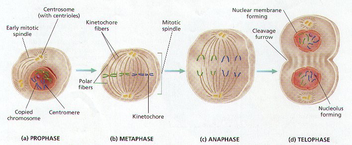
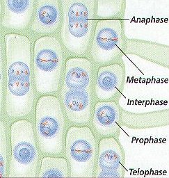
  • Mitosis consists of 4 stages — Prophase, Metaphase, anaphase, & Telophase
  • Prophase:
    • Chromosomes become visible when they condense into sister chromatids
    • Sister chromatids attach to each other by the centromere
    • Centrioles in animal cells move to opposite ends of cell
    • Spindle forms from centriole (animals) or microtubules (plants)
    • Kinetochore fibers of spindle attach to centromere
    • Polar fibers of spindle extend across cell from pole to pole
    • Nuclear membrane dissolves
    • Nucleolus disintegrates
  • Metaphase:
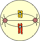
    • Chromosomes line up in center or equator of the cell attached to kinetochore fibers of the spindle
  • Anaphase:
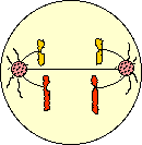
    • Kinetochore fibers attached to the centromere pull the sister chromatids apart
    • Chromosomes move toward opposite ends of cell
  • Telophase:
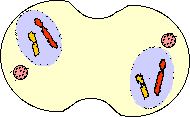
    • Nuclear membrane forms at each end of the cell around the chromosomes
    • Nucleolus reform
    • Chromosomes become less tightly coiled & appear as chromatin again
    • Cytokinesis begins

Cytokinesis:

  • Cytoplasm of the cell and its organelles separate into 2 new daughter cells
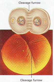
  • In animals, a groove called the cleavage furrow forms pinching the parent cell in two
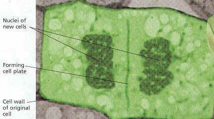
  • In plants, a cell plate forms down the middle of the cell where the new cell wall will be
Summary of Mitosis:
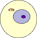


 Interphase


  1. Cell matures & carries on normal activities
  2. DNA copied & appears as chromatin
  3. Nucleolus visible
 Early Prophase  


  1. Chromosomes condense & become visible
  2. Centrioles separate & spindle starts forming
 
 Late Prophase

  1. Spindle forms with aster at each pole
  2. Nuclear membrane & nucleolus disintegrate
  3. Centromere of chromosomes attaches to spindle fibers
 Metaphase

  1. Chromosomes line up at the equator of the cell attached to kinetochore fibers of spindle
 Anaphase

  1. Centromeres split apart
  2. Homologs move to opposite poles of the cell
 Telophase/Cytokinesis  

  1. Nuclear membrane & nucleolus reform
  2. Cell pinches into 2 cells in animals
  3. In plants, a cell plate separates the 2 new cells


Cancer is Uncontrolled Mitosis:

  • Mitosis must be controlled, otherwise growth will occur without limit (cancer)
  • Control is by special proteins produced by oncogenes
  • Mutations in control proteins can cause cancer

Meiosis & Sexual Reproduction

  • Reduces the number of chromosomes in new cells to half the number in the original cell
  • New cells have a single copy of chromosomes (23 total) but are not identical to each other or the original parent cell
  • Used for making gametes ( sperm and eggs) with the haploid or n number
  • In meiosis, cells divide twice after a single DNA duplication
  • Meiosis I separates homologs & the Meiosis II separates sister chromatids
  • Meiosis I stages are Prophase I, Metaphase I, Anaphase I, & Telophase I
  • Meiosis II stages are Prophase II, Metaphase II, Anaphase II, & Telophase II
  • Produces 4 haploid cells or gametes
  • When a sperm fertilizes an egg to form a zygote, the diploid number of chromosomes is restored (23 + 23 = 46)
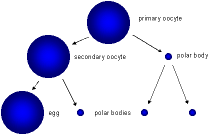
  • Egg cells or ova (ovum, singular) are larger , nonmotile cells
  • Gametoogenesis is meiosis producing eggs & occurs in the female’s ovaries


Oogenesis

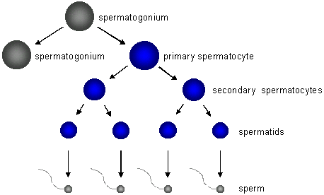
  • Sperms contain less cytoplasm so they’re smaller & have a flagellum to swim to the egg
  • Spermatogenesis is meiosis producing sperm cells & occurs in the testes


Spermatogenesis

Meiosis I:

  • The cell that undergoes Meiosis I is a primary spermatocyte or oocyte
  • Prophase I:
    • Chromosomes coil tightly & are visible
    • Nuclear membrane & nucleolus disintegrate
    • Spindle forms
    • Synapsis (joining) of homologous chromosomes occurs making tetrads
    • Kinetochore fiber forms on each chromosome
    • Chromosomes in tetrad exchange fragments by a process called crossing over

  • Metaphase I:
    • Tetrads become aligned in the center of the cell attached to spindle fibers
  • Anaphase I:
    • Homologous chromosomes separate
  • Telophase I:
    • May not occur in all species
    • Cytokinesis occurs producing 2 cells
    • In females,  2nd cell in females is called the 1st Polar Body
    • 1st Polar Body dies due to uneven splitting of the cytoplasm

  • Prophase II:
    • Cells called Secondary Spermatocytes or oocytes
    • DNA is not copied before cell divides
    • Chromatids attach to spindle fiber
  • Metaphase II:
    • Chromosomes become aligned in the center of the cell attached to spindle fibers
  • Anaphase II:
    • Sister chromatids separate randomly
    • Called independent assortment
  • Telophase I:
    • Cytokinesis occurs producing 4 cells in males called spermatids
    • Spermatids mature & form flagellum to become sperm
    • Cytokinesis in females produces a 2nd Polar Body that dies and an ootid 
    • Ootids mature to become ovum or egg

Asexual & Sexual reproduction:

  • Evolution is the slow process of change in living populations over time
  • Variations are differences that occur due to crossing-over among members of a sexually reproducing population
  • Variations are important to the survival of individuals in a population (some must survive to reproduce)
  • Asexually reproducing organisms rarely show variations because the organisms have identical genes

Cell Drawings HRWch4

Cell Drawings

Holt, Rinehart, Winston    Modern Biology

Draw on separate sheets of unlined paper, label drawing & each part, color, and tell the function of EACH LABELED PART (FUNCTION MUST BE WRITTEN NEXT TO THE LABEL) for the following cell drawings:

Page 72    Figure 4.4         Cell Shapes

Page 74    Figure 4.6         Animal Cell

Page 75    Figure 4.7         Bacterial cell (Prokaryote)

Page 76    Figure 4.9         Cell Organization

Page 77    Figure 4.10       Phospholipid

Page 78    Figure 4.11        Cell Membrane

Page 79    Figure 4.12        Nucleus & Nucleolus

Page 80    Figure 4.13        Mitochondria

Page 80    Figure 4.14        Ribosome

Page 81    Figure 4.15        Endoplasmic Reticulum

Page 82    Figure 4.16        Golgi

Page 84    Figure 4.18        Cytoskeleton

Page 85    Figure 4.19        Microtubule

Page 87    Figure 4.21        Plant Cell

Page 89    Figure 4.23        Chloroplast

When all drawings are complete — drawn, colored, labeled, and all functions written — then make a cover sheet with your name and a title and staple this to the top of your drawings. Number the pages in the lower right hand corner.

NO FUNCTIONS; NO GRADE!

Campbell Problem 6

Molecular Genetics Problem 6
6. A space probe discovers a planet inhabited by creatures who reproduce with the same hereditary patterns as those in humans. Three phenotypic characters are height (T = tall, t = dwarf), hearing appendages (A = antennae, a = no antennae), and nose morphology (S = upturned snout, s = downturned snout). Since the creatures were not “intelligent” Earth scientists were able to do some controlled breeding experiments, using various heterozygotes in testcrosses. For a tall heterozygote with antennae, the offspring were tall-antennae, 46; dwarf-antennae 7; dwarf-no antennae 42; tall-no antennae 5. For a heterozygote with antennae and an upturned snout, the offspring were antennae-upturned snout 47; antennae-downturned snout, 2; no antennae-downturned snout, 48: no antennae-upturned snout 3. Calculate the recombination frequencies for both experiments.

Experiment 1 (Frequency/Distance between T and A).

Determine the recombination frequency for the genes controlling Tallness and Antennae:

46 tall-antennae = 46% expected
42 dwarf-no antennae = 42% expected
7 dwarf-antennae = 7% recombinant
5 tall-no antennae = 5% recombinant

Total = 100

Therefore this recombination frequency between genes T and A is 12%

Experiment 2. (Frequency/Distance between A and S)

Determine the recombination frequency for the genes controlling Antennae and Snout:

47 antennae-upturned snout = 47% expected
48 no antennae-downturned snout = 48% expected
2 antennae-downturned snout = 2% recombinant
3 no antennae-upturned snout = 3% recombinant

Total = 100

Therefore this recombination frequency between genes A and S is 5%

BACK 

Cell Division Study Guide BI

Cell Division Study Guide

What molecule contains the information needed to direct all the activities of a cell?
Where in a cell are prokaryotic chromosomes found? eukaryotic chromosomes?
A human somatic cell contains how many homologous chromosomes?
How many chromosomes are in an human egg cell? sperm cell?
What is a karyotype?
Are gametes diploid or haploid?
Zygotes will have what chromosome number?
Does cell division in bacteria take place in the same way as it does in eukaryotes? Explain.
In what stage do cells spend most of their life cycle?
Is mitosis asexual or sexual reproduction?
A new nuclear envelope develops during cell division in what stage?
In what stage do chromatids separate from each other?
How does the number of chromosomes in newly divided cells compare with the number of chromosomes in the original cell?
During what type of cell division do haploid cells develop from diploid cells?
In order for DNA to fit into a cell, what must be done to compact it?
What is a centromere?
How many chromosomes are in a human skin cell? a human ovum?
Bacteria reproduce by a method known as _____________  ______________.
What is the shape of a bacterial chromosome?
Chromosomes are arranged along the equator of a cell during which stage of cell division?
Spindle fibers are made of ________________.
Be able to recognize sketches of the stages of mitosis.
What happens during cytokinesis in a plant cell?
Homologs separate during ________________.
BACK


Cell Exploration Webquest


CELL EXPLORATION WEBQUEST


INTRODUCTION

Every living thing is composed of at least one cell. Bacteria, amoebae, and paramecia are made of one cell and are capable of the activities of life. Organisms made of one cell are unicellular. Most living things are made of more than one cell and are called multicellular. Cells of these organisms function together to accomplish life activities. How many cells do you think make up your body? The human body is made of trillions of cells.

In order to understand how the cell functions in your body, we have to take a look at how your body is organized. Since you are made of matter, and all matter is made of atoms, your body is a collection of atoms. These atoms combined in specific ways to form molecules. Some of the important molecules in your body are proteins, carbohydrates, lipids, salts, water, and nucleic acids. These molecules combined to form the structures that make up a cell. Since each cell is capable of the activities of life, it is the smallest unit of life.

Cells that are similar can function together. These collections of cells are called tissues. Some tissues that you may be familiar with are the muscle tissue that makes up your heart, epithelial tissue that makes up your skin, and connective tissue that holds your body together. Different groups of tissues can be arranged to form organs. Some organs that you may be familiar with are the stomach, intestines, heart, and lungs. For example, the stomach has epithelium to line the outside and inside surfaces for protection and the muscle tissue allows your stomach to squeeze and churn. Groups of organs can work together as an organ system to perform a specific function. The digestive system functions to breakdown and absorb food so that our bodies can use the energy. The pancreas, stomach, intestines, gall bladder, and esophagus are some of the organs that make up the digestive system. There are 13 systems in the human body that function together to produce an organism – YOU!

To review:

ATOMS ——> MOLECULES ——-> CELLS ——-> TISSUES ——-> ORGANS ——> SYSTEMS ——> ORGANISM
The focus of this activity is to learn more about the cell and how it functions in your body.

CELL SIZE

Cells are very small and you must use a microscope to look at them. Watch this video (click on “start animation”), then look at the size of cells and answer the following questions. To give you an idea about size, the length of a key on the keyboard is about 1 cm.

Question:
Answer:
A. Is a bacterium larger or smaller than an animal cell?
B. How many bacteria can fit into an animal cell?
C. Are plant cells larger or smaller than animal cells?


EUKARYOTIC CELL ORGANELLES

Since the cell is the fundamental unit of life, it must be capable of independent existence. Some of the necessary life activities are communication, metabolism, protection, and waste disposal. In order to carry out these jobs, the cell has different organs inside of it just like your body has organs. These “tiny organs” are called organelles. Different organs have different jobs and they need the proper supplies of ATP (cellular energy), proteins, oxygen, and other nutrients to carry out their jobs.
There are different types of cells that have different functions, but all cells have some common features. The things common to all cells are a cell membrane (plasma membrane), cytoplasm, and organelles. Take a look at a drawing of an animal cell. (Hold cursor over organelle to identify it.)

To understand how the cell carries out its functions, you should know more about the cytoplasm, cell membrane, and organelles. Click on each structure given in the table below to learn more about each cell part. Complete the table by writing a brief description and function for each part.

The things common to all cells are a cell membrane (plasma membrane), cytoplasm, and organelles. Remember that plant cells have three structures that animal cells don’t.  Now look at a drawing of a plant cell.  (Hold cursor over organelle to identify it.)

COMPLETE THE TABLE BELOW:

Structure
Description
Function
 CHLOROPLAST
 CELL WALL
CENTRAL VACUOLE 

 
PROKARYOTIC CELLS


        Remember that prokaryotic cells are only found in bacteria!  They’re simpler than eukaryotic cells.  Look at the bacterial cell, and complete the table below:

Structure
Description
Function

After you have read about  cells, take the cell quiz. Check your answer after you answer each question.


BACK